Patient-Derived Xenografts and Organoids of Prostate Cancer: History
Please note this is an old version of this entry, which may differ significantly from the current revision.
Contributor: , , , , , , ,

Several models of prostate cancer (PCa) have been developed, each with their unique applications, advantages, and disadvantages. The shortage of clinically relevant, in vivo models is particularly a large barrier to comprehending the tumor progression observed in PCa. Patient-derived xenografts (PDXs) are models that are derived from biopsy specimens and metastatic lesions from human patients, and allow researchers to understand in vivo physiology as well as tumor heterogeneity. Despite the clinical utility of PDXs, they are also met with limited availability, higher cost, and advanced technical expertise required for use. Organoids, or "mini organs", are clusters of cells grown in vitro that self-organize and differentiate into functional cell types. Organoids referred to as patient-derived organoids (PDOs) can be derived from primary tissue materials, and have also been demonstrated to be derived from PDXs themselves. Due to their lower cost and ease of use, they work well for molecular and mechanistic studies, while still maintaining appropriate tumor heterogeneity and disease modeling. 

  • prostate cancer
  • knockout mouse models
  • genetically-engineered mouse models
  • xenografts
  • PDXs
  • organoids

1. Introduction

Historical Timeline of PCa Models
In the United States, prostate cancer is estimated to be the most commonly diagnosed cancer in men in 2022 [1]. The level of genetic information people have obtained over past decades of research has guided the development of models that attempt to study PCa. When cancer cell lines were first developed in the late 1940s through the 1970s, information about their genetic makeup or mutations present in the malignant tumor cells was limited. Cancer cells were simply taken from either the primary tumor or metastasis and were grown in vitro, allowing researchers to obtain ‘immortal’ cells by passaging them over time. The first cell line, or the L929 cell line, was established by Dr. Wilton R. Earle in 1948 and was derived from fibroblasts in subcutaneous mouse tissue [2]. The first immortalized human cell line, the HeLa cell line developed by Dr. George O. Gay in 1951, used cells taken from epithelial cervix tissue from Henrietta Lacks [3]. Many more cell lines soon followed throughout the 1950s and 1960s that were taken from hamsters, canines, monkeys, and humans [4]. Important PCa cell lines—LNCaP cells that were taken from a lymph node metastasis [5], DU145 cells that were taken from a central nervous system metastasis [6], and PC3 cells that were taken from a bone metastasis [7]—were developed in 1977, 1978, and 1979, respectively. Despite the cancer researcher’s ability to finally grow malignant, immortalized cells in vitro, little was known about the cells’ gene expression or the mutations that promoted their tumorigenicity.
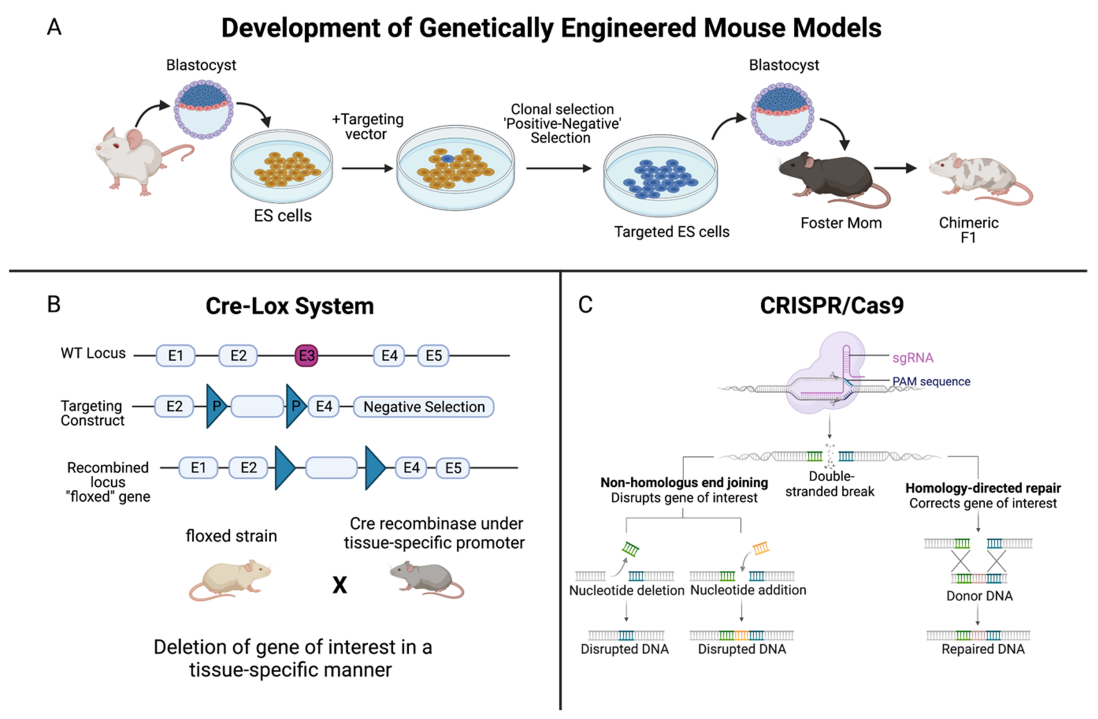
PCa models, as well as the understanding of several aspects of mammalian physiology, were significantly enhanced thanks to the research by Mario R. Capecchi, Sir Martin J. Evans, and Oliver Smithies in the late 1980s. In 2007, these scientists were jointly awarded The Nobel Prize in Physiology or Medicine for discovering how to introduce specific gene modifications in mice using embryonic stem cells [8]. Through homologous recombination between introduced DNA and endogenous DNA in embryonic stem cells, pure populations of cells carrying the target gene could be grown and injected into blastocysts [8]. The injected blastocysts are implanted into a surrogate mother where they develop into mosaic embryos, and then mosaic and normal mice mate to produce both gene targeted and normal offspring [8]. This powerful technique, known as gene targeting in mice, is often used to inactivate, or knock out, specific genes and thus elucidate the function of those genes (Figure 1A) [8]. The current knowledge of PCa is owed to the numerous mouse knockout models made available through gene targeting.
Figure 1. Summary of the development of genetically-engineered mouse models. (A) Genetically-engineered (GE) mice are used extensively in research as models of human disease. In the context of cancer, by deleting, or “knocking out”, the function of a specific gene, one can determine the significance of that gene in either promoting or halting the progression of tumor development. The method to develop GE models begins with extracting blastocysts from the mouse, and culturing embryonic stem (ES) cells. A targeting vector, which is a DNA construct containing DNA sequences homologous to the gene of interest, is added. ES cells that successfully recombine with the genomic DNA are selected for, and these targeted ES cells are injected into the mouse blastocysts. (B) The Cre-Lox system is used to control site specific recombination events in genomic DNA, allowing one to control expression of a specific gene or to delete undesired sequences. Cre recombinase is an enzyme that is derived from the bacteriophage P1, and it catalyzes a site-specific recombination event between two DNA recognition sites known as LoxP sites [9]. The result is a recombined locus in which the gene of interest is deleted, creating a “floxed” gene. (C) In the CRISPR/Cas9 system, a foreign single guide RNA (sgRNA) seeks, matches, and binds a specific DNA sequence, and the nuclease Cas9 cuts the sequence at a precise binding site near a protospacer adjacent motif (PAM) sequence [10]. The double-stranded DNA break induced by Cas9 is repaired by the cell’s DNA repair machinery. One method is via non-homologous end joining, either by deleting or adding a nucleotide, which does not require a template but disrupts the gene of interest. Or, homology-directed repair can be performed, which corrects the gene of interest, but a single stranded (ss) DNA template is required, which is provided by the CRISPR/Cas9 system [10]. Created with BioRender.com.
Critical for the development of conditional knockout models is the site-specific recombinase technology known as Cre-Lox recombination (Figure 1B). This technology allows DNA modification—deletions, insertions, translocations, and inversions—at specific sites to be targeted to a particular cell type, or to be triggered by a particular external stimulus [9]. Derived from the bacteriophage P1, Cre recombinase is an enzyme that catalyzes a site-specific recombination event between two DNA recognition sites known as LoxP sites [9]. In 1994, the laboratories of Dr. Jamey Marth and Dr. Klaus Rajewsky reported that this system could be used for conditional gene targeting [9]. Since then, Cre-Lox recombination has been widely used to manipulate genes and chromosomes, creating genetic knock-out or knock-in mouse models [9]. In addition, the use of CRISPR (clustered regularly interspaced short palindromic repeats)/Cas 9 genome editing has further strengthened the understanding of the role of genes in cancer (Figure 1C). This technology can be designed to introduce RNA-directed, double strand DNA breaks or single strand ‘nicks’ using a mutated form of Cas9, a DNA endonuclease [10]. CRISPR is a simple and effective mechanism to introduce mutations both in vitro and in vivo that mimic somatic mutations, revolutionizing mouse models of cancer [10].
Thanks to DNA sequencing and gene expression data in the 21st century, PCa research models and the understanding of PCa as a whole have expanded tremendously. In 2002, The International Mouse Genome Sequencing Consortium, which was part of the Human Genome Project, achieved a high-quality draft sequence of the mouse genome [11]. Drafts for the Human Genome Project were already established a couple of years prior to the draft of the mouse genome, and the final sequence mapping of the human genome was finished in 2003 [11]. Although mice and humans are separated by 75 million years of evolution, their genomes are about 90% similar and both share almost 30 thousand genes [11]. Thanks to their similarities, the field known as comparative genomics can explore how mouse genes and their human counterparts contribute to health and disease.

2. Patient-Derived Xenografts and Organoids of PCa

The shortage of clinically relevant, in vivo models is a large barrier to the understanding of tumor progression seen in PCa. Generating models that are derived from biopsy specimens and metastatic lesions from human patients is one means of mitigating this dilemma. One form of patient-derived models is xenografts (PDXs), which allow researchers to study and appreciate the tumor heterogeneity of prostatic diseases. It has been shown that populations of PDX mice upon passaging preserve most of the genetics of the original human tumors [12]. Extensive copy number alterations between human tumors and those in the PDX models were not found [12]. One type of PDX model, LuCaP PDXs, which harbor genetic alterations such as androgen receptor amplification, PTEN deletion, and TP53 deletion, demonstrate molecular heterogeneity in response to androgen deprivation therapy (ADT) and docetaxel treatment [13]. In PDXs studying hormone-naive prostate cancer, the Growth Factor Receptor Bound Protein 10 (GRB10) gene was found to be the most significantly upregulated, showing increased expression during and before the development of castration resistant prostate cancer [14]. Furthermore, PDXs were also established that are based on subrenal capsule grafting of patients’ tumor tissue into nonobese diabetic/severe combined immunodeficient (NOD/SCID) mice [15]. These PDXs preserve crucial properties of the original malignancies, including histopathological and molecular characteristics, reactions to androgen suppression, and tumor heterogeneity [15]. Consequently, PDXs represent valuable models not only for comprehending prostate tumor progression but also establishing drug screenings and therapies [15].
Despite their advantages, xenograft models are costly, time consuming, and require the use of immunocompromised mice. Three-dimensional (3D) patient derived organoids (PDOs) are perhaps a more efficient alternative—one that still maintains tumor heterogeneity and appropriate disease modeling. Organoids, or “mini-organs”, are clusters of cells grown in vitro that self-organize and differentiate into functional cell types [16]. Like PDXs, PDOs can be derived from primary tissue materials such as needle biopsies [16], but it has also been demonstrated that they can be derived from PDXs themselves [17]. These PDOs can conserve the genotypic and phenotypic characteristics of the LuCaP PDX cells they are derived from and represent an effective model to study mechanisms of resistance to ADT in PCa [17]. Organoids can also be derived from stem cells, including those expressing Lgr5, which is a genetic marker of Wnt-dependent stem cells found in tissues including the prostate [18]. In addition, organoids have also been used to study uncommon variants of PCa, including neuroendocrine prostate cancer (NEPC) derived from four human patients [19]. In these PDOs, the epigenetic modifier histone methyltransferase enhancer of zeste 2 (EZH2) was found to show increased expression in NEPC compared to adenocarcinomas [19]. Overall, PDOs share a similar mutational landscape with PCa, and they recapitulate in situ histology both in vitro and in vivo [20].

This entry is adapted from the peer-reviewed paper 10.3390/cancers14215321

References

  1. Siegel, R.L.; Miller, K.D.; Fuchs, H.E.; Jemal, A. Cancer statistics, 2022. CA Cancer J. Clin. 2022, 72, 7–33.
  2. Sanford, K.K.; Hobbs, G.L.; Earle, W.R. The tumor-producing capacity of strain L mouse cells after 10 years in vitro. Cancer Res. 1956, 16, 162–166.
  3. Scherer, W.F.; Syverton, J.T.; Gey, G.O. Studies on the propagation in vitro of poliomyelitis viruses. IV. Viral multiplication in a stable strain of human malignant epithelial cells (strain HeLa) derived from an epidermoid carcinoma of the cervix. J. Exp. Med. 1953, 97, 695–710.
  4. Jedrzejczak-Silicka, M. History of Cell Culture. In New Insights into Cell Culture Technology; Gowder, S.J.T., Ed.; IntechOpen: London, UK, 2017.
  5. Horoszewicz, J.S.; Leong, S.S.; Kawinski, E.; Karr, J.P.; Rosenthal, H.; Chu, T.M.; Mirand, E.A.; Murphy, G.P. LNCaP model of human prostatic carcinoma. Cancer Res. 1983, 43, 1809–1818.
  6. Stone, K.R.; Mickey, D.D.; Wunderli, H.; Mickey, G.H.; Paulson, D.F. Isolation of a human prostate carcinoma cell line (DU 145). Int. J. Cancer. 1978, 21, 274–281.
  7. Kaighn, M.E.; Narayan, K.S.; Ohnuki, Y.; Lechner, J.F.; Jones, L.W. Establishment and characterization of a human prostatic carcinoma cell line (PC-3). Investig. Urol. 1979, 17, 16–23.
  8. Press Release. Nobel Prize Outreach AB. 2022. Available online: https://www.nobelprize.org/prizes/medicine/2007/press-release/ (accessed on 25 October 2022).
  9. Nagy, A. Cre recombinase: The universal reagent for genome tailoring. Genesis 2000, 26, 99–109.
  10. Dow, L.E.; Fisher, J.; O’Rourke, K.P.; Muley, A.; Kastenhuber, E.R.; Livshits, G.; Tschaharganeh, D.F.; Socci, N.D.; Lowe, S.W. Inducible in vivo genome editing with CRISPR-Cas9. Nat. Biotechnol. 2015, 33, 390–394.
  11. Waterston, R.H.; Lindblad-Toh, K.; Birney, E.; Rogers, J.; Abril, J.F.; Agarwal, P.; Agarwala, R.; Ainscough, R.; Alexandersson, M.; An, P.; et al. Initial sequencing and comparative analysis of the mouse genome. Nature 2002, 420, 520–562.
  12. Woo, X.Y.; Giordano, J.; Srivastava, A.; Zhao, Z.M.; Lloyd, M.W.; de Bruijn, R.; Suh, Y.S.; Patidar, R.; Chen, L.; Scherer, S.; et al. Conservation of copy number profiles during engraftment and passaging of patient-derived cancer xenografts. Nat. Genet. 2021, 53, 86–99.
  13. Nguyen, H.M.; Vessella, R.L.; Morrissey, C.; Brown, L.G.; Coleman, I.M.; Higano, C.S.; Mostaghel, E.A.; Zhang, X.; True, L.D.; Lam, H.M.; et al. LuCaP Prostate Cancer Patient-Derived Xenografts Reflect the Molecular Heterogeneity of Advanced Disease and Serve as Models for Evaluating Cancer Therapeutics. Prostate 2017, 77, 654–671.
  14. Hao, J.; Ci, X.; Xue, H.; Wu, R.; Dong, X.; Choi, S.Y.C.; He, H.; Wang, Y.; Zhang, F.; Qu, S.; et al. Patient-derived Hormone-naive Prostate Cancer Xenograft Models Reveal Growth Factor Receptor Bound Protein 10 as an Androgen Receptor-repressed Gene Driving the Development of Castration-resistant Prostate Cancer. Eur. Urol. 2018, 73, 949–960.
  15. Lin, D.; Xue, H.; Wang, Y.; Wu, R.; Watahiki, A.; Dong, X.; Cheng, H.; Wyatt, A.W.; Collins, C.C.; Gout, P.W.; et al. Next generation patient-derived prostate cancer xenograft models. Asian J. Androl. 2014, 16, 407–412.
  16. Corro, C.; Novellasdemunt, L.; Li, V.S.W. A brief history of organoids. Am. J. Physiol. Cell Physiol. 2020, 319, C151–C165.
  17. Beshiri, M.L.; Tice, C.M.; Tran, C.; Nguyen, H.M.; Sowalsky, A.G.; Agarwal, S.; Jansson, K.H.; Yang, Q.; McGowen, K.M.; Yin, J.; et al. A PDX/Organoid Biobank of Advanced Prostate Cancers Captures Genomic and Phenotypic Heterogeneity for Disease Modeling and Therapeutic Screening. Clin. Cancer Res. 2018, 24, 4332–4345.
  18. Karthaus, W.R.; Iaquinta, P.J.; Drost, J.; Gracanin, A.; van Boxtel, R.; Wongvipat, J.; Dowling, C.M.; Gao, D.; Begthel, H.; Sachs, N.; et al. Identification of multipotent luminal progenitor cells in human prostate organoid cultures. Cell 2014, 159, 163–175.
  19. Puca, L.; Bareja, R.; Prandi, D.; Shaw, R.; Benelli, M.; Karthaus, W.R.; Hess, J.; Sigouros, M.; Donoghue, A.; Kossai, M.; et al. Patient derived organoids to model rare prostate cancer phenotypes. Nat. Commun. 2018, 9, 2404.
  20. Gao, D.; Vela, I.; Sboner, A.; Iaquinta, P.J.; Karthaus, W.R.; Gopalan, A.; Dowling, C.; Wanjala, J.N.; Undvall, E.A.; Arora, V.K.; et al. Organoid cultures derived from patients with advanced prostate cancer. Cell 2014, 159, 176–187.
More
This entry is offline, you can click here to edit this entry!
Video Production Service