Green Synthesis of Platinum Nanoparticles Utilizing Plant Extracts: History
Please note this is an old version of this entry, which may differ significantly from the current revision.

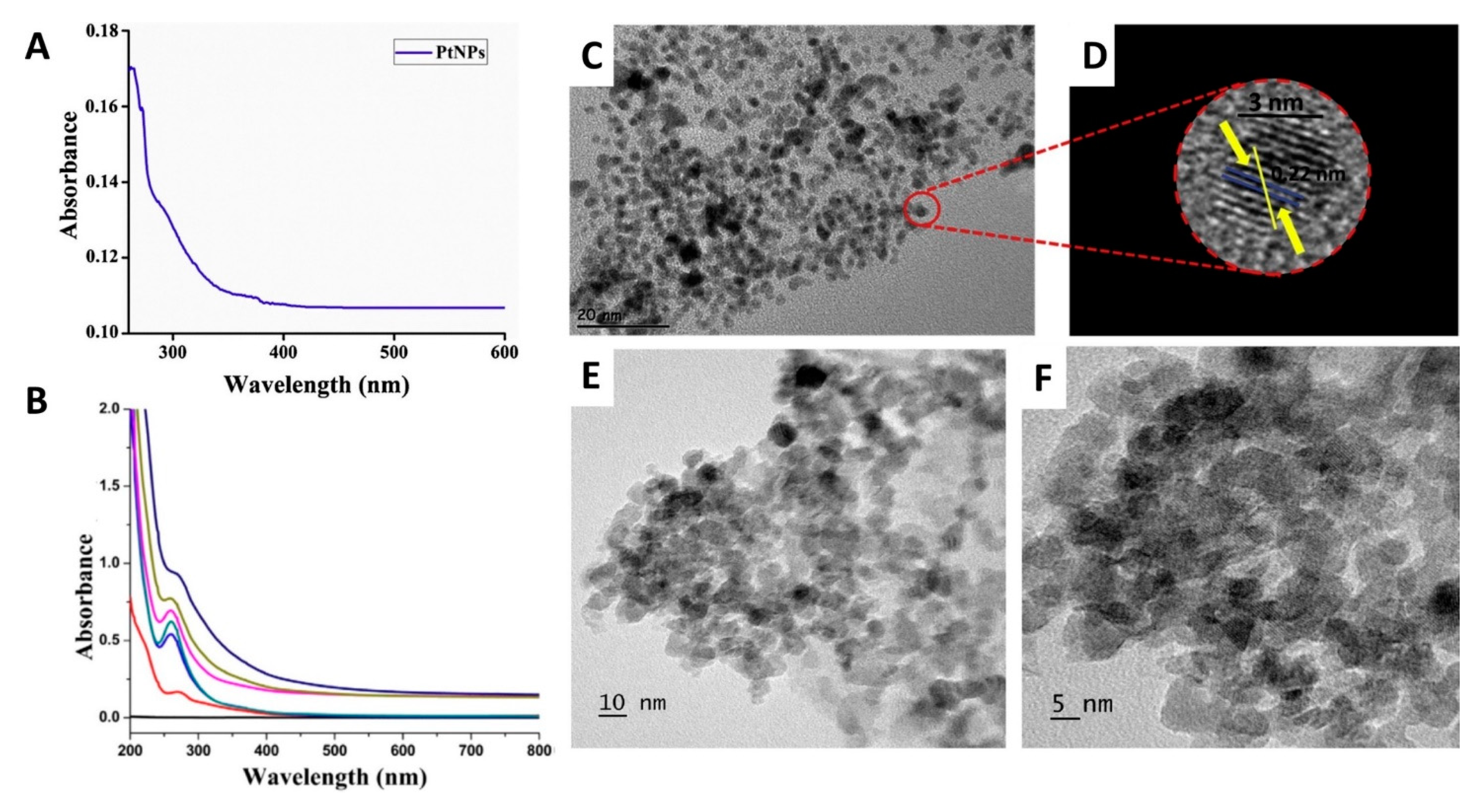
Platinum nanoparticles (PtNPs) have superior physicochemical properties and great potential in biomedical applications. Eco-friendly and economic approaches for the synthesis of PtNPs have been developed to overcome the shortcomings of the traditional physical and chemical methods. Various biogenic entities have been utilized in the green synthesis of PtNPs, including mainly plant extracts, algae, fungi bacteria, and their biomedical effects were assessed. Other biological derivatives have been used in the synthesis of PtNPs such as egg yolk, sheep milk, honey, and bovine serum albumin protein. The green approaches for the synthesis of PtNPs have reduced the reaction time, the energy required, and offered ambient conditions of fabrication. Plant extracts contain diverse primary and secondary metabolites, which could serve as natural reducing and capping agents. The plant-mediated biosynthesis of MNPs is a simple and rapid process involving mixing the plant extract with the metal ions solution at an optimized temperature and pH. The nanoparticle generation is indicated by the change in color of the reaction medium.

  • green synthesis
  • biosynthesis
  • platinum nanoparticles
  • anticancer

1. Green Synthesis of PtNPs Using Plant Extracts

Plant extracts have been considered a green route and a reliable approach for safe, eco-friendly, and biocompatible PtNPs. The phytosynthesis of PtNPs has been used to replace the current multi-step hazardous synthetic methods. Few studies reported the use of green approaches involving plants and plant extracts. In one study, Azadirachta indica leaf broth was incubated with Pt(IV) ions for 1 h at 100 °C. The terpenoids of the Azadirachta indica leaf acted as the reducing and capping agents. The generated PtNPs were sonicated for 30 min to enhance the monodispersity of the NPs [1] (Table 1). A recent study reported on the phytosynthesis of PtNPs using Nigella sativa (black cumin) seed extract. Pt(IV) ions were stirred with the black cumin extract for two days at 200 rpm and 75 °C [2]. Kumar et al. created a single and simple step for the biosynthesis of PtNPs, employing the fruit extract of Terminalia chebula. Many researchers have considered using Terminalia chebula in the biosynthesis of MNPs due to its abundance in nature and polyphenolic content. The reaction temperature was maintained at 100 °C for 10 min. The reduction of Pt(IV) ions was mediated by the polyphenols present in the fruit extract [3].
Table 1. Green synthesis of PtNPs using plant extracts.
Alshatwi et al. reported on the biosynthesis of PtNPs using tea polyphenols that act as natural reducing agents. Besides, their potential to form chelating complexes with various metal ions allowed them to be used as effective capping agents. The tea polyphenols were mixed with Pt(IV) in a ratio of 1:5 via magnetic stirring and then incubated for 1 h at room temperature [4].
Vinod et al. obtained PtNPs Cochlospermum gossypium gum mixed with the metal ions solution at 120 °C using an autoclave (15 psi) and pH 8. This is the only study that involved autoclave use to achieve the rapid biosynthesis of PtNPs. Heating and adjusting the pH were involved in activating the glucose, a mild reducing agent, present in the gum extract to achieve controllable bioreduction kinetics [11]. Ghosh et al. employed the tuber extract of Dioscorea bulbifera in biosynthesizing PtNPs by conducting the bio-reaction at 100 °C for 5 h [12]. A recent study by Anyik et al. reported the synthesis of PtNPs using the leaf extract of Eichhornia crassipes (Water hyacinth). The reaction was carried out at 100 °C for 1 h [19]. In a recent study, PtNPs were biosynthesized by mixing green tea powder extract with Pt (II) ions for 4 h using a magnetic stirrer at 50 °C. The flavonoids present in green tea played a significant role in the bioreduction of Pt(IV) ions owing to their hydroxyl groups [13].
Dobrucka et al. reported on the green synthesis of PtNPs employing Ononis spinosa radix extract. Patient ions’ reaction was maintained at 80 °C for 10 h with continuous stirring [14]. Ullah et al. reported the green synthesis of PtNPs utilizing leaf extract of Maytenus royleanus. The flavonoids and phenolic compounds present in the leaf extract are responsible for reducing Pt(IV) ions into PtNPs. The reaction temperature was maintained at 90 °C for 3 h with continuous stirring [15].
Yang et al. used the leaf extract of Mentha piperita (Peppermint) in biosynthesizing spherical PtNPs by conducting the bio-reduction at 60 °C for 90 min [16]. Tahir et al. developed a facile method for the biosynthesis of spherical PtNPs, employing the plant extract of Taraxacum laevigatum. Bioreduction was carried out at 90 °C for 10 min [17]. Finally, eco-friendly PtNPs were fabricated using gum extract of Prunus x yedoensis. The reaction conditions were optimized at pH 8 and gum extract concentrations of 7% and 8% for 30 min [18].

2. Characterization and Biological Activities of PtNPs Prepared Using Plant Extracts

Various analytical tools are utilized in the characterization of PtNPs such as Fourier transform infrared spectroscopy (FT-IR), X-ray diffraction (XRD), transmission electron microscopy (TEM), scanning electron microscopy (SEM), dynamic lights scattering (DLS), and thermal gravimetric analysis (TGA) [1][2][3][4][5][6][7][8][9][10][11][12][13][14][15][16][17][18][19][20][21][22][23][24]. However, dynamic light scattering (DLS), UV/Vis spectrophotometry, and TEM can be used for facile confirmation of the formation of PtNPs (Figure 1).
Figure 1. Characterization of PtNPs phytosynthesized utilizing plant extracts using UV/Vis spectrophotometry and TEM. (A,B) are the UV/Vis spectra of the biosynthesized PtNPs using Nigella sativa and Mentha piperita, respectively. (C,D) are the TEM and the HR-TEM images of the biosynthesized PtNPs using Nigella sativa. (E,F) are the TEM images of the biosynthesized PtNPs using Mentha piperita at a scale of 10 and 5 nm, respectively [2][16].
The formation of Azadirachta indica mediated PtNPs was confirmed qualitatively by converting the initial light-yellow color into black, and quantitatively by UV/Vis spectrophotometry. A strong surface Plasmon resonance (SPR) absorption band at 241 nm indicated the formation of the reduced PtNPs. TEM investigations indicated the production of small to large spherical nanoparticles in a size range of 5–50 nm [1].
The PtNPs generated using Nigella sativa (black cumin) seed extract were shown to have a spherical shape and an average size of 3.47 nm. Additionally, the formation of PtNPs was confirmed by the appearance of a significant SPR peak at 263 nm (Figure 1A,C,D). The biosynthesized PtNPs were found to have significant anticancer activity against human breast (MDA-MB-231) and cervical (HeLa) cancer cells (IC50: 36.86 µg/mL and 19.83 µg/mL; respectively). In addition, the produced NPs showed a pronounced bactericidal activity against Gram-negative and Gram-positive bacteria at concentrations of 100 µg and 500 µg/mL [2].
The Terminalia chebula mediated PtNPs were spherical, having an average size of < 4 nm. Besides, the disappearance of the UV/Vis absorption band at 262 nm corresponding to Pt(IV) ions suggested the reduction of Pt(IV) ions [3].
PtNPs prepared using tea polyphenols had flower-shaped morphology and an average size of 30 to 60 nm. In addition, the biosynthesized PtNPs had potent cytotoxic activity, attributed to induction of apoptosis, against cervical human cancer cells (SiHa). The IC50 and IC75 were 18.34 µg/mL and 11.4 µg/mL, respectively [4].
Two studies reported the green synthesis of PtNPs using Ocimum sanctum (Tulsi) leaves extract yielding irregular structured NPs with sizes 23 and 2 nm, respectively [5][6]. PtNPs were characterized by FT-IR, XRD, energy dispersive absorption X-Ray, SEM, and TEM. Additionally, the UV/Vis spectrophotometry was involved as an extra tool to confirm the production of PtNPs. An absorption band was observed at 400 nm, which indicated the formation of PtNPs [6].
The phytosynthesis of monodispersed spherical and cubical PtNPs with an average size of 20 nm was done using ethanolic extract of (Punica granatum) pomegranate crusts. The biosynthesized PtNPs were found to have significant cytotoxicity against MCF-7 cancer cells with an IC50 of 17.84 µg/mL after 48 h of incubation [7].
PtNPs biosynthesized utilizing the leaf extract of Diopyros kaki (Persimmon) had a size range of 2–12 nm. Furthermore, a UV/Vis absorption band was observed at 477 nm, which increased with increasing the Pt concentration, indicating the generation of PtNPs [8]. PtNPs prepared using Anacardium occidentale (Cashew) showed irregular rod-shaped morphology. The synthesis of PtNPs was confirmed by the disappearance of the UV/Vis absorption band at 259 nm, corresponding to Pt(IV) ions, and the observation of a continuum at 200 nm suggested the reduction of Pt(IV) ions and the formation of PtNPs. The UV/Vis data showed that the absorbance increases by increasing the quantity of dried leaf powder and decreasing pH [9].
Spherical PtNPs, 5–20 nm, were prepared using the leaf extract of Bacopa monnieri (Water hyssop). The formation of the PtNPs was confirmed by the appearance of a UV/Vis absorbance band at 330–380 nm. The biosynthesized nanoparticles were found to have pronounced antioxidant and neuroprotective activities, making them promising candidates for the potential treatment of Parkinson’s disease [10].
Vinod et al. obtained spherical PtNPs of 2.4 nm size using Cochlospermum gossypium gum [11]. Ghosh et al. used the tuber extract of Dioscorea bulbifera in biosynthesizing spherical PtNPs (2–5 nm). The generated nanoparticles exhibited anticancer activity against human cervical (HeLa) cancer cells. Besides, PtNPs showed antioxidant and pronounced free radical scavenging activity when tested using 2,2-diphenyl-1-picrylhydrazyl, superoxide, nitric oxide, and hydroxyl radicals [12].
The DLS and TEM sizes of the Eichhornia crassipes mediated PtNPs were 73.3 and 3.74 nm, respectively, with spherical shapes (it is of note that TEM measures the actual diameter of the PtNPs while DLS measures the diameter in the hydrated state). The disappearance of the UV/Vis absorption band observed at 261 nm corresponding to Pt(IV) ions and the observation of a continuum at 200–300 nm suggested the generation of PtNPs [19]. In another study, the photothermal properties of green tea mediated PtNPs were exploited in cancer therapy. The synthesized PtNPs were found to have a spherical morphology and an average size of 2 nm. The disappearance of the bio-reducer’s absorption band at 320 nm suggested the involvement of the bio-reducer, present in green tea, in reduction of Pt(IV) ions into Pt0.
Additionally, the appearance of UV bands in the range of 270–280 suggested the formation of PtNPs. Two human colon cancer cell lines (SW480 and SW620) were used to investigate the anticancer activity of the biosynthesized PtNPs combined with photothermal treatment for 5 min using low-intensity lasers operating at 650 and 808 nm. High cytotoxicity was observed for the biosynthesized PtNPs combined with laser irradiation. The % viability of cancer cells cultured with PtNPs and irradiated with 650 nm laser was found to be 21% and 18% for SW480 and SW620, respectively. While the % viability for cancer cells cultured with PtNPs and irradiated with 808 nm laser was 25% and 22% for SW480 and SW620, respectively. These findings may support future applications of biosynthesized PtNPs in photothermal cancer therapy [13].
Dobrucka et al. reported the green synthesis of spherical and hexagonal PtNPs (4 nm) employing Ononis spinosa radix extract. The anticancer activity of the biosynthesized PtNPs was evaluated against A549 cancer cell lines. The maximum cell mortality of 8.8% was observed at an incubation time of 72 h and a concentration of 100 µg/mL of PtNPs [14]. Ullah et al. reported the synthesis of spherical PtNPs of size 5 nm utilizing leaf extract of Maytenus royleanus. The appearance of the UV/Vis absorption SPR band at 282 nm suggested the generation of PtNPs. The cytotoxic activity of the prepared PtNPs was evaluated against the A549 cancer cell line. Exposure of A549 cancer cells to increasing concentrations of PtNPs for 24 h has resulted in reduced cell viability, damage to cellular morphology, and reduction in cell number in a dose-dependent manner [15]. Additionally, the PtNPs were found to be biocompatible with normal cells. These findings support the possible development of potent and selective anticancer drugs at low cost [15].
Yang et al. used the leaf extract of Mentha piperita (Peppermint) in biosynthesizing spherical PtNPs (54 nm) with surface charge of −50.1 mV, indicating high stability. The appearance of a UV/Vis absorption band at 272 nm indicated the formation of the PtNPs (Figure 1B,E,F). The anticancer activity of the biosynthesized PtNPs was tested against the human colon cancer cell line (HCT116). PtNPs reduced tumor cells’ viability at lower concentrations with an IC50 value of 20 µg/mL [16]. The PtNPs created by Tahir et al. employing the plant extract of Taraxacum laevigatum were spherical in size range of 2–7 nm. The UV/Vis absorption band’s appearance at 283 nm suggested the formation of PtNPs [17]. The bactericidal activity of the greenly synthesized PtNPs was evaluated against Gram-positive bacteria (Bacillus subtilis) and Gram-negative bacteria (Pseudomonas aeruginosa). The findings revealed that the PtNPs exhibited significant antibacterial activity against both strains (zones of inhibitions were 15 (± 0.5) mm and 18 (± 0.8) mm for P. aeruginosa and B. subtilis, respectively), making them promising antibiotics that could overcome bacterial resistance [17]. Finally, spherical PtNPs (10–50 nm) were fabricated using gum extract of Prunus x yedoensis. The appearance of the UV/Vis absorption band at 277 nm suggested the creation of the PtNPs. The antifungal activity of the biosynthesized PtNPs was investigated against Colletotrichum acutatum and Cladosporium fulvum exhibiting 15 mm and 18 mm zones of inhibition at concentrations of 4 and 8 µg/well, respectively [18].

This entry is adapted from the peer-reviewed paper 10.3390/molecules25214981

References

  1. Thirumurugan, A.; Aswitha, P.; Kiruthika, C.; Nagarajan, S.; Christy, A.N. Green synthesis of platinum nanoparticles using Azadirachta indica—An eco-friendly approach. Mater. Lett. 2016, 170, 175–178.
  2. Ayguna, A.; Gülbagcaa, F.; Ozerb, L.Y.; Ustaogluc, B.; Altunogluc, Y.C.; Balogluc, M.C.; Atalard, M.N.; Almae, M.H.; Sena, F. Biogenic platinum nanoparticles using black cumin seed and their potential usage as antimicrobial and anticancer agent. J. Pharm. Biomed. Anal. 2020, 179, 112961.
  3. Kumar, K.M.; Mandal, B.K.; Tammina, S.K. Green synthesis of nano platinum using naturally occurring polyphenols. RSC Adv. 2013, 3, 4033.
  4. Alshatwi, A.A.; Periasamy, J.A.; Subbarayan, V. Green synthesis of platinum nanoparticles that induce cell death and G2/M-phase cell cycle arrest in human cervical cancer cells. J. Mater. Sci. Mater. Med. 2015, 26, 7.
  5. Soundarrajan, C.; Sankari, A.; Dhandapani, P.; Maruthamuthu, S.; Ravichandran, S.; Sozhan, G.; Palaniswamy, N. Rapid biological synthesis of platinum nanoparticles using Ocimum sanctum for water electrolysis applications. Bioprocess Biosyst. Eng. 2012, 35, 827–833.
  6. Prabhu, N.; Gajendran, T. Green Synthesis of Noble Metal of Platinum Nanoparticles from Ocimum sanctum (Tulsi) Plant- Extracts. J. Biotechnol. Biochem. 2017, 3, 107–112.
  7. Sahin, B.; Aygün, A.; Gündüz, H.; Sahin, K.; Demir, E.; Akocak, S.; Sen, F. Cytotoxic effects of platinum nanoparticles obtained from pomegranate extract by the green synthesis method on the MCF-7 cell line. Colloids Surf. B Biointerfaces 2018, 163, 119–124.
  8. Song, J.Y.; Kwon, E.Y.; Kim, B.S. Biological synthesis of platinum nanoparticles using Diopyros kaki leaf extract. Bioprocess Biosyst. Eng. 2010, 33, 159–164.
  9. Sheny, D.S.; Philip, D.; Mathew, J. Synthesis of platinum nanoparticles using dried Anacardium occidentale leaf and its catalytic and thermal applications. Spectrochim. Acta A Mol. Biomol. Spectrosc. 2013, 114, 267–271.
  10. Nellore, J.; Pauline, C.; Amarnath, K. Bacopa monnieri Phytochemicals Mediated Synthesis of Platinum Nanoparticles and Its Neurorescue Effect on 1-Methyl 4-Phenyl 1,2,3,6 Tetrahydropyridine-Induced Experimental Parkinsonism in Zebrafish. J. Neurodegener. Dis. 2013, 2013, 1–8.
  11. Vinod, V.; Saravanan, P.; Sreedhar, B.; Devi, D.K.; Sashidhar, R. A facile synthesis and characterization of Ag, Au and Pt nanoparticles using a natural hydrocolloid gum kondagogu (Cochlospermum gossypium). Colloids Surf. B Biointerfaces 2011, 83, 291–298.
  12. Ghosh, S.; Nitnavare, R.; Dewle, A.; Tomar, G.B.; Chippalkatti, R.; More, P.; Chopade, B.A. Novel platinum–palladium bimetallic nanoparticles synthesized by Dioscorea bulbifera: Anticancer and antioxidant activitiesInt. J. Nanomed. 2015, 10, 7477–7490.
  13. Depciuch, J.; Stec, M.; Maximenko, A.; Drzymała, E.; Pawlyta, M.; Baran, J.; Parlinska-Wojtan, M. Synthesis method-dependent photothermal effects of colloidal solutions of platinum nanoparticles used in photothermal anticancer therapy. Appl. Organometal. Chem. 2020, 34, e5401.
  14. Dobrucka, R.; Romaniuk-Drapała, A.; Kaczmarek, M. Evaluation of biological synthesized platinum nanoparticles using Ononidis radix extract on the cell lung carcinoma A549. Biomed. Microdevices 2019, 21, 75.
  15. Ullah, S.; Ahmad, A.; Wang, A.; Raza, M.; Jan, A.U.; Tahir, K.; Rahman, A.U.; Qipeng, Y. Bio-fabrication of catalytic platinum nanoparticles and their in vitro efficacy against lungs cancer cells line (A549). J. Photochem. Photobiol. B Biol. 2017, 173, 368–375.
  16. Yang, C.; Wang, M.; Zhou, J.; Chi, Q. Bio-synthesis of peppermint leaf extract polyphenols capped nano-platinum and their in-vitro cytotoxicity towards colon cancer cell lines (HCT 116). Mater. Sci. Eng. C 2017, 77, 1012–1016.
  17. Tahir, K.; Nazir, S.; Ahmad, A.; Li, B.; Khan, A.U.; Khan, Z.U.H.; Khan, F.U.; Khan, Q.U.; Khan, A.; Rahman, A.U. Facile and green synthesis of phytochemicals capped platinum nanoparticles and in vitro their superior antibacterial activity. J. Photochem. Photobiol. B Biol. 2017, 166, 246–251.
  18. Velmurugan, P.; Shim, J.; Kim, K.; Oh, B.T. Prunus × yedoensis tree gum mediated synthesis of platinum nanoparticles with antifungal activity against phytopathogens. Mater. Lett. 2016, 174, 61–65.
  19. Anyik, J.L.; Oluwafemi, O.S. Plant-mediated synthesis of platinum nanoparticles using water hyacinth as an efficient biomatrix source—An eco-friendly development. Mater. Lett. 2017, 196, 141–144.
  20. Nadaroglu, H.; Gungor, A.A.; Ince, S.; Babagil, A. Green synthesis and characterisation of platinum nanoparticles using quail egg yolk. Spectrochim. Acta Part A Mol. Biomol. Spectrosc. 2017, 172, 43–47.
  21. Baskaran, B.; Muthukumarasamy, A.; Chidambaram, S.; Sugumaran, A.; Ramachandran, K.; Rasu Manimuthu, T. Cytotoxic potentials of biologically fabricated platinum nanoparticles from Streptomyces sp. on MCF-7 breast cancer cells. IET Nanobiotechnol. 2017, 11, 241–246.
  22. Gaidhani, S.V.; Yeshvekar, R.K.; Shedbalkar, U.U.; Bellare, J.H.; Chopade, B.A. Bio-reduction of hexachloroplatinic acid to platinum nanoparticles employing Acinetobacter calcoaceticus. Process. Biochem. 2014, 49, 2313–2319.
  23. Gholami-Shabani, M.; Gholami-Shabani, Z.; Shams-Ghahfarokhi, M.; Akbarzadeh, A.; Riazi, G.; Razzaghi-Abyaneh, M. Biogenic Approach using Sheep Milk for the Synthesis of Platinum Nanoparticles: The Role of Milk Protein in Platinum Reduction and Stabilization. Int. J. Nanosci. Nanotechnol. 2016, 12, 199–206.
  24. Venu, R.; Ramulu, T.S.; Anandakumar, S.; Rani, V.S.; Kim, C.G. Bio-directed synthesis of platinum nanoparticles using aqueous honey solutions and their catalytic applications. Colloid Surf. A 2011, 384, 733–738.
More
This entry is offline, you can click here to edit this entry!
Video Production Service