Biological and Therapeutical Properties of Silica-Based Nanoparticles: History
Please note this is an old version of this entry, which may differ significantly from the current revision.

Nanoscience and nanotechnology explore the properties and application of particulate systems at the nanometric size. At this scale, materials exhibit properties and characteristics different from their bulk form due to the surface and quantum confinement effects. These effects are related to the increase in the area/volume ratio, which can improve textural properties, such as specific surface area and porosity. Additionally, quantum effects are involved with electronic and optical modifications. When considering their unique properties and characteristics, nanoparticles and nanomaterials have been widely used in water remediation, pesticide detection, and especially in biomedical applications. Despite their excellent properties, the toxicity of nanoparticles (NPs) is a common concern for the scientific community.

  • magnetic nanoparticles
  • multifunctional systems
  • nanocarriers

1. Biomolecule Purification

Microbiological degradation is a common issue that affects the preservation of foods, beverages, and shelf-time. By considering antibiotic resistance, numerous strategies have been developed to prevent the deterioration and damage caused by pathogenic strains [1]. Among these, antimicrobial peptides (AMPs) show excellent activity against different microbial strains. AMPs are the host-defense peptides found in animals, plants, and microorganisms [2]. Although peptides have promising applications, purification and separation processes are difficult and time-consuming as they involve several methods and steps. Additionally, the magnetic separation and immobilization techniques present some advantages, such as simplicity, easy operation, and low cost [3][4]. Recently, Niu and coauthors [5] developed a new strategy (one step) employing silica-decorated Fe3O4 nanoparticles for AMPs separation. Magnetic nanoparticles were synthesized through the typical co-precipitation method, following silanization and amidation reactions. After the optimization of the purification procedure, it was verified that the adsorption of the AMPs occurred satisfactorily and easily (1 h of contact with the cell-free supernatant).
Furthermore, Benelmekki et al. [6] captured the protein GFP-H6 by adsorption process employing Ni2+/Co2+ decorated porous magnetic silica spheres (PMS-Ni and PMS-Co). Subsequentially, from the purification step, it was possible to observe that the capacity of protein capture was enhanced for both doped magnetic nanocomposites. Liu and co-workers [7] synthesized Cu2+ modified magnetic mesoporous silica microspheres (Fe3O4@SiO2Cu2+) by employing the sol–gel method to capture and enrich the peptides. The results showed that magnetic nanoparticles and Cu2+ ions improved the selective peptides and enrichment efficiency due to the affinity between these inorganic compounds and biomolecules.

2. Food Packaging

The considerable increase in fish consumption and its low shelf-life leads to a substantial need for packaging with properties that make it possible to reduce the degradation caused by deteriorating bacteria, maintaining the flavor and nutritional properties [8]. Essential oils are natural compounds, derivatives of the secondary metabolism of plants with numerous properties and applications [9]. By considering the antimicrobial and antioxidant activities of turmeric essential oil (TEO), Surendhiran et al. [8] synthesized a magnetic silica-based composite with film food packaging for the preservation of Surimi fish. The antimicrobial activity of nanocomposites was evaluated using the Bacillus cereus as the deteriorating bacteria model. According to the results, the impregnation of TEO onto magnetic nanocomposite improved bacterial activity due to its slow and controlled release. In addition, the pH of Surimi was maintained, avoiding the oxidation and proliferation of bacteria.

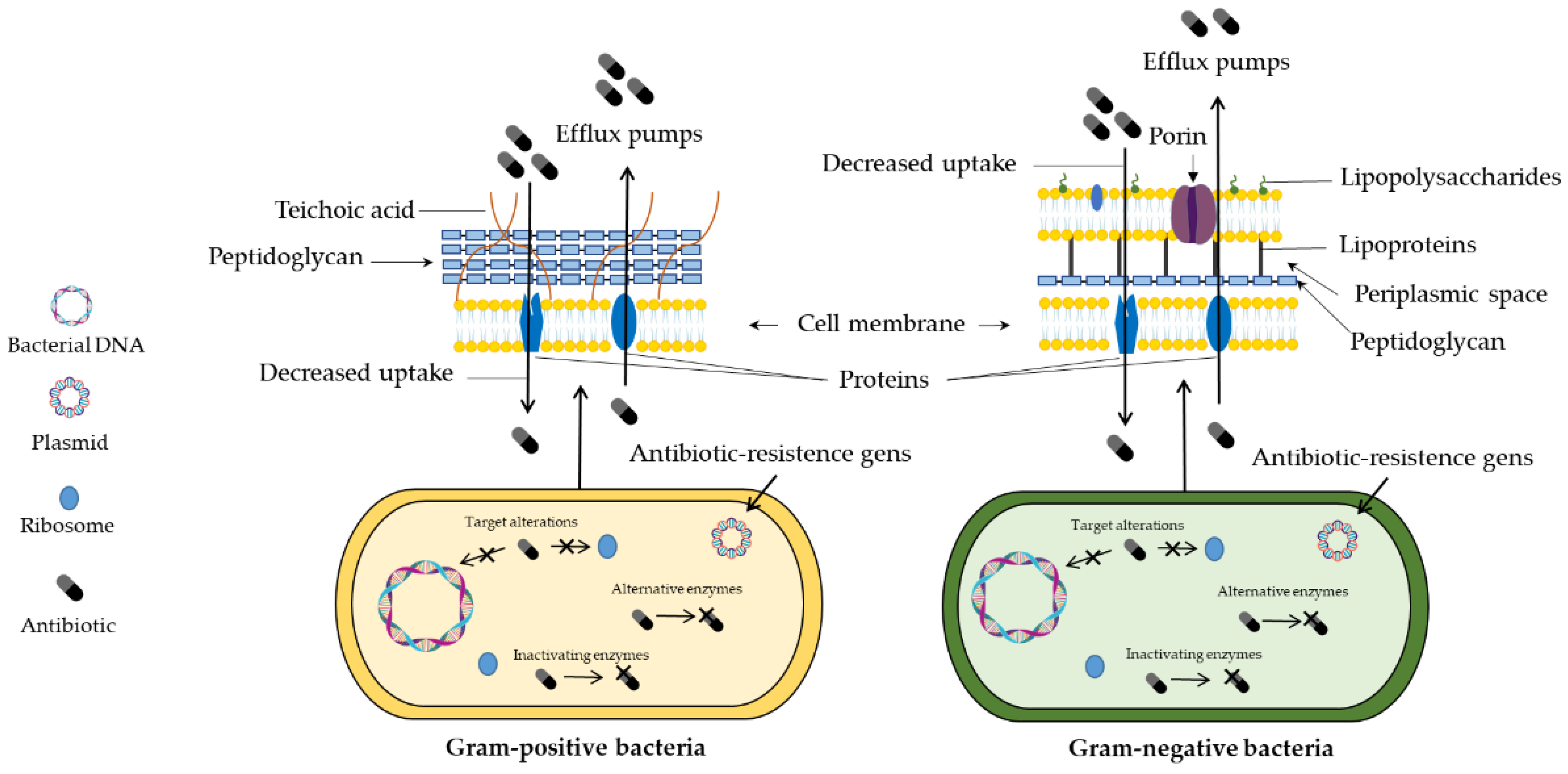
3. Antimicrobial Activity

Antimicrobial resistance has been considered an emerging health problem due to the ability of microorganisms to resist the pharmacological effect of many drugs [10]. Through gene hypermutation, different pathways may be associated with decreased antimicrobial activity, such as efflux pumps, enzymatic inactivation, decreased drug uptake, and pharmacological target changes (Figure 1) [11]. Thus, the potential advances in nanoscience and nanotechnology have contributed to the development of multifunctional materials that exhibit different properties and applications [12].
Figure 1. Bacterial structure and resistance mechanisms.
The replacement of an anti-inflammatory drug was evaluated by Follmann et al. [13]. Nanohybrids loaded with dexamethasone were used in an antibacterial study to verify their biological activity. The results of the susceptibility assay showed important bactericidal action against E. coli, S. aureus, Bacillus subtilis, and P. aeruginosa.
Nayeem et al. [14] developed cationic magnetic nanocomposites containing vancomycin (Van). According to the antimicrobial activity studies, it was verified that the Van impregnated into the nanomaterial showed higher inhibitory capacity compared to the free drug. Similarly, Zhang et al. [15] also immobilized Van onto magnetic hybrid nanoparticles to improve its pharmacological action. Corroborating with Nayeem and coauthors [14], the positive effects observed were the lower dosage to inhibit bacterial growth and the enhanced antibacterial activity.
Chemically modified silica nanoparticles with essential oils also were employed to improve the antibacterial activity. Shahabadi et al. [16] developed magnetic silica nanoparticle-based composites coated with eugenol (EUG). The bacterial activity was evaluated against four food pathogens. The results showed that among the strains tested, Klebsiella pneumoniae (Gram-negative) was the most sensitive bacterium. Additionally, the nanoparticles presented higher activity than free essential oil.
Silica nanoparticles present not only high biocompatibility but also exhibit high surface area and reactive sites that enable the conjugation with biocides compounds. Chen and coworkers [17] synthesized core–shell–like (Fe3O4/SiO2) magnetic nanoparticles containing a quaternary ammonium salt (QAS) site and three N-halamine sites. The antibacterial assay revealed that the QAS and N-halamine sites act in a synergic manner. Both bacterial strains (Staphylococcus aureus and Escherichia coli) showed susceptibility to the magnetic compound, with bacterial inactivation of 100% after 30 min of incubations with treatments.
Cationic dendrimers with terminal amine display antimicrobial properties. Nazari et al. [18] also modified the surface of Fe3O4/SiO2 nanoparticles to investigate bacterial effects against S. aureus and E. coli. The inhibitory performance of Fe3O4/SiO2 nanoparticles functionalized with dendrimers demonstrated that the minimum inhibitory concentration (MIC) for E. coli and S. aureus was 4 and 8 µg mL−1, respectively. The higher susceptibility of Gram-negative strain to treatments can be associated with the thickness of the cell wall peptidoglycan.
Transition metals have also been incorporated into the surface of magnetic silica nanoparticles. Shatan and coauthors [19] reported the coating of magnetite with silica nanoparticles and its functionalization with thiol groups to facilitate the impregnation of silver clusters. The antibacterial activity was evaluated using clinical isolates of S. aureus and E. coli strains. The results clearly express that the decorated Ag+ ions on the surface of the magnetic nanoparticle contribute to the inhibitory effect, considering that no significant difference was observed without the magnetic nanoparticles. In addition, the E. coli strain demonstrates higher susceptibility to treatments.
Mesoporous silica-coated magnetic nanocomposites were synthesized for the controlled release of Ag+ ions. Wang et al. [20] reported a novel silica-based core–shell compound for drinking water decontamination. The study to evaluate the antibacterial activity was carried out using E. coli as the model. The results showed that the etching time on the nanocomposite structure influenced the disinfectant action.
The copper ions–Schiff base complex was anchored in mesoporous silica-based magnetic nanoparticles. Ahmadi et al. [21] synthesized different magnetic nanoparticles containing streptomycin and investigated the antimicrobial activity against two bacterial strains (S. aureus and E. coli) in the presence and absence of a magnetic field (H). The results demonstrated that the bacterial strains treated with nanoparticles together with the magnetic field exhibited higher sensitivity to all treatments. Moreover, the nanoparticles containing copper and streptomycin showed the most significant effects. At the same time, the compounds presented antibacterial action more expressively against E. coli.
Platinum-doped core–shell magnetic nanoparticles (Fe3O4@SiO2-Pt) were developed to evaluate the inhibitory effect against S. aureus [22]. The results showed that the antiproliferative effects of nanoparticles occur in a dose-dependent manner. Additionally, the antibacterial activity is improved with the presence of hydrogen peroxide. Similarly, titanium–silica–iron oxide (TSI) nanocomposites were prepared to explore the potential bactericidal activity. The effects of TSI combined with UV light were also studied. The experimental results indicated that the nanocomposites showed bactericidal performance only under the presence of ultraviolet light [23].
The synthesis of a novel magnetic nanocomposite containing different metallic elements and quantities of Ni2+ was reported by El Nahrawy et al. [24]. These nanocomposites were tested in diverse bacterial strains. The in vitro data revealed that the compounds with higher proportions of Ni2+ ions presented the best inhibitory capacity.
Iron oxide was also impregnated into silica-based microporous materials for antibacterial studies against methicillin-resistant S. aureus (MRSA) and Pseudomonas aeruginosa [25]. The MIC values and antibiofilm assay showed clearly that the nanocomposite exhibited antibacterial activity and inhibited biofilm formation.

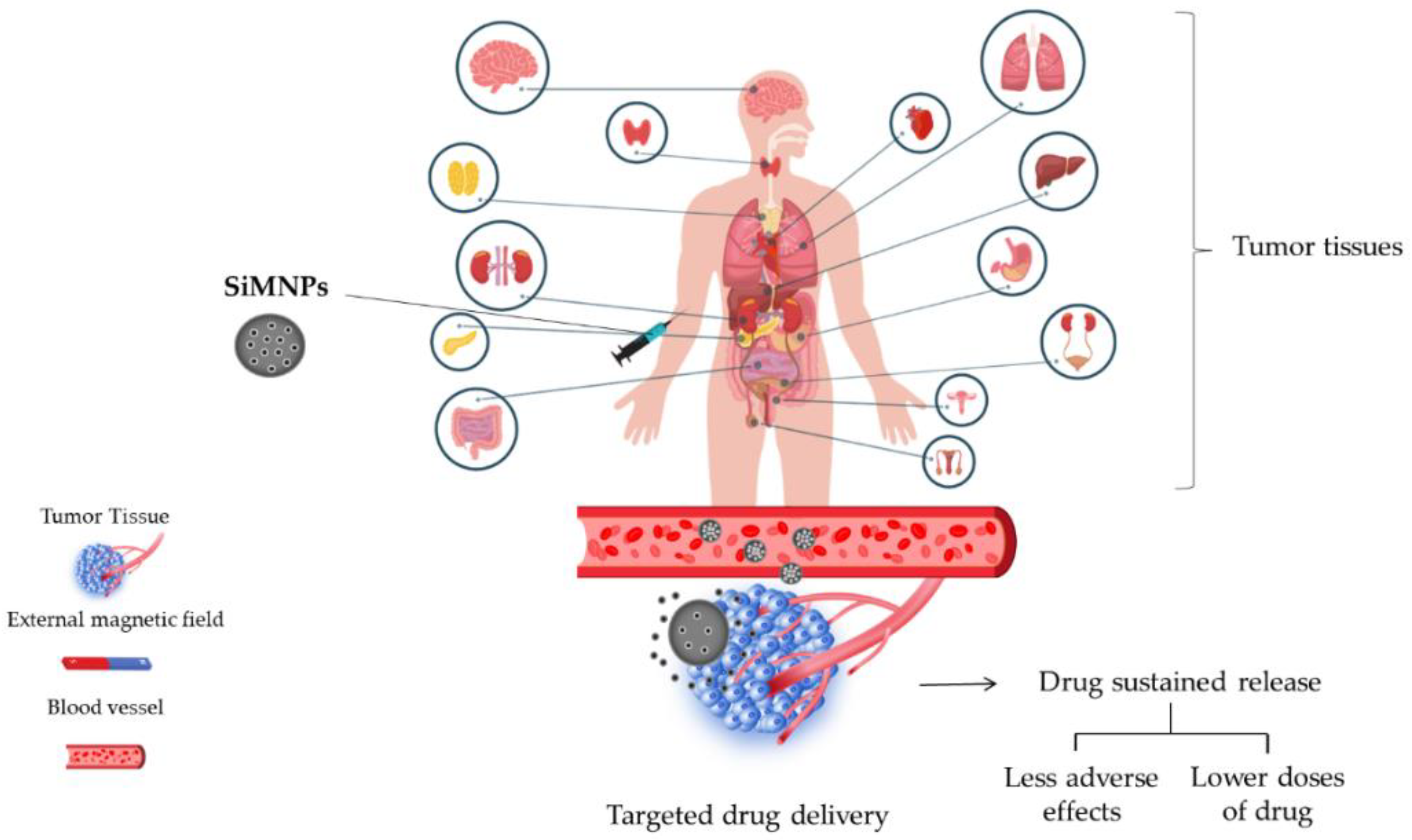
4. Drug Delivery and Release Studies

Although conventional treatments are effective in controlling and curing numerous diseases, pharmacological therapies still present diverse limitations, such as nonspecific action, several side effects, toxicity, and instability to pH and light [26][27]. Thus, nanostructured systems have been widely used to improve the biocompatibility and systemic circulation of drugs (Figure 2) as well as decrease immunogenicity [28].
Figure 2. Drug delivery systems using silica-based magnetic nanoparticles.
Some investigations indicated that silica nanoparticles could enhance drug delivery and improve treatment response. In this scenario, Pinna et al. [29] conducted a study using MIL-88A (Fe) MOF crystals nucleated and grown around a polymer core containing superparamagnetic nanoparticles focused on the magnetophoretic drug release of dopamine. The safety profile of MOF was measured using PC12 cells as the neuronal cell model. The findings indicated that dopamine could be delivered into the intracellular compartment using the PMP@MIL-88A carrier as well as avoid side effects.
Moreover, Balasamy et al. [25] reported the action of nanoformulation focusing on biocompatible metal–organic frameworks (MOFs) as well as magnetic nanocarriers for drug delivery and tumor imaging to reveal new cancer therapies. It was developed a nanocomposite comprising Fe/SBA-16 and ZIF-8 (Fe/S-16/ZIF-8) via ultrasonic irradiation.
An automated diffusion cell system equipped with a flow-type Franz cell was used analyzed cisplatin (Cis) drug delivery. The results of the drug release profile established the nanocarrier as an excellent platform for cisplatin delivery, enabling a slow and sustained release.
Follmann et al. [13] performed a synthesis of novel hybrid organic–inorganic aerogel materials with one-dimensionally aligned pores to conduct an extended delivery system for a hydrophobic drug. Given the fact that these compounds present hydrophobic pores, the hybrid aerogels exhibited a high drug-loading ability and excellent release pattern for dexamethasone, which was used as a model drug. The results showed a prolonged drug delivery of more than 50 days.
Chitosan-modified magnetic silica nanoparticles and N-isopropylacrylamide (MagSi@Chi-g-NIPAAm) was used as a doxorubicin (Dox) carrier [30]. The drug release study was performed at different pHs (4.0 and 7.4, respectively) to simulate the physiological and tumor environment. In addition, two temperatures were also used to mimic body temperature and hyperthermia conditions (37 and 45 °C), respectively. Regarding the in vitro results, it was verified that the drug release is highly pH/thermos-responsive, i.e., the highest drug release rate was reached at the acidic medium and 45 °C. This behavior is sustained by swelling of the polymers at pH 4.0 (easy protonation of amine groups) and higher rupture of nanoparticles. Similar behavior was observed by Tran et al. [31].
The magnetic nanocarrier containing cisplatin showed higher drug release at pH 5.5 than at pH 7.4. These findings were also attributed to the protonation of NH2 groups in an acidic environment.
Silica-based magnetic nanoparticles were used for targeted delivery and cellular recognition of doxorubicin and miRNA-21 [32]. The effect of the amount of doxorubicin on the loading efficiency (%), as well as the influence of the surface modification with DNA and miRNA-21, was investigated. In vitro results revealed that a gradual increase in the amount of Dox significantly improved drug loading efficiency. Furthermore, the presence of DNA and miRNA-21 in the nanocarrier structure determined the drug release.
Espinoza et al. [33] employed nanocomposite for carrier doxorubicin, MNPs-SiO2-F127-2%, and MNPs-SiO2-F127-4%. The assays were accomplished at two different pHs, 5.4 and 7.4, miming physiological and tumoral conditions, respectively. Drug release data revealed a significant influence on pH. At acidic conditions, the drug release is higher than the pH of 7.4, which may occur by breaking amide bonds. The Pluronic F127 content also affected the drug release rate. Along with this, Wu and co-workers [34] used a nanocomposite (Fe3O4@mSiO2 and CS/Fe3O4@mSiO2) to deliver the same drug (Dox) and noted that under acidic conditions (pH 4.0), a higher drug release percentage (%) was reached. Both nanocarriers exhibit a time-dependent behavior; initially, a high concentration was released to the medium, followed by a slower release. The nanocarrier containing chitosan (CS) showed the highest drug release rate due to breaking the “gatekeeper” faster than Fe3O4@mSiO2.
Moorthy et al. [35] studied the fucoidan-coated core–shell magnetic mesoporous silica drug carrier (FeNP@SiOH@Fuc NPs) system action in human breast adenocarcinoma cell line (MDA-MB-231). This system contains a magnetic iron oxide (FeNP) core, a mesoporous silica shell (SiOH), and a marine biopolymer known as fucoidan (Fuc), which focuses on Dox drug delivery involving pH and hyperthermia.
In a similar manner, a novel Dox nanocarrier based on nanoparticles loaded into multi-walled carbon nanotubes with mesoporous silica (mSiO2) MWCNTs@CoFe2O4@mSiO2 was developed. The results showed a remarkable pH-responsive drug delivery character within 48 h, targeting cancer therapy improvement [36]. Wang et al. [37] produced Fe2B@SiO2 nanoparticles covalently grafted from graphene oxide containing 90% entrapped epirubicin (EPI). The drug delivery was pH-dependent, with higher EPI release achieved at pH 5.7.
Wang et al. [38] investigated the synergistic antitumor effect of the hydrogel-based nanocomposite (MMSN-RBITCs) for Dox delivery by MTT assay. The free Dox showed a low cytotoxicity capacity for the 4T1 cell line over 24 h of contact. In contrast, the Dox-loaded hydrogel was able to decrease cell viability. Moreover, the cytotoxicity increased dramatically when the hydrogel was exposed to magnetic hyperthermia therapy (MHT), indicating that heat potentiates the cytotoxic effects of nanoparticles.

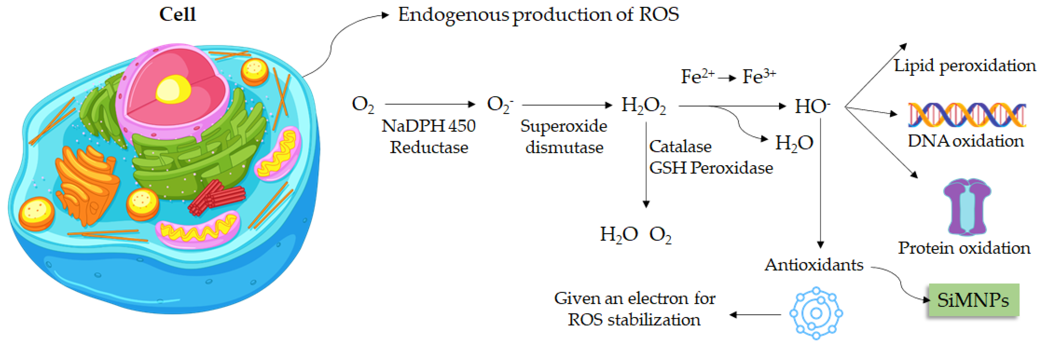
5. Antioxidant Activity

Oxidative stress is known as an organism imbalance between the production of oxidative substances and antioxidant compounds [39]. Free radicals are atoms or molecules that have unpaired electrons in the valence shell. This characteristic enables those free radicals the ability to react with macromolecules (Figure 3) and trigger several diseases [40].
Figure 3. Silica-based magnetic nanoparticles focusing on antioxidant activity.
Mesoporous silica nanoparticles are widely used in biological studies due to their distinct properties. A recent report showed that quercetin conjugated with silica-based magnetic nanoparticles was able to significantly reduce Aβ40-induced ROS generation in primary hippocampal neurons [41].
The antioxidant activity of silica-based magnetic nanoparticles functionalized with chitosan and tannic acid (γ-Fe2O3@SiO2-CS-TA) and also amine groups (γ-Fe2O3@SiO2-NH2-TA) was reported by Świętek et al. [42]. DPPH assay results showed that nanoparticles modified with chitosan exhibit a slightly higher antioxidant capacity. At the same time, the amount of TA incorporated into the magnetic nanoparticles and the concentration of treatments also influenced the ability to scavenge free radicals.
Zare and Sarkati [43] synthesized iron oxide nanoparticles coated with silica and chitosan immobilized into the surface (Cs-f-SiO2@Fe3O4 MNPs). Magnetic nanoparticles were used as a carrier for silymarin. The antioxidant activity assay showed that silymarin incorporated into magnetic nanoparticles presented higher DPPH radical scavenging ability compared to pure nanoparticles. Corroborating with results reported by Świętek et al. [41], the inhibitory capacity increases with the concentration of compounds.
Patsula et al. [44] investigated the antioxidant activity of γ-Fe2O3@SiO2-PEI-PEG nanoparticles coated with poly(l-lysine) (PLL) and polyethyleneimine (PEI) + PLL (PLLL). The antioxidant power of magnetic nanoparticles at the cell level was investigated by flow cytometry. According to the results, the treatments showed the capacity to reduce the endogenous production of ROS in human glioblastoma cells (U87MG cell line).

6. Silica-Based Nanoparticles Biocompatibility and Safe Profile Assessment

Investigations that focus on discovering new therapeutic agents that exhibit low side effects are significant to the nanomedicine field. In this scenario, studies that analyze the biocompatibility of these compounds are essential. Keshavarz et al. [45] reported that silica nanoparticles improved drug release using Tamoxifen (anticancer treatment) by hyperthermia. This investigation used poly (carboxybetaine methacrylate) (pCBMA) coating for magnetite mesoporous silica nanoparticles (MMSNs) to inhibit protein uptake and avoid the protein corona effect. The findings herein are indicated that nanogels are biocompatible and present no cytotoxic effects since it was tested on L929 cells at different concentrations and distinct treatment times of 24, 48, and 72 h.
Ledda et al. [46] assessed the biocompatibility of sub-5 nm silica-coated superparamagnetic iron oxide nanoparticles in human stem cells and mice for possible use in nanomedicine therapies. The results showed that this system did not alter any of the stem cells’ tested parameters, such as growth, viability, morphology, cytoskeletal organization, cell cycle progression, immunophenotype, and the expression of pro-angiogenic and immunoregulatory paracrine factors. Neither the osteogenic nor myogenic differentiation markers suffered any modification. Moreover, in vivo analyses using a mice model suggested no acute or chronic cytotoxicity, exhibiting short- and long-term biocompatibility and safe profile via analyses of histopathology, hematology, serum pro-inflammatory response, body weight, and mortality.
The biocompatibility and bioaccumulation of nanoparticles were investigated by employing mice as an animal model. The use of magnetic nanoporous silica nanoparticles (MNPSNPs) in an in vivo study was reported by Janßen et al. [47]. By considering the difficulty for magnetic nanoparticles to accumulate in deeper regions, a magnetic implant was inserted subcutaneously to improve circulation. The analysis of biological tissues showed that the nanoparticles accumulated mainly in the lung, liver, and spleen. However, depletion occurred quickly, and no significant tissue changes were observed. In a similar manner, Nasiri et al. [48] also reported that silica-coated iron oxide nanoparticles caused insignificant changes in histological tissues.
Mesoporous silica nanoparticles with magnetic core (Fe3O4) coated with polyethylene glycol-phospholipids were used in in vitro and in vivo studies to evaluate biocompatibility. The cellular uptake was explored using hepatic cells as the model (HepG2 and HepaRG cells). There was aggregation due to its high ionic strength, protein adsorption, and accumulation in the liver and spleen. Cell internalization was slower with coating nanoparticles than with native ones. Despite the accumulation, the nanoparticles did not cause toxicity in these tissues [49].
Navarro-Palomares et al. [50] reported that dye-doped biodegradable nanoparticle SiO2 coating on zinc- and iron-oxide nanoparticles enhanced biocompatibility and can be used for in vivo imaging investigations via supplying luminescent functionality to zinc and iron oxide multifunctional nanoparticles.
With the aim of increasing biocompatibility and avoiding protein adsorption and ion aggregation in the biological environment, SiMNPs were conjugated with PEG (MFO/SiO2-PEG). The cytotoxicity of MFO/SiO2-PEG was investigated in NIH/3T3 cell line (MFO/SiO2-PEG concentration from 2 μg·mL−1 to 200 μg·mL−1) by CCK-8 assay. In this range, the complex showed high biocompatibility (cell viability remained above 80% for all treatments) [51].

7. Silica Nanoparticles Carriers Focusing on Cardiovascular Treatment

Huang et al. [52] used silica nanoparticles to improve stroke treatment. This study was conducted using a novel surface imprinted magnetic mesoporous silica nNOS–PSD-95 (nitric oxide synthase–postsynaptic 12 density protein-95) as artificial antibodies. This complex showed a significant adsorbing performance and high recyclability and can be used as a sorbent to catch uncouplers from natural compounds. Biological action and interaction were measured in vitro and in vivo. This compound showed neuroprotective action on glutamate-injured PC12 cells (rat adrenal medulla cell line) and uncoupling activity targeting nNOS–PSD-95. Moreover, nNOS–PSD-95 uncouplers improving neurological deficit and decreased infarct volume of MCAO/R (middle cerebral artery occlusion and reperfusion) in rats, indicating significant potential for ischemic stroke treatment.

8. Silica Nanoparticles against Cancer Cells

The development of nanoparticles with potential anticancer activity is a very important aspect to be investigated, considering the chemoresistance, recurrence, and several side effects [53]. Previous investigations reported that silica nanoparticles present action against different cancer cell lines [35][54][55][56]. In addition, silica nanoparticles are widely explored due to their inert characteristic and high biocompatibility (Figure 4). Moreover, they can be easily obtained from sustainable sources such as rice husks by a top-down approach [57]. Chemically modified silica nanoparticles are synthesized for carrier drugs and other organic compounds aiming to explore their antitumor activity. Due to responsiveness to an external magnetic field and biocompatibility, magnetic nanoparticles are commonly used in modification reactions [15].
Figure 4. Interaction of silica-based nanoparticles with healthy cells.
In this scenario, Abbasi Kajani et al. [58] synthesized gold/silver decorated silica-coated iron oxide nanoparticles and evaluated the anticancer activity using human breast (MCF-7) and cervical cancer (HeLa) cell lines. According to the results, it was possible to verify a dose-dependent anticancer activity. In addition, higher effects were exhibited on the MCF-7 cell line than on HeLa. The nanostructures containing gold/silver showed high results when compared to silica–iron oxide nanoparticles (FeSi-NPs), indicating that metal ions improve the cytotoxicity against tumor cells. Moreover, nanoparticles showed good biocompatibility with healthy cells, significantly affecting cell viability only at the highest concentrations.
Taher et al. [59] synthesized magnetosome mimics nanoparticles with a lipid-covered magnetite core synthesized by the bacterium Magnetospirillum magneticum (called magnetosome). Cytotoxic effects of mimetic magnetosomes were evaluated using MDA-MB-231 (human breast adenocarcinoma) cells by flow cytometry. In vitro results showed that magnetosomes exhibited higher toxicity than nanoparticles. Among them, the silica-containing mimetic magnetosomes (Si@rMNP) showed a higher capacity for cellular internalization.
Iron oxide-loaded hollow silica spheres were synthesized and evaluated by MTT on HCT-166 cells (colon-rectal carcinoma). The cellular viability decreased in a dose and iron-dependent manner. These data suggest that the coupling with magnetic nanoparticles could increase the anticancer capacity of the iron oxide-loaded hollow silica spheres [60].
Foglia et al. [61] developed silica-coated magnetic iron oxide fluorescent nanoparticles. Confocal microscopy confirmed internalization in human colon cancer cells (Caco-2). However, no interference with cytoskeleton organization or cell cycle progression was observed.
Solak and coauthors [62] employed magnetic mesoporous silica nanoparticles modified with acid-linked polyethyleneimine (mMDPF) to loaded Disulfiram (DSF) against MCF-7 and MCF-10A cell lines. The results of cytoviability showed that MCF-7 cells were the most sensitive line to treatments. The higher selectivity of nanoparticles modified with folic acid is attributed to the expression of fatty acid (FA) receptors by MCF-7 cells [63].
The combined therapy of Cu2+ ions and/or sodium nitroprusside (SNP) improved the cytotoxicity of the nanoparticle-loaded DSF, although it did not enhance the effects of free disulfiram. Further, the magnetic field also contributed to higher cellular uptake and drug release.

9. Point-of-Care Detection of Silica-Based Nanoparticles

Silica nanoparticles are highlighted in point-of-care detection to enhance imaging assessment and improve the diagnosis in nanomedicine [50][64][65]. In this scenario, a recent review summarized the importance of silica nanoparticles for improving the sensitivity of lateral flow assays (LFAs), which is a rapid and affordable point-of-care device for health diagnostics. Silica nanomaterials have been used to enhance the signal of label materials or offer stability and subsequent detection performance improvement [66]. Moreover, Monaco et al. [64] developed a smart assembly of Mn-ferrites/silica core–shell with fluorescein and gold nanorods that exhibit robust and stable nanomicelles for in vivo triple modality imaging. The results herein was indicated a contrast-to-noise ratio (CNR) as high as 60 in a mouse leg injected subcutaneously with the nanosystem. Moreover, biocompatibility analysis did not show hemolytic effects, highlighting this investigation’s medical diagnosis. Additionally, Navarro-Palomares et al. [50] developed a dye-doped biodegradable nanoparticle SiO2 coating on zinc- and iron-oxide nanoparticles to enhance biocompatibility and in vivo imaging investigations. In addition, Cabrera-García et al. [65] reported the synthesis of engineered contrast agents in a single structure for T1–T2 dual magnetic resonance imaging using a thin and homogeneous silica coating through hydrolysis and polymerization of silicate at neutral pH. They produced Gd(H2O)4[Fe(CN)6]@SiO2 magnetic nanoparticles that are very stable in biological fluids. Thus, this product exhibits an important action as MRI CA, improving positive and negative contrasts in T1- and T2-weighted MR images, which can be applied for both in vitro and in vivo models. Moreover, it is showed that biosafety profile and excellent capacity to add in organic molecules, showing remarkable potential for medical diagnostic tools.

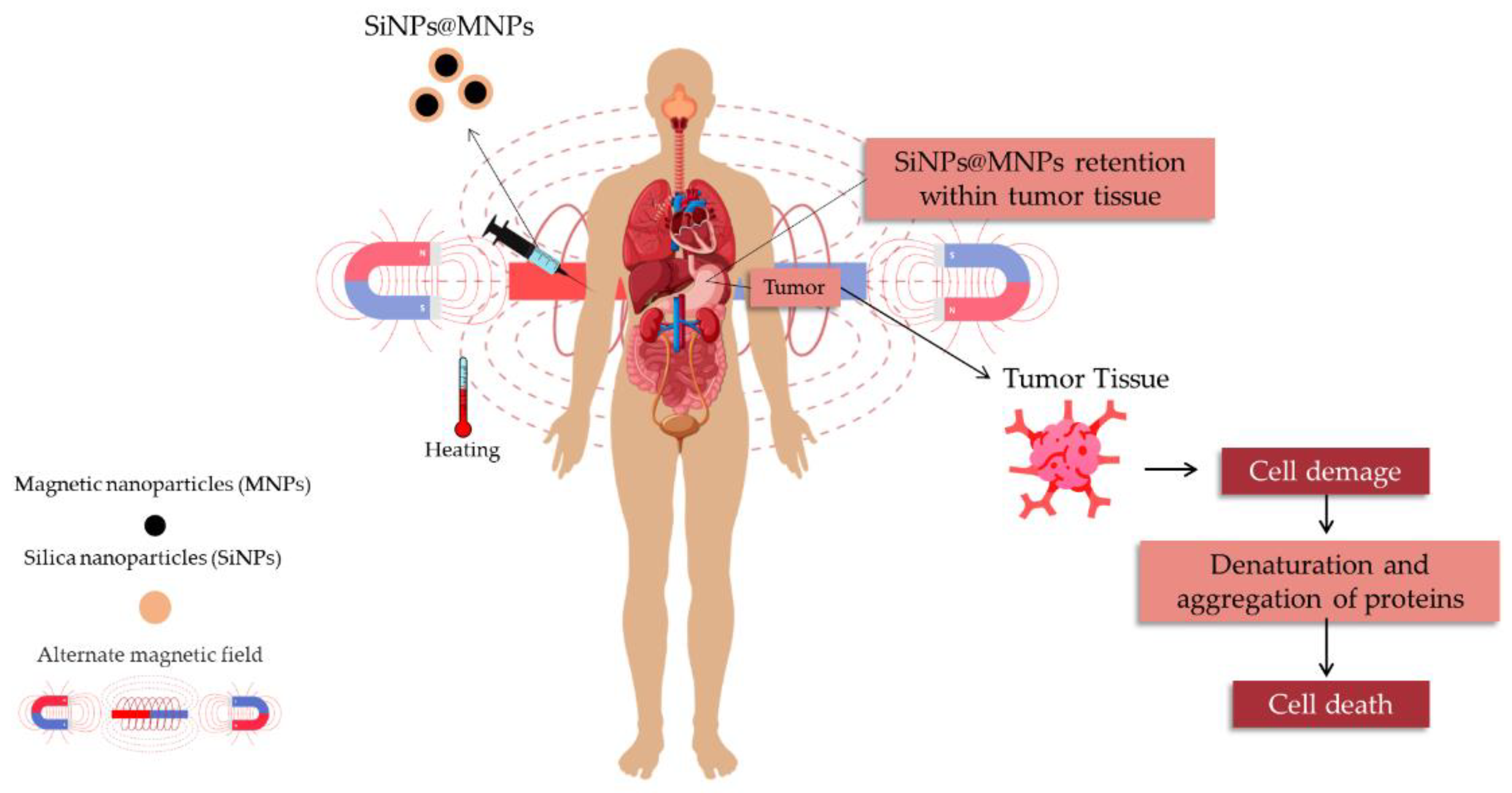
Magnetic Hyperthermia Therapy and Magnetic Resonance Imaging

Magnetic nanoparticles are commonly studied for magnetic hyperthermia therapy (MHT) and as contrast agents for magnetic resonance imaging (MRI). The responsiveness of MNPs to an external magnetic field (MF) is useful in cancer treatment due to the ability to target the drug to the organs and tissues of interest, decreasing the non-specific toxicity of chemotherapeutic agents [67]. Iron oxide nanoparticles (IONPs) are a class of nanoparticles widely explored [68][69]. These nanoparticles are known to exhibit magnetic properties only in the presence of the external magnetic field. Moreover, IONPs can be easily synthesized and chemically modified [42]. However, considering the critical size and high surface energy, magnetic nanoparticles tend to aggregate [3]. Thus, surface modification with silica-based nanoparticles can not only increase stability but also become magnetic nanoparticles even more functional and biocompatible [70].
The advantage of the use of magnetic hyperthermia therapy (Figure 5) is the higher sensitivity of tumor cells to heat compared to healthy cells. The increase in local tumor tissue temperature (41–46 °C) causes cell damage for different pathways as well as sensibilize cells to drug chemotherapy [71]. Cell damage can occur by diverse pathways: i: through conversion of energy to heat (Neel relaxation); ii: mechanical damage caused by MNPs rotation in response to the external changing MF direction (Brownian relaxation) [38]; iii: production of heat shock proteins; iv: increase in immune response (recognition by cytotoxic T lymphocytes) [67].
Figure 5. Magnetic nanoparticles as the therapeutic agents for MHT.
The MRI-based conventional diagnosis uses gadolinium derivatives as contrast agents. Gd is rare earth from the lanthanide series that shows high toxicity in its inorganic form. Therefore, this metal is employed in chelate forms to increase stability and consequently reduce its toxicity [72]. However, gadolinium-based contrast agents (GdBCAs) still trigger side effects such as gadolinium deposition disease, skin changes, neurotoxicity, nephrotoxicity, and pancreatitis [73]. Iron oxide nanoparticles have been widely explored in MRI, considering their superparamagnetic properties, high biocompatibility, and relaxivity [74].
Nanotheranostic is a novel concept involving nanoscience and nanotechnology that associates therapy and diagnostics. In the past, magnetic nanoparticles were employed for cell migrating detection through MRI, and the results obtained revealed an improvement in the resonance signal [75]. Similarly, Benyettou and coauthors [76] reported an interesting study regarding the use of organic framework-based magnetic nanoparticles for multitasking applications. According to the results, it was possible to verify a linear correlation between the nanoparticles concentration and contrast, improving the relaxivity values. The theranostic proposal allows targeting antitumor drugs using nanocarriers as well as verifying tissue accumulation, toxicity, and cell processes [76][77]. Aiming to overcome the limitations of chemotherapy, such as high toxicity and poor specificity, Wang et al. [38] developed an injectable self-healing hydrogel (Mn-Znferrite@mesoporous silica) for doxorubicin delivery in the tumor site. Doped ferrite with Zn2+ and Mn2+ can convert the H2O2 into ·OH radical (·OH radicals cause tumor cell death) through ion-mediate Fenton reaction. Moreover, the superparamagnetic properties of this self-healing hydrogel allowed its use in MHT and MRI. The results showed the excellent effect of magnetic hyperthermia and MRI. An increase in the cytotoxicity of doxorubicin against the 4T1 cell line was observed due to higher cell uptake triggered by the nanoencapsulation and association with an alternate magnetic field (AMF). The in vitro and in vivo MRI results demonstrated that the increase in iron concentration improves transverse relaxation. Moreover, magnetic nanoparticles can enhance image resolution due to their high relaxivity (303.9 mM−1 s−1). Magnetic silica-based nanoparticles with high relaxation values (175 mM−1 s−1) were also reported by Sheng et al. [51]. These results display the potential application of nanoparticles as a theranostic platform.
Effects on the relaxation time caused by iron concentration were reported by Kubíčková et al. [78]. The MRI experiment revealed that an increase in iron content caused a reduction in the signal, which can be explained by the high relaxivity. Furthermore, MnFe2O4 and MnFe2O4@mSiO2 nanoparticles were also used as contrast agents (CAs) [79]. The results of the MRI showed that pristine manganese ferrite exhibits higher capacity than the CAs (transverse relaxation-T2) due to relaxivity value (1.04 μg mL−1 s−1). Regarding the MHT study, the results revealed that the coating with silica nanoparticles decreased the specific absorption rate (SAR). The heat dissipation mechanisms reported are the Neel and Brown relaxation due to the critical size of magnetic nanoparticles.
Effects of the alternate magnetic field were also evidenced by Pon-on et al. [30] when employing Dox-carried into MagSi@Chi-g-NIPAAm. The internal stimulation increased the Dox release. Under the same experimental conditions (pH 4.0), it was observed that the drug release rate improved by around 30% with AMF application.
Moorthy et al. [54] produced a mesoporous silica nanocarrier system (FeNP@SiOH@CET) modified by a crown ether triad unit (CET) focused on drug carriers, especially chemotherapy and heat therapy. This system presents a potential application for cancer therapy to reduce the side effects of pH-responsive drug delivery and magnetic hyperthermia. The surface-modified CET gating units protect the encapsulated drug inside the mesopores of the FeNP@SiOH@CET NPs system under a physiological environment and exhibit a pH-responsive release in acidic conditions promoted by an alternating magnetic field (AMF). This system presents action against MDA-MB-231 considering magnetic hyperthermic heating induced by the exposed AMF. Therefore, FeNP@SiOH@CET NPs present an important potential for cancer treatment combined with magnetic hyperthermia therapy applications.
Iliasov et al. [80] investigated the in vitro cytotoxicity of PEGylated MNPs and bioinert silica-coated MNPs on tumor cells (PC3-human prostate cancer) using a low-frequency alternating magnetic field (LF AMF). After 48 h of incubation, it was verified that the cytotoxic effects occurred in a dose-dependent manner and increased with coating. The amplitude of LF AMF and preincubation time had a significant contribution to the cytotoxicity, while the frequency did not affect cell proliferation. Similarly, Yang et al. [81] reported that the FDPOMs complex improved MR tumor imaging ability and showed excellent anticancer action considering the intense magnetic targeting capacity under an external magnetic field.

This entry is adapted from the peer-reviewed paper 10.3390/magnetochemistry8100131

References

  1. Rai, M.; Pandit, R.; Gaikwad, S.; Kövics, G. Antimicrobial peptides as natural bio-preservative to enhance the shelf-life of food. J. Food Sci. Technol. 2016, 53, 3381–3394.
  2. Tang, S.S.; Prodhan, Z.H.; Biswas, S.K.; Le, C.F.; Sekaran, S.D. Antimicrobial peptides from different plant sources: Isolation, haracterization, and purification. Phytochemistry 2018, 154, 94–105.
  3. Rhoden, C.R.B.; da Silva Bruckmann, F.; da Rosa Salles, T.; Junior, C.G.K.; Mortari, S.R. Study from the influence of magnetite onto removal of hydrochlorothiazide from aqueous solutions applying magnetic graphene oxide. J. Water Process Eng. 2021, 43, 102262.
  4. Sánta-Bell, E.; Molnár, Z.; Varga, A.; Nay, F.; Hornyánsky, G.; Paizs, C.; Balogh, D.; Poppe, L. “Fishing and Hunting”—Selective Immobilization of a Recombinant Phenylalanine Ammonia-Lyase from Fermentation Media. Molecules 2019, 24, 4146.
  5. Niu, C.; Song, X.; Zhang, Y.; Dai, L.; Wei, J.; Yue, T.; Song, Z. A rapid one-step process for the isolation of antibacterial peptides by silica-decorated Fe3O4 nanoparticles. LWT 2022, 155, 112858.
  6. Benelmekki, M.; Xuriguera, E.; Caparros, C.; Rodríguez-Carmona, E.; Mendoza, R.; Corchero, J.L.; Lanceros-Mendez, S.; Martinez, L.M. Design and characterization of Ni2+ and Co2+ decorated Porous Magnetic Silica spheres synthesized by hydrothermal-assisted modified-Stöber method for His-tagged proteins separation. J. Colloid Interf. Sci. 2012, 365, 156–162.
  7. Liu, S.; Chen, H.; Lu, X.; Deng, C.; Zhang, X.; Yang, P. Facile synthesis of copper (II) immobilized on magnetic mesoporous silica microspheres for selective enrichment of peptides for mass spectrometry analysis. Angew. Chem. Int. Ed. 2010, 49, 7557–7561.
  8. Surendhiran, D.; Roy, V.C.; Park, J.S.; Chun, B.S. Fabrication of chitosan-based food packaging film impregnated with turmeric essential oil (TEO)-loaded magnetic-silica nanocomposites for surimi preservation. Int. J. Biol. Macromol. 2022, 203, 650–660.
  9. Da Silva Bruckmann, F.; Viana, A.R.; Lopes, L.Q.S.; Santos, R.C.V.; Muller, E.I.; Mortari, S.R.; Rhoden, C.R.B. Synthesis, characterization, and biological activity evaluation of magnetite-functionalized eugenol. J. Inorg. Organomet. Polym. Mater. 2022, 32, 1459–1472.
  10. Deon, M.; Morawski, F.M.; Passaia, C.; Dalmás, M.; Laranja, D.C.; Malheiros, P.S.; Arenas, L.T.; Costa, T.M.H.; de Menezes, E.W.; Benvenutti, E.V. Chitosan-stabilized gold nanoparticles supported on silica/titania magnetic xerogel applied as antibacterial system. J. Sol-Gel Sci. Technol. 2019, 89, 333–342.
  11. Reygaert, W.C. An overview of the antimicrobial resistance mechanisms of bacteria. AIMS Microbiol. 2018, 4, 482.
  12. Da Rosa Salles, T.; da Silva Bruckamann, F.; Viana, A.R.; Krause, L.M.F.; Mortari, S.R.; Rhoden, C.R.B. Magnetic nanocrystalline cellulose: Azithromycin adsorption and in vitro biological activity against melanoma cells. J. Polym. Environ. 2022, 30, 2695–2713.
  13. Follmann, H.D.; Oliveira, O.N.; Lazarin-Bidóia, D.; Nakamura, C.V.; Huang, X.; Asefa, T.; Silva, R. Multifunctional hybrid aerogels: Hyperbranched polymer-trapped mesoporous silica nanoparticles for sustained and prolonged drug release. Nanoscale 2018, 10, 1704–1715.
  14. Nayeem, J.; Al-Bari, M.A.A.; Mahiuddin, M.; Rahman, M.A.; Mefford, O.T.; Ahmad, H.; Rahman, M.M. Silica coating of iron oxide magnetic nanoparticles by reverse microemulsion method and their functionalization with cationic polymer P (NIPAm-co-AMPTMA) for antibacterial vancomycin immobilization. Colloids Surf. A Physicochem. Eng. Asp. 2021, 611, 125857.
  15. Zhang, W.; Taheri-Ledari, R.; Hajizadeh, Z.; Zolfaghari, E.; Ahghari, M.R.; Maleki, A.; Hamblin, M.R.; Tian, Y. Enhanced activity of vancomycin by encapsulation in hybrid magnetic nanoparticles conjugated to a cell-penetrating peptide. Nanoscale 2020, 12, 3855–3870.
  16. Shahabadi, N.; Akbari, A.; Karampour, F.; Falsafi, M. Cytotoxicity and antibacterial activities of new chemically synthesized magnetic nanoparticles containing eugenol. J. Drug Deliv. Sci. Technol. 2019, 49, 113–122.
  17. Chen, Y.; Feng, C.; Zhang, Q.; Luo, M.; Xu, J.; Han, Q. Engineering of antibacterial/recyclable difunctional nanoparticles via synergism of quaternary ammonia salt site and N-halamine sites on magnetic surface. Colloids Surf. B Biointerfaces 2020, 187, 110642.
  18. Nazari, S.; Gholami, M.; Farzadkia, M.; Dourbash, F.A.; Arzanlou, M.; Kalantary, R.R. Synthesis and evaluation of the antibacterial effect of silica-coated modified magnetic poly-(amidoamine) G5 nanoparticles on E. coli and S. aureus. J. Mol. Liq. 2019, 276, 93–104.
  19. Shatan, A.B.; Venclíková, K.; Zasońska, B.A.; Patsula, V.; Pop-Georgievski, O.; Petrovský, E.; Horák, D. Antibacterial silver-conjugated magnetic nanoparticles: Design, synthesis and bactericidal effect. Pharm. Res. 2019, 36, 147.
  20. Wang, X.; Sun, W.; Yang, W.; Gao, S.; Sun, C.; Li, Q. Mesoporous silica-protected silver nanoparticle disinfectant with controlled Ag+ ion release, efficient magnetic separation, and effective antibacterial activity. Nanoscale Adv. 2019, 1, 840–848.
  21. Ahmadi, A.; Sedaghat, T.; Motamedi, H.; Azadi, R. Anchoring of Cu (II)-Schiff base complex on magnetic mesoporous silica nanoparticles: Catalytic efficacy in one-pot synthesis of 5-substituted-1H-tetrazoles, antibacterial activity evaluation and immobilization of α-amylase. Appl. Organomet. Chem. 2020, 34, e5572.
  22. Han, Q.; Wang, X.; Liu, X.; Xiao, W.; Cai, S.; Wang, C.; Yang, R. Controllable fabrication of magnetic core–shell nanocomposites with high peroxide mimetic properties for bacterial detection and antibacterial applications. J. Mater. Chem. B 2019, 7, 1124–1132.
  23. Chanhom, P.; Charoenlap, N.; Tomapatanaget, B.; Insin, N. Colloidal titania-silica-iron oxide nanocomposites and the effect from silica thickness on the photocatalytic and bactericidal activities. J. Magn. Magn. Mater. 2017, 427, 54–59.
  24. El Nahrawy, A.M.; Mansour, A.M.; Elzwawy, A.; Abou Hammad, A.B.; Hemdan, B.A. Spectroscopic and magnetic properties of Co0.15Al0.25−xNi0.6+xFe2O4 nanocomposites aided by silica for prohibiting pathogenic bacteria during sewage handling. Environ. Nanotechnol. Monit. Manag. 2022, 18, 100672.
  25. Balasamy, R.J.; Ravinayagam, V.; Alomari, M.; Ansari, M.A.; Almofty, S.A.; Rehman, S.; Dafalla, H.; Marimuthu, P.R.; Akhtar, S.; Al Hamad, M. Cisplatin delivery, anticancer and antibacterial properties of Fe/SBA-16/ZIF-8 nanocomposite. RSC Adv. 2019, 9, 42395–42408.
  26. González-Sarrías, A.; Tomé-Carneiro, J.; Bellesia, A.; Tomás-Barberán, F.A.; Espín, J.C. The ellagic acid-derived gut microbiota metabolite, urolithin A, potentiates the anticancer effects of 5-fluorouracil chemotherapy on human colon cancer cells. Food Funct. 2015, 6, 1460–1469.
  27. Jiang, A.; Song, B.; Ji, X.; Peng, F.; Wang, H.; Su, Y.; He, Y. Doxorubicin-loaded silicon nanoparticles impregnated into red blood cells featuring bright fluorescence, strong photostability, and lengthened blood residency. Nano Res. 2018, 11, 2285–2294.
  28. Suk, J.S.; Xu, Q.; Kim, N.; Hanes, J.; Ensign, L.M. PEGylation as a strategy for improving nanoparticle-based drug and gene delivery. Adv. Drug Deliv. Rev. 2016, 99, 28–51.
  29. Pinna, A.; Migheli, R.; Rocchitta, G.; Serra, P.A.; Falcaro, P.; Malfatti, L.; Innocenzi, P. A MOF-based carrier for in situ dopamine delivery. RSC Adv. 2018, 8, 25664–25672.
  30. Pon-On, W.; Tithito, T.; Maneeprakorn, W.; Phenrat, T.; Tang, I.M. Investigation of magnetic silica with thermoresponsive chitosan coating for drug controlled release and magnetic hyperthermia application. Macromol. Mater. Eng. 2019, 97, 23–30.
  31. Tran, V.A.; Shim, K.; Lee, S.W.; An, S.S.A. Multimodal mesoporous silica nanocarriers for dual stimuli-responsive drug release and excellent photothermal ablation of cancer cells. Int. J. Nanomed. 2020, 15, 7667.
  32. Liu, J.; Liu, W.; Zhang, K.; Shi, J.; Zhang, Z. A magnetic drug delivery system with “OFF–ON” state via specific molecular recognition and conformational changes for precise tumor therapy. Adv. Healthc. Mater. 2020, 9, 1901316.
  33. Espinoza, M.J.; Lin, K.-S.; Weng, M.-T.; Kunene, S.C.; Wang, S.-S.S. In Vitro Studies of Pluronic F127 Coated Magnetic Silica Nanocarriers for Drug Delivery System Targeting Liver Cancer. Eur. Polym. J. 2021, 153, 110504.
  34. Wu, J.; Jiang, W.; Shen, Y.; Jiang, W.; Tian, R. Synthesis and Characterization of Mesoporous Magnetic Nanocomposites Wrapped with Chitosan Gatekeepers for PH-Sensitive Controlled Release of Doxorubicin. Mater. Sci. Eng. C Mater. Biol. Appl. 2017, 70, 132–140.
  35. Moorthy, M.S.; Subramanian, B.; Panchanathan, M.; Mondal, S.; Kim, H.; Lee, K.D.; Oh, J. Fucoidan-coated core–shell magnetic mesoporous silica nanoparticles for chemotherapy and magnetic hyperthermia-based thermal therapy applications. New J. Chem. 2017, 41, 15334–15346.
  36. Fan, H.; Xing, X.; Yang, Y.; Li, B.; Wang, C.; Qiu, D. Triple function nanocomposites of porous silica-CoFe2O4-MWCNTs as a carrier for pH-sensitive anti-cancer drug controlled delivery. Dalton Trans. 2017, 46, 14831–14838.
  37. Wang, X.; Yin, H.; Guan, Y.; Yang, Y.; Huang, Y.; Yuan, H.; Ni, C. Graphene Oxide Covalently Grafted Nanoparticles for Epirubicin Loading and Releasing. J. Nanosci. Nanotechnol. 2020, 20, 2104–2113.
  38. Wang, C.; Zhao, N.; Huang, Y.; He, R.; Xu, S.; Yuan, W. Coordination of injectable self-healing hydrogel with Mn-Zn mesoporous silica nanospheres for tumor MR imaging and efficient synergistic magnetothermal-chemo-chemodynamic therapy. Chem. Eng. Technol. 2020, 401, 126100.
  39. Pizzino, G.; Irrera, N.; Cucinotta, M.; Pallio, G.; Mannino, F.; Arcoraci, V.; Squadrito, F.; Altavilla, D.; Bitto, A. Oxidative stress: Harms and benefits for human health. Oxid. Med. Cell. Longev. 2017, 2017, 8416763.
  40. Forman, H.J.; Zhang, H. Targeting oxidative stress in disease: Promise and limitations of antioxidant therapy. Nat. Rev. Drug Discov. 2021, 2, 689–709.
  41. Halevas, E.; Mavroidi, B.; Nday, C.M.; Tang, J.; Smith, G.C.; Boukos, N.; Litsardakis, G.; Pelecanou, M.; Salifoglou, A. Modified magnetic core-shell mesoporous silica nano-formulations with encapsulated quercetin exhibit anti-amyloid and antioxidant activity. J. Inorg. Biochem. 2020, 213, 111271.
  42. Świętek, M.; Ma, Y.H.; Wu, N.P.; Paruzel, A.; Tokarz, W.; Horák, D. Tannic Acid Coating Augments Glioblastoma Cellular Uptake of Magnetic Nanoparticles with Antioxidant Effects. Nanomaterials 2022, 12, 1310.
  43. Zare, M.; Sarkati, M.N. Chitosan-functionalized Fe3O4 nanoparticles as an excellent biocompatible nanocarrier for silymarin delivery. Polym. Adv. Technol. 2021, 32, 4094–4100.
  44. Patsula, V.; Moskvin, M.; Siow, W.X.; Konefal, R.; Ma, Y.H.; Horák, D. Antioxidant polymer-modified maghemite nanoparticles. J. Magn. Magn. Mater. 2019, 473, 517–526.
  45. Keshavarz, H.; Khavandi, A.; Alamolhoda, S.; Naimi-Jamal, M.R. Magnetite mesoporous silica nanoparticles embedded in carboxybetaine methacrylate for application in hyperthermia and drug delivery. New J. Chem. 2020, 44, 8232–8240.
  46. Ledda, M.; Fioretti, D.; Lolli, M.G.; Papi, M.; Di Gioia, C.; Carletti, R.; Lisi, A. Biocompatibility assessment of sub-5 nm silica-coated superparamagnetic iron oxide nanoparticles in human stem cells and in mice for potential application in nanomedicine. Nanoscale 2020, 12, 1759–1778.
  47. Janßen, H.C.; Angrisani, N.; Kalies, S.; Hansmann, F.; Kietzmann, M.; Warwas, D.P.; Behrens, P.; Reifenrath, J. Biodistribution, biocompatibility and targeted accumulation of magnetic nanoporous silica nanoparticles as drug carrier in orthopedics. J. Nanobiotechnol. 2020, 18, 14.
  48. Nasiri, R.; Dabagh, S.; Meamar, R.; Idris, A.; Muhammad, I.; Irfan, M.; Nodeh, H.R. Papain grafted into the silica coated iron-based magnetic nanoparticles ‘ SiO2-PPN’as a new delivery vehicle to the HeLa cells. Nanotechnology 2020, 31, 195603.
  49. Rascol, E.; Pisani, C.; Dorandeu, C.; Nyalosaso, J.L.; Charnay, C.; Daurat, M.; Da Silva, A.; Devoisselle, J.-M.; Gaillard, J.-C.; Armengaud, J.; et al. Biosafety of mesoporous silica nanoparticles. Biomimetics 2018, 3, 22.
  50. Navarro-Palomares, E.; González-Saiz, P.; Renero-Lecuna, C.; Martín-Rodríguez, R.; Aguado, F.; González-Alonso, D.; Valiente, R. Dye-doped biodegradable nanoparticle SiO 2 coating on zinc-and iron-oxide nanoparticles to improve biocompatibility and for in vivo imaging studies. Nanoscale 2020, 12, 6164–6175.
  51. Sheng, Y.; Li, S.; Duan, Z.; Zhang, R.; Xue, J. Fluorescent magnetic nanoparticles as minimally-invasive multi-functional theranostic platform for fluorescence imaging, MRI and magnetic hyperthermia. Mater. Chem. Phys. 2018, 204, 388–396.
  52. Huang, J.; Sun, C.; Yao, D.; Wang, C.Z.; Zhang, L.; Zhang, Y.; Yuan, C.S. Novel surface imprinted magnetic mesoporous silica as artificial antibodies for efficient discovery and capture of candidate nNOS–PSD-95 uncouplers for stroke treatment. J. Mater. Chem. 2018, 6, 1531–1542.
  53. Chatterjee, K.; Sarkar, S.; Jagajjanani Rao, K.; Paria, S. Core/Shell Nanoparticles in Biomedical Applications. Adv. Colloid Interface Sci. 2014, 209, 8–39.
  54. Moorthy, M.S.; Bharathiraja, S.; Manivasagan, P.; Lee, K.D.; Oh, J. Crown ether triad modified core–shell magnetic mesoporous silica nanocarrier for pH-responsive drug delivery and magnetic hyperthermia applications. New J. Chem. 2017, 41, 10935–10947.
  55. Itatahine, A.; Mehdi, Y.A.; Fizir, M.; Qi, M.; Dramou, P.; He, H. Multifunctional carbon nanomateriels for camptothecine low-water soluble anticancer drug delivery. New J. Chem. 2018, 42, 1326–1336.
  56. Gao, Q.; Xie, W.; Wang, Y.; Wang, D.; Guo, Z.; Gao, F.; Cai, Q. A theranostic nanocomposite system based on radial mesoporous silica hybridized with Fe 3 O 4 nanoparticles for targeted magnetic field responsive chemotherapy of breast cancer. RSC Adv. 2018, 8, 4321–4328.
  57. da Silva Bruckmann, F.; Pimentel, A.C.; Viana, A.R.; da Rosa Salles, T.; Krause, L.M.F.; Mortari, S.R.; Silva, I.Z.; Rhoden, C.R.B. Synthesis, characterization and cytotoxicity evaluation of magnetic nanosilica in L929 cell line. Discip. Sci. Sér. Ciên. Nat. Tecnol. 2020, 21, 1–14.
  58. Abbasi Kajani, A.; Bordbar, A.-K.; Zarkesh-Esfahani, S.H.; Razmjou, A.; Hou, J. Gold/Silver Decorated Magnetic Nanostructures as Theranostic Agents: Synthesis, Characterization and in-Vitro Study. J. Mol. Liq. 2017, 247, 238–245.
  59. Taher, Z.; Legge, C.; Winder, N.; Lysyganicz, P.; Rawlings, A.; Bryant, H.; Staniland, S. Magnetosomes and magnetosome mimics: Preparation, cancer cell uptake and functionalization for future cancer therapies. Pharmaceutics 2021, 13, 367.
  60. Akhtar, S.; Gunday, S.T.; Alqosaibi, A.I.; Aldossary, H.; Bozkurt, A.; Khan, F.A. Template-free preparation of iron oxide loaded hollow silica spheres and their anticancer proliferation capabilities. RSC Adv. 2022, 12, 6791–6802.
  61. Foglia, S.; Ledda, M.; Fioretti, D.; Iucci, G.; Papi, M.; Capellini, G.; Lisi, A. In vitro biocompatibility study of sub-5 nm silica-coated magnetic iron oxide fluorescent nanoparticles for potential biomedical application. Sci. Rep. 2017, 7, 1–13.
  62. Solak, K.; Mavi, A.; Yılmaz, B. Disulfiram-loaded functionalized magnetic nanoparticles combined with copper and sodium nitroprusside in breast cancer cells. Mater. Sc. Eng. C 2021, 119, 111452.
  63. Soleymani, J.; Hasanzadeh, M.; Somi, M.H.; Shadjou, N.; Jouyban, A. Probing the Specific Binding of Folic Acid to Folate Receptor Using Amino-Functionalized Mesoporous Silica Nanoparticles for Differentiation of MCF 7 Tumoral Cells from MCF 10A. Biosens. Bioelectron. 2018, 115, 61–69.
  64. Monaco, I.; Armanetti, P.; Locatelli, E.; Flori, A.; Maturi, M.; Del Turco, S.; Franchini, M.C. Smart assembly of Mn-ferrites/silica core–shell with fluorescein and gold nanorods: Robust and stable nanomicelles for in vivo triple modality imaging. J. Mater. Chem. 2018, 6, 2993–2999.
  65. Cabrera-García, A.; Checa-Chavarria, E.; Pacheco-Torres, J.; Bernabeu-Sanz, Á.; Vidal-Moya, A.; Rivero-Buceta, E.; Sastre, G.; Fernández, E.; Botella, P. Engineered contrast agents in a single structure for T1–T2 dual magnetic resonance imaging. Nanoscale 2018, 10, 6349–6360.
  66. Zhuang, H.; Xu, C.; Gao, F.; Li, Y.; Lei, C.; Yu, C. Recent Advances in Silica-Nanomaterial-Assisted Lateral Flow Assay. Bioengineering 2022, 9, 266.
  67. Liao, S.H.; Liu, C.H.; Bastakoti, B.P.; Suzuki, N.; Chang, Y.; Yamauchi, Y.; Lin, F.L.; Wu, K.C. Functionalized magnetic iron oxide/alginate core-shell nanoparticles for targeting hyperthermia. Int. J. Nanomed. 2015, 10, 3315.
  68. Nunes, F.B.; Da Silva Bruckmann, F.; Da Rosa Salles, T.; Rhoden, C.B.R. Study of phenobarbital removal from the aqueous solutions employing magnetite-functionalized chitosan. Environ. Sci. Pollut. Res. 2022, 1–14.
  69. Da Silva Bruckmann, F.; Ledur, C.M.; Da Silva, I.Z.; Dotto, G.L.; Rhoden, C.R.B. A DFT theoretical and experimental study about tetracycline adsorption onto magnetic graphene oxide. J Mol. Liq. 2022, 353, 118837.
  70. Bruckmann, F.D.S.; Rossato Viana, A.; Tonel, M.Z.; Fagan, S.B.; Garcia, W.J.D.S.; Oliveira, A.H.D.; Rhoden, C.R. B Influence of magnetite incorporation into chitosan on the adsorption of the methotrexate and in vitro cytotoxicity. Environ. Sci. Pollut. Res. 2022, 29, 70413–70434.
  71. Healy, S.; Bakuzis, A.F.; Goodwill, P.W.; Attaluri, A.; Bulte, J.W.; Ivkov, R. Clinical magnetic hyperthermia requires integrated magnetic particle imaging. Wiley Interdiscip. Rev. Nanomed. Nanobiotechnol. 2022, 14, e1779.
  72. Rodríguez-Galván, A.; Rivera, M.; García-López, P.; Medina, L.A.; Basiuk, V.A. Gadolinium-containing carbon nanomaterials for magnetic resonance imaging: Trends and challenges. J. Cell. Mol. Med. 2020, 24, 3779–3794.
  73. Ramalho, J.; Ramalho, M.; Jay, M.; Burke, L.M.; Semelka, R.C. Gadolinium toxicity and treatment. Magn. Reson. Imaging 2016, 34, 1394–1398.
  74. Avasthi, A.; Caro, C.; Pozo-Torres, E.; Leal, M.P.; García-Martín, M.L. Magnetic nanoparticles as MRI contrast agents. Top. Curr. Chem. 2020, 378, 49–91.
  75. Weissleder, R.; Cheng, H.C.; Bogdanova, A.; Bogdanov, A., Jr. Magnetically labeled cells can be detected by MR imaging. J. Magn. Reson. Imaging 1997, 7, 258–263.
  76. Benyettou, F.; Das, G.; Nair, A.R.; Prakasam, T.; Shinde, D.B.; Sharma, S.K.; Whelan, J.; Lalatonne, Y.; Traboulsi, H.; Pasricha, R.; et al. Covalent organic framework embedded with magnetic nanoparticles for MRI and chemo-thermotherapy. J. Am. Chem. Soc. 2020, 142, 18782–18794.
  77. Rümenapp, C.; Gleich, B.; Haase, A. Magnetic nanoparticles in magnetic resonance imaging and diagnostics. Pharm. Res. 2012, 29, 1165–1179.
  78. Kubíčková, L.; Kaman, O.; Veverka, P.; Herynek, V.; Brázda, P.; Vosmanská, M.; Kohout, J. The ε-AlxFe2-xO3 nanomagnets as MRI contrast agents: Factors influencing transverse relaxivity. Colloids Surf. 2020, 589, 124423.
  79. Kavkhani, R.; Hajalilou, A.; Abouzari-Lotf, E.; Ferreira, L.P.; Cruz, M.M.; Yusefi, M.; Ismail, U.N. CTAB assisted synthesis of MnFe2O4@ SiO2 nanoparticles for magnetic hyperthermia and MRI application. Mater. Today Commun. 2022, 31, 103412.
  80. Iliasov, A.R.; Nizamov, T.R.; Naumenko, V.A.; Garanina, A.S.; Vodopyanov, S.S.; Nikitin, A.A.; Abakumov, M.A. Non-magnetic shell coating of magnetic nanoparticles as key factor of toxicity for cancer cells in a low frequency alternating magnetic field. Colloids. Surf. B 2021, 206, 111931.
  81. Yang, T.; Niu, D.; Chen, J.; He, J.; Yang, S.; Jia, X.; Li, Y. Biodegradable organosilica magnetic micelles for magnetically targeted MRI and GSH-triggered tumor chemotherapy. Biomater. Sci. 2019, 7, 2951–2960.
More
This entry is offline, you can click here to edit this entry!