Your browser does not fully support modern features. Please upgrade for a smoother experience.
Fluorescence Biosensor for SARS-CoV-2 Diagnosis: History
View Latest Version
Please note this is an old version of this entry, which may differ significantly from the current revision.
Contributor: , , , , , , Yong Yang

The outbreak of Corona Virus Disease 2019 (COVID-19) has again emphasized the significance of developing rapid and highly sensitive testing tools for quickly identifying infected patients. The current reverse transcription polymerase chain reaction (RT-PCR) diagnostic techniques can satisfy the required sensitivity and specificity, the inherent disadvantages with time-consuming, sophisticated equipment and professional operators limit its application scopes. Compared with traditional detection techniques, optical biosensors based on nanomaterials/nanostructures have received much interest in the detection of SARS-CoV-2 due to the high sensitivity, high accuracy, and fast response.

  • optical biosensors
  • SARS-CoV-2 detection
  • point-of-care diagnostics

1. Introduction

In December 2019, an unprecedented pneumonia case was first reported in Wuhan, a city in China, and quickly spread around the world [1]. The Coronavirus Study Group (CSG) of the International Committee on Taxonomy of Viruses (ICTV) officially named the novel coronavirus as severe acute respiratory syndrome coronavirus 2 (SARS-CoV-2) on 11 February 2020 [2]. The surface of SARS-CoV-2 is mainly composed of four structure proteins, including spike glycoprotein (S), which specifically binds to host cell receptors, membrane protein (M), which is related to envelope formation, envelope small membrane protein (E), and nucleoprotein, which is related to viral assembly (N). SARS-CoV-2 enters body tissues rapidly and initiates infection when the S protein binds to the angiotensin-converting enzyme 2 (ACE2) on the surface of the host cell [3][4]. To date, more than 0.6 billion infections have been reported around the world, and the surprisingly fast propagation is responsible for the pandemic. The virus can be transmitted primarily through three routes: (1) respiratory droplets and close contact; (2) contact with virus-contaminated items; and (3) exposure to high concentrations of aerosols in a relatively closed environment. According to reports, SARS-CoV-2 can not only damage various organs, including the lung, heart, kidney, and brain, but can also cause serious health and mental problems after recovery [5][6][7]. Particularly, the recent outbreaks of novel coronavirus pneumonia (COVID-19) in various countries and the emergence of variants have increased infectivity, posing an enormous threat to people’s lives and health, and have brought immeasurable pressure to the global medical system. Therefore, the early diagnosis of infection and cutting off the transmission route are the keys to effectively controlling the epidemic. At the same time, the development of rapid, sensitive, convenient, and accurate virus detection technology is imminent [8][9][10].
Current clinical diagnostic methods for COVID-19 mainly include molecular (ribonucleic acid (RNA)), antigen, antibody, or imaging tests [2][11]. The reverse transcription polymerase chain reaction (RT-PCR) for the detection of the RNA of SARS-CoV-2 is regarded as the gold standard in epidemic prevention and control because of its high sensitivity and specificity. However, professional operators and equipment are required, and the entire process takes more than 2 h. Furthermore, false negative signals are prone to occur due to varying viral loads during different infection periods [12][13]. Enzyme-Linked Immunosorbent Assay (ELISA) is another commonly used immunological experimental technology and offers the merits of semi-quantitative or qualitative analysis skills based on the color depth but still needs complicated operations, and the accuracy can be easily affected by interference [3]. Likewise, lateral flow immunochromatography (LFIA) based on colloidal gold color reaction has attracted much attention owing to the simple operation, low cost, and strong specificity [14]. As a paradigm, Ge et al. developed the “COVID-19 IgM Antibody Detection Kit”, which has become one of the first batches of rapid detection reagents that have passed the verification of statutory testing institutions. The test kit shows a specificity of 99.6%, as well as an accuracy of 98.1%, and it is considered to be a point-of-care (POC) diagnostics technology that can be used for large-scale screening [15]. Nonetheless, the LFIA is poor in terms of quantitative analysis ability and sensitivity, as it only relies on visual detection.
In light of the tremendous pressure of COVID-19 prevention and control, scientists have been working hard to explore more convenient, rapid, and accurate POC diagnosis technologies. It is worth pointing out that the emergence of nanobiosensors has greatly promoted the development of POC technology. The nanobiosensors are mainly composed of three parts, namely, the identification element (analyte to be measured), the sensitive unit, and the transducer that can convert the biological response into a visual physical or chemical signal. According to the type of output signal, it can be divided into electrochemical biosensors, optical biosensors, etc. [16][17][18]. Among them, optical biosensors exhibit broad application prospects in the field of POC diagnosis in virtue of their high sensitivity, low cost, simple operation, and rapid analysis abilities [19][20]. Recently, a few teams have summarized and prospected the technologies for detecting SARS-CoV-2. For example, Naikoo et al. reviewed the current strategies for the diagnosis of SARS-CoV-2 [21]. The review introduces the merits and shortcomings of traditional detection techniques such as PCR and ELISA in detail and also discusses the research progress of current biosensors including surface plasmon resonance biosensors, multifunctional gene sensors, and electrochemical sensors, which have certain guiding significance for researchers. Thapa et al. discussed smart biosensors for SARS-CoV-2 diagnosis according to the types of nanomaterials, including metal and metal oxide-based nanobiosensors, carbon-based nanobiosensors, and polymer-based biosensors [22]. It provides a foundation for the development of new materials for nanobiosensors. However, there is no systematic discussion of optical sensors in the review. Although Lukose et al. described the development and prospects of optical technologies for virus detection, such as Raman spectroscopy, Fourier transforms infrared spectroscopy, and fluorescence technology for virus detection, which can help people understand the current development of optical sensors to a certain extent, the discussion of fluorescent sensors in the research mainly focuses on their applications in the detection of HPV, HIV, and HBV viruses [23]. To the best of the knowledge, at present, the review of optical sensors—particularly, fluorescence, Raman, Surface Plasmon Resonance (SPR), and additional biosensors—in the detection of SARS-CoV-2 is relatively rare.
It mainly concentrates on the research progress of novel near-infrared fluorescence biosensors, fluorescence resonance energy transfer biosensors, dual-mode optical biosensors, and surface-enhanced Raman scattering biosensors, with examples in SARS-CoV-2 diagnosis, as shown in Scheme 1. The promising strategies to improve optical biosensors are also discussed.
Scheme 1. Schematic illustration of optical biosensors for SARS-CoV-2 detection.

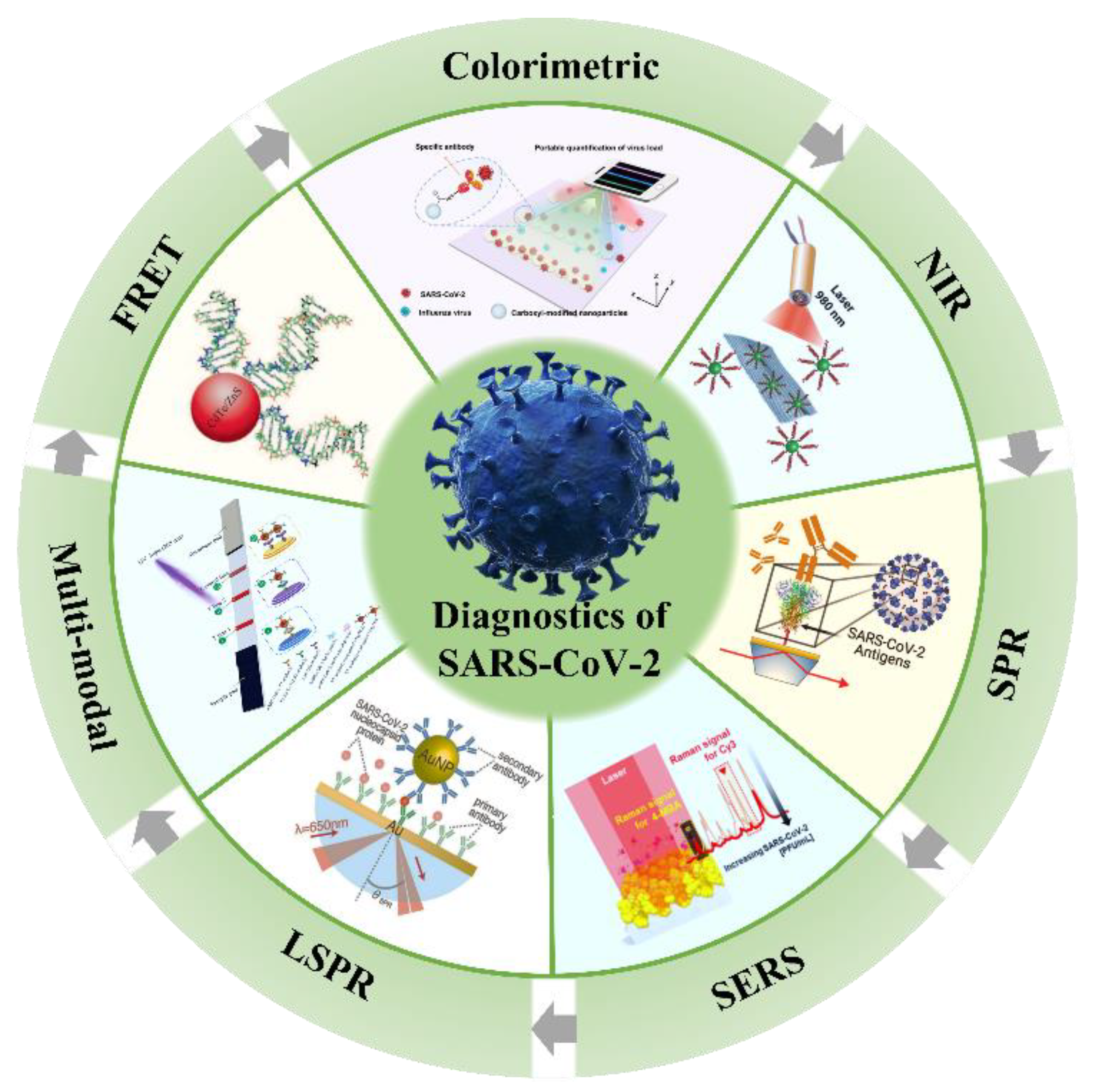
2. Potential Optical Biosensors

The optical sensors mainly analyze the optical signals (absorption, polarization, intensity, wavelength, or refractive index) generated during the combination of the target and the identification element and directly convert or amplify them via the transducer in real time to achieve a qualitative and quantitative analysis of the target. The characteristics of high sensitivity, good selectivity, simple operation, integration, and fast response make them widely used in environmental detection, biotechnology, food safety, medical diagnosis, and so on [24]. In the past decade, optical biosensors have developed rapidly, and many optical biosensing platforms have been used for the detection of nucleic acids, proteins, and exosomes, including but not limited to colorimetric biosensors, fluorescence biosensors, surface-enhanced Raman scattering biosensors (SERS), and surface plasmon resonance biosensors (SPR or LSPR). Notably, the optical sensors do not require complex nucleic acid amplification steps and post-processing in virus detection, which is considered a potential POC diagnostic tool and is expected to play a role in the large-scale screening of COVID-19, especially in low and middle-income regions and countries [25].
Colorimetric sensing technology can perform colorimetric analysis of the target through the visible color change of the solution, without the help of sophisticated instruments. The method is a semi-quantitative analysis and is pretty simple and convenient but has a low accuracy and can only be measured in a limited range. Moitra’s team designed a colorimetric assay based on AuNPs that can diagnose isolated RNA samples within 10 min [26]. The thiol-modified antisense oligonucleotides of SARS-CoV-2 were modified on the surface of Au NPs. The single Au NPs will aggregate when the RNA of SARS-CoV-2 is present in the detection sample, and the precipitate is observable after using ribonuclease to cut the RNA chain in the solution. The limit of detection (LOD) of the sensing assay reaches 0.18 ng/μL, providing a reliable method for disease diagnosis (Figure 1a).
Figure 1. Schematic diagram of optical sensors in biological detection: (a) colorimetric sensor for the detection of RNA from SARS-CoV-2. Reprinted with permission from ref. [26]. (b) Fluorescent sensor for the identification of SARS-CoV-2 in wastewater. Reprinted with permission from ref. [27]. Copyright 2022, American Chemical Society. (c) SERS sensor for the detection of tumor cell exosomes. Reprinted with permission from ref. [28]. Copyright 2022, Elsevier. (d) Plasma sensor for the detection of SARS-CoV-2. Reprinted with permission from ref. [29]. Copyright 2020, American Chemical Society.
Fluorescence sensing mainly conducts a qualitative and quantitative analysis of the target by determining the excitation and emission wavelengths and detecting the changes of the fluorescence intensity, which is commonly accompanied by the enhancement, quenching, or change in the emission signal. The presence of asymptomatic infections has brought enormous pressure to epidemic prevention and control. At present, relevant studies have shown that the detection of viruses in community sewage, namely, wastewater-based epidemiology (WBE), may be an effective strategy for the rapid diagnosis of the COVID-19 epidemic. Alafeef et al. reported a novel fluorescent sensing array based on three different lanthanum-doped carbon nanoparticles (LnCNPs), as shown in Figure 1b [27]. The LnCNPs were prepared by a hydrothermal process. The SARS-CoV-2 can be distinguished from other viruses or bacteria through machine learning according to differential fluorescence response patterns due to the counterion–ligand interactions. The LOD of the sensing array was 1.5 copies/μL, showing great promise in preventing the spread of the epidemic.
SERS refers to the phenomenon of an enhanced Raman signal generated on the surface of some rough nanomaterials. The technology is provided with high sensitivity, and the detection ability of some SERS-active substrates can even reach the single molecule level. Therefore, the active substrates are essential to achieving excellent SERS performance. The properties of nanomaterials, such as high surface energy, agglomeration and dispersion states, surface plasmon resonance, etc., can affect the activity of SERS substrates. Their group reported a novel nano-porous gold SERS chip functionality with CP05 polypeptide, which can specifically capture exosomes, as depicted in Figure 1c [28]. The lung and colon cell exosomes can be distinguished from normal plasma at the single vesicle level by combining Raman spectroscopy and machine learning methods without any labels or the purification of exosomes, providing a new idea for studying exosomes at the spectral level.
The principle of surface plasmon resonance (SPR) biosensors is to detect the target substances through the change in the refractive index caused by the interaction between the plasma resonance wave and the target molecules on the metal surface. The bifunctional plasmonic system developed by Qiu’s team possesses highly sensitive, rapid, and reliable diagnostic potential for the detection of SARS-CoV-2 [29]. The dual-function biosensor integrates the PPT effect and LSPR-sensing transduction onto a cost-effective gold nanoislands (AuNIs) chip. The PPT and LSPR effects can be excited at different wavelengths by using two different angles of incidence, significantly enhancing sensing stability, sensitivity, and reliability (Figure 1d). More importantly, the in situ PPT photothermal enhancement on the AuNI chip significantly improves the hybridization kinetics and nucleic acid detection specificity. The elaborated dual-function LSPR biosensor could provide a reliable and easily implementable diagnostic platform to improve the diagnostic accuracy of clinical tests.
The rapid development of nano-biotechnology has opened up infinite possibilities for the development of new detection technologies, especially the emergence of nanofluorescent materials, providing a wide range of prospects for the application of novel fluorescence detection techniques. As mentioned above, fluorescence sensors, colorimetric sensors, SERS sensors, and SPR sensors exhibit fast and high-sensitivity properties in biological detection, which are of great significance for realizing the timely detection of SARS-CoV-2.

3. Fluorescence Biosensor for SARS-CoV-2 Diagnosis

Fluorescence biosensors are a common method for the on-site detection of infectious diseases, which possess the advantages of high sensitivity, low cost, and simple operation [30][31]. Fluorescent molecules are the most important component in the construction of fluorescent biosensors, including fluorescent dyes and fluorescent nanomaterials. The Fluorescence Resonance Energy Transfer (FRET) mechanism is often introduced in designing fluorescence biosensors, involving the energy transfer from a fluorescent donor to a fluorescent acceptor. Additionally, some nanomaterials can generate fluorescence on their own, which is widely used in fluorescent biosensors due to their unique physical, chemical, and electron transport properties [32].

3.1. FRET Biosensors for SARS-CoV-2 Detection

Fluorescence Resonance Energy Transfer (FRET) was first proposed by Föster in 1948, so it is also called Föster energy transfer, which is a typical non-radiative energy transfer process [33]. FRET probes with multiple fluorescence analysis, high sensitivity, and easy operation have been rapidly developed in the field of biosensing and have become a powerful tool for the detection of the target molecule in the biosensing field [34][35][36]. The preconditions of FRET are that the emission spectrum of one fluorescent molecule (also known as the donor molecule) overlaps with the excitation light of the other fluorescent molecule (also known as the acceptor molecule), and the distance between them is less than 10 nm. Then, the excitation energy of the donor is used to induce fluorescence emission from the acceptor, while the fluorescence intensity of the donor is reduced. In particular, the selection of the donor and acceptor is crucial for achieving an efficient FRET process.
Among the candidate materials, quantum dots (QDs) with the features of good dispersion, high quantum yield, stable optical activity, and excellent photoluminescence performance have garnered much interest [37]. For example, Bardajee et al. designed CdTe-ZnS QDs functioned with DNA (QDs-DNA) to specifically recognize the DNA or RNA of the COVID-19 virus via the FRET method [38]. The QDs-DNA acts as a donor molecule, and the BHQ2-DNA was prepared to act as an acceptor molecule in the FRET process, as can be seen in Figure 2a. When combined with target RNA, the fluorescence of QDs-DNA can be extremely quenched by BHQ2-DNA in 25 min under an excitation of 325 nm, and the limit of detection was evaluated to be 0.000823 μM. The presence of thiolate-captured DNA oligonucleotides on the surface of QDs is responsible for the high sensitivity, because it can facilitate the interactions between QDs and target DNA. In another study, Gorshkov et al. provided a FRET-based biosensor to monitor the interactions of the spike proteins receptor binding domain to the host cell’s ACE2 receptor (Figure 2b). In the system, the QDs were conjugated with the recombinant spike receptor binding domain as a donor (QD-RBD), and ACE2-conjugated gold nanoparticles were regarded as an acceptor (AuNP-ACE2). It was shown that the QD-RBD and ACE2 are tightly bound and can enter the cell via the receptor-mediated endocytosis that is dependent on clathrin. This assay can be used for the high-throughput screening and detection of SARS-CoV-2 virus particles [39]. In addition, organic dyes are often used in the FRET system. Recently, Bardajee et al. established a novel cyanine 3 (Cy3)-based bio-conjugated sensor to rapidly and sensitively identify the specific DNA of COVID -19 via the FRET method. During the detection process, the target DNA, Cy3-DNA probe (donor), and BHQ2-DNA (acceptor) can form a sandwiched hybrid structure. The LOD of the Cy3 probe was calculated to be 0.09945 μM. Although the detection limit is higher than that of QDs-based sensors, it can still be used to analyze real samples [40].
Figure 2. The schematics of the DNA-conjugated CdTe/ZnS QDs nanoprobe for the detection of the target DNA derived from the COVID-19 virus genome(a). Reprinted with permission from ref. [38]. Copyright 2022, Elsevier. The energy transfer process from QD-RBD to AuNP-ACE2 and the corresponding cellular assay (b). Reprinted with permission from ref. [39].

3.2. NIR Biosensors for SARS-CoV-2 Detection

Both the excitation and emission peaks of conventional biosensors based on organic dyes, semiconductor quantum dots, etc. locate in the UV-visible region [41]. However, most biomolecules have autofluorescence in the range, which can interfere with the fluorescence signal of the biosensor and reduce the detection sensitivity. Compared with ultraviolet-visible light, biomolecules have ultra-low absorption and scattering in the near-infrared region (700–1700 nm), and the chance of background signal interference is rare. Therefore, the NIR biosensors will enable more accurate and sensitive diagnosis [42][43].
Lanthanide ions (Ln3+) possess inherent advantages such as narrow emission peaks, long fluorescence lifetimes, low biological toxicity, and the easy regulation of luminescence properties owing to the shielding effect of the outermost 5s electrons, attracting extensive attention in the field of biological detection [44][45][46]. Guo’s team reported a 5G fluorescent biosensor based on NaYF4: Yb, Er@SiO2 upconversion luminescence nanoparticles combined with LIFA to quantitatively detect S and N proteins of SARS-CoV-2. The detection platform has a detection limit of 1.6 and 2.2 ng/mL for S and N proteins, respectively. Additionally, the sensor can transmit personal medical data to a computer or smartphone via Bluetooth. Then, the medical centers can master the situation of patients in time and give professional medical advice on the Internet of Medical Things [47], as depicted in Figure 3. The sensing platform is of great significance for the early detection and treatment of patients infected with SARS-CoV-2.
Figure 3. (a) The working principle of the detection of S and N proteins from SARS-CoV-2 based on NaYF4: Yb, Er@SiO2 nanoparticles; (b) 5G-enabled NIR-fluorescence sensor used for SARS-CoV-2 diagnosis. Reprinted with permission from ref. [47]. Copyright 2021, Elsevier.
Lanthanide-doped polyethylene (LNP) nanoparticles are also an excellent candidate for NIR biosensing. For instance, Feng et al. constructed a sensitive immunofluorescence analysis method using the Eu fluorescent microsphere that can rapidly detect the IgM and IgG of COVID-19 in human serum or plasma within 10 min, solving the problem of the rapid quantification of serum antibodies [48]. By testing dozens of positive and negative serum or plasma samples, the results showed that the sensitivity and specificity of the biosensor were 98.72% and 100% (IgG) and 98.68%, and 93.10% (IgM), respectively. Chen et al. prepared lanthanide-doped polysterene nanoparticles via the miniemulsion polymerization method to detect anti-SARS-CoV-2 IgG in human serum within 10 min. The validation of clinical samples showed that the nanoplatform can detect anti-SARS-CoV-2 IgG in human serum rapidly and sensitively and can analyze suspicious cases [49]. Compared with NIR-I, the fluorescence emission in NIR-II can also improve the signal to background auto-fluorescence. Hu and his colleagues fabricated NIR-II nanoparticles by encapsulating organic dyes in polyethylene and used them as a sensing platform for the rapid detection of the SARS-CoV-2 antigen. The biosensor possesses a wide analytical range of 0.02–120 ng/mL, and the LOD is 0.01 ng/mL, which cannot be compared with traditional colloidal gold technology [50]. These results suggest that the NIR-II biosensor with enhanced performances is suitable for mass screening.
Carbon materials, as an emerging class of luminescence nanoprobes, have been extensively developed and studied due to their tunable emission, controllable morphology, and good biocompatibility. Several excellent recent articles have highlighted the essential role of carbon-based materials in responding to the COVID-19 pandemic [51]. Single-walled carbon nanotubes (SWCNT) are also considered promising materials for the preparation of fluorescent biosensors due to their intrinsic NIR fluorescence, easy functionalization, and excellent photostability. Pinals et al. developed an optical sensing method based on SWCNT (Figure 4a). The S protein of SARS-CoV-2 can be identified by linking ACE2 to SWCNT through non-covalent bonding [52]. The non-covalent bonding is beneficial to maintaining the inherent luminescent properties of carbon nanotubes. The constructed sensor can rapidly image the S protein in several seconds, with a detection limit of 12.6 nM. In the latest reports, an upconversion nanoparticles/graphene-based biosensor was constructed for the rapid identification of viral oligonucleotide by Alexaki’s team [53]. The upconversion material is functionalized by an oligonucleotide. The oligonucleotide aromatic bases will interact with the graphene oxide (GO) when graphene is present, resulting in the quenching of fluorescence. In the presence of the target virus, the functional upconversion nanoparticles will preferentially bind to the target RNA, thereby reducing fluorescence quenching (Figure 4b). The whole detection process takes 30 min, and the minimum detection limit for this system is 5 fM.
Figure 4. (a) Construction of the ACE2-SWCNT nanosensor formation to sense protein ACE2. Reprinted with permission from ref. [52]. Copyright 2021, American Chemical Society. (b) Detection of the RdRp/Hel gene of SARS-CoV-2 via an upconversion nanoparticles/graphene-based biosensor. Reprinted with permission from ref. [53].

3.3. Multi-Modal Biosensors for SARS-CoV-2 Detection

Single-modality detection techniques such as colorimetric, fluorescence, SPR, and SERS techniques—although each of them has its own unique merits—still fail to meet the requirements of practical applications. To overcome the issues that arise in SARS-CoV-2 diagnosis, multimodal detection methods have become a focus for researchers [54][55]. Because the multifunctional biosensors combine the advantages of two or more working units, they can greatly reduce false negative results and improve the accuracy of diagnosis. Additionally, novel biosensors can be used in various scenarios, expanding the application scope. Many groups have also been working on the development of multi-modal biosensors since the outbreak of COVID-19.
For example, Han et al. proposed a dual-functional LFIA biosensor relying on the SiO2@Au/QD nanoparticles to identify the spike 1 (S1) protein of SARS-CoV-2. The performance of the colorimetric and fluorescent was evaluated in real samples, and the detection limits of the detected S1 proteins were 1 ng/mL and 33 pg/mL within 30 min, respectively, indicating that the novel biosensor has brilliant application prospects [56]. Antibodies and RNA are also significant indexes for diagnosing the SARS-CoV-2 infection. Recently, Liang et al. reported a colorimetric and SERS dual-mode lateral flow immunoassay for the qualitative and quantitative detection of SARS-CoV-2 IgG, with a detection limit of 0.52 pg/mL in 15 min (Figure 5a). The excellent performance is related to the delicate structure of AgMBA@Au, prepared by a ligand-assisted epitaxial growth method [57]. Gao et al. constructed a triple-mode biosensor to detect RNA in SARS-CoV-2 by using the colorimetric, SERS, and fluorescence properties of Au nanoparticles [58]. The system realizes a limit detection of the femtomole level in all modes, which is 160 fM in the colorimetric mode, 259 fM in the fluorescence mode, and 395 fM in the SERS mode (Figure 5b).
Figure 5. (a) The schematic diagram of the colorimetric and SERS dual-mode detection of SARS-CoV-2 based on LFIA. Reprinted with permission from ref. [57]. Copyright 2022, American Chemical Society. (b) the preparation process of AuNPs and the tri-mode detection of target RNA. Reprinted with permission from ref. [58]. Copyright 2021, Elsevier. (c) Dual-mode LFIA for the simultaneous detection of the S and NP of SARS-CoV-2 by Fe3O4 nanocomposites with a multilayer QD-Shell. Reprinted with permission from ref. [59]. Copyright 2021, American Chemical Society.
It is worth noting that the combined detection of multiple markers of SARS-CoV-2 can further improve the detection sensitivity and accuracy, and some related studies have been published. As a paradigm, Wang et al. provided a rapid and sensitive colorimetric-fluorescent biosensor to detect SARS-CoV-2-specific IgM and IgG by using SiO2@Au@QD labels [60]. The biosensor can complete detection within 15 min with 100 times more sensitivity than colloidal gold-based LFIA and only needs 1 μL of serum sample. Likewise, as can be seen from Figure 5c, Wang et al. developed a dual-mode and high-sensitivity fluorescence lateral flow immunoassay system based on Fe3O4 nanocomposites with a multilayer QD-Shell that can simultaneously detect the S and NP antigens of SARS-CoV-2. The system can achieve rapid direct detection in 10 min, and high-quality detection can be achieved in 35 min via enrichment. The detection limits were 1 and 0.5 pg/mL, respectively, which can meet the requirements of rapid screening and accurate identification [59]. The rapid development of multi-modal biosensors will make the diagnosis more convenient and efficient.
At present, the reported fluorescence sensors can specifically identify and quickly detect the antibodies, proteins, DNA, or RNA of SARS-CoV-2. Particularly, the fastest response time of the biosensor based on fluorescence intensity variations is about 10 min, and the detection limit for the S protein can be as high as 1.6 ng/mL, which can meet the requirements of clinical detection. In addition, the fluorescence biosensor based on imaging technology can rapidly image SARS-CoV-2 in a few seconds, and POC detection can be achieved by combining them with microfluidic techniques. Meanwhile, researchers found that, compared with traditional fluorescent materials, NIR-responsive fluorescent materials have more advantages in virus detection. Therefore, it is expected to further improve the detection sensitivity and accuracy by developing new near-infrared materials and optimizing the structure and composition of existing materials.

This entry is adapted from the peer-reviewed paper 10.3390/bios12100862

References

  1. Zhou, P.; Yang, X.L.; Wang, X.G.; Hu, B.; Zhang, L.; Zhang, W.; Si, H.R.; Zhu, Y.; Li, B.; Huang, C.L.; et al. Addendum: A pneumonia outbreak associated with a new coronavirus of probable bat origin. Nature 2020, 588, E6.
  2. Gilmutdinova, I.R.; Yakovlev, M.Y.; Eremin, P.S.; Fesun, A.D. Prospects of plasmapheresis for patients with severe COVID-19. Eur. J. Transl. Myol. 2020, 30, 9165.
  3. Orooji, Y.; Sohrabi, H.; Hemmat, N.; Oroojalian, F.; Baradaran, B.; Mokhtarzadeh, A.; Mohaghegh, M.; Karimi-Maleh, H. An overview on SARS-CoV-2 (COVID-19) and other human coronaviruses and their detection capability via amplification assay, chemical sensing, biosensing, immunosensing, and clinical assays. Nano-Micro Lett. 2021, 13, 18.
  4. Walls, A.C.; Park, Y.-J.; Tortorici, M.A.; Wall, A.; McGuire, A.T.; Veesler, D. Structure, function, and antigenicity of the SARS-CoV-2 spike glycoprotein. Cell 2020, 181, 281–292.e6.
  5. Xue, X.; Ball, J.K.; Alexander, C.; Alexander, M.R. All surfaces are not equal in contact transmission of SARS-CoV-2. Matter 2020, 3, 1433–1441.
  6. Gupta, A.; Madhavan, M.V.; Sehgal, K.; Nair, N.; Mahajan, S.; Sehrawat, T.S.; Bikdeli, B.; Ahluwalia, N.; Ausiello, J.C.; Wan, E.Y.; et al. Extrapulmonary manifestations of COVID-19. Nat. Med. 2020, 26, 1017–1032.
  7. Fakhoury, H.M.A.; Kvietys, P.R.; Shakir, I.; Shams, H.; Grant, W.B.; Alkattan, K. Lung-Centric Inflammation of COVID-19: Potential Modulation by Vitamin D. Nutrients 2021, 13, 2216.
  8. Song, F.; Shen, Y.; Wei, Y.; Yang, C.; Ge, X.; Wang, A.; Li, C.; Wan, Y.; Li, J. Botulinum toxin as an ultrasensitive reporter for bacterial and SARS-CoV-2 nucleic acid diagnostics. Biosens. Bioelectron. 2021, 176, 112953.
  9. Roda, A.; Cavalera, S.; Di Nardo, F.; Calabria, D.; Rosati, S.; Simoni, P.; Colitti, B.; Baggiani, C.; Roda, M.; Anfossi, L. Dual lateral flow optical/chemiluminescence immunosensors for the rapid detection of salivary and serum IgA in patients with COVID-19 disease. Biosens. Bioelectron. 2021, 172, 112765.
  10. Torres, M.D.T.; de Araujo, W.R.; de Lima, L.F.; Ferreira, A.L.; de la Fuente-Nunez, C. Low-cost biosensor for rapid detection of SARS-CoV-2 at the point of care. Matter 2021, 4, 2403–2416.
  11. Duan, X.; Shi, Y.; Zhang, X.; Ge, X.; Fan, R.; Guo, J.; Li, Y.; Li, G.; Ding, Y.; Osman, R.A.; et al. Dual-detection fluorescent immunochromatographic assay for quantitative detection of SARS-CoV-2 spike RBD-ACE2 blocking neutralizing antibody. Biosens. Bioelectron. 2022, 199, 113883.
  12. Chen, W.; Cai, B.; Geng, Z.; Chen, F.; Wang, Z.; Wang, L.; Chen, X. Reducing False Negatives in COVID-19 Testing by Using Microneedle-Based Oropharyngeal Swabs. Matter 2020, 3, 1589–1600.
  13. Li, Z.; Yi, Y.; Luo, X.; Xiong, N.; Liu, Y.; Li, S.; Sun, R.; Wang, Y.; Hu, B.; Chen, W.; et al. Development and clinical application of a rapid IgM-IgG combined antibody test for SARS-CoV-2 infection diagnosis. J. Med. Virol. 2020, 92, 1518–1524.
  14. Bahadır, E.B.; Sezgintürk, M.K. Lateral flow assays: Principles, designs and labels. TrAC Trends Anal. Chem. 2016, 82, 286–306.
  15. Li, H.; Liu, Z.; He, Y.; Qi, Y.J.; Chen, J.; Ma, Y.Y.; Liu, F.J.; Lai, K.S.; Zhang, Y.; Jiang, L.; et al. A new and rapid approach for detecting COVID-19 based on S1 protein fragments. Clin. Transl. Med. 2020, 10, e90.
  16. Zhang, Z.; Zhao, M.; Su, M.; Sun, Y.; Ponkratova, E.; Tan, S.-J.; Pan, Q.; Chen, B.; Li, Z.; Cai, Z.; et al. Self-assembled 1D nanostructures for direct nanoscale detection and biosensing. Matter 2022, 5, 1865–1876.
  17. Sheervalilou, R.; Shirvaliloo, M.; Sargazi, S.; Shirvalilou, S.; Shahraki, O.; Pilehvar-Soltanahmadi, Y.; Sarhadi, A.; Nazarlou, Z.; Ghaznavi, H.; Khoei, S. Application of nanobiotechnology for early diagnosis of SARS-CoV-2 infection in the COVID-19 pandemic. Appl. Microbiol. Biotechnol. 2021, 105, 2615–2624.
  18. Saylan, Y.; Erdem, O.; Unal, S.; Denizli, A. An Alternative Medical Diagnosis Method: Biosensors for Virus Detection. Biosensors 2019, 9, 65.
  19. Soler, M.; Estevez, M.C.; Cardenosa-Rubio, M.; Astua, A.; Lechuga, L.M. How nanophotonic label-free biosensors can contribute to rapid and massive diagnostics of respiratory virus infections: COVID-19 case. ACS Sens. 2020, 5, 2663–2678.
  20. Wang, C.; Liu, M.; Wang, Z.; Li, S.; Deng, Y.; He, N. Point-of-care diagnostics for infectious diseases: From methods to devices. Nano Today 2021, 37, 101092.
  21. Naikoo, G.A.; Arshad, F.; Hassan, I.U.; Awan, T.; Salim, H.; Pedram, M.Z.; Ahmed, W.; Patel, V.; Karakoti, A.S.; Vinu, A. Nanomaterials-based sensors for the detection of COVID-19: A review. Bioeng. Transl. Med. 2022, 7, e10305.
  22. Thapa, S.; Singh, K.R.; Verma, R.; Singh, J.; Singh, R.P. State-of-the-art smart and intelligent nanobiosensors for SARS-CoV-2 diagnosis. Biosensors 2022, 12, 637.
  23. Lukose, J.; Chidangil, S.; George, S.D. Optical technologies for the detection of viruses like COVID-19: Progress and prospects. Biosens. Bioelectron. 2021, 178, 113004.
  24. Khansili, N.; Rattu, G.; Krishna, P.M. Label-free optical biosensors for food and biological sensor applications. Sens. Actuators B Chem. 2018, 265, 35–49.
  25. Zhou, X.; Xue, Z.; Wang, T. A point-of-care biosensor for rapid and ultra-sensitive detection of SARS-CoV-2. Matter 2022, 5, 2402–2404.
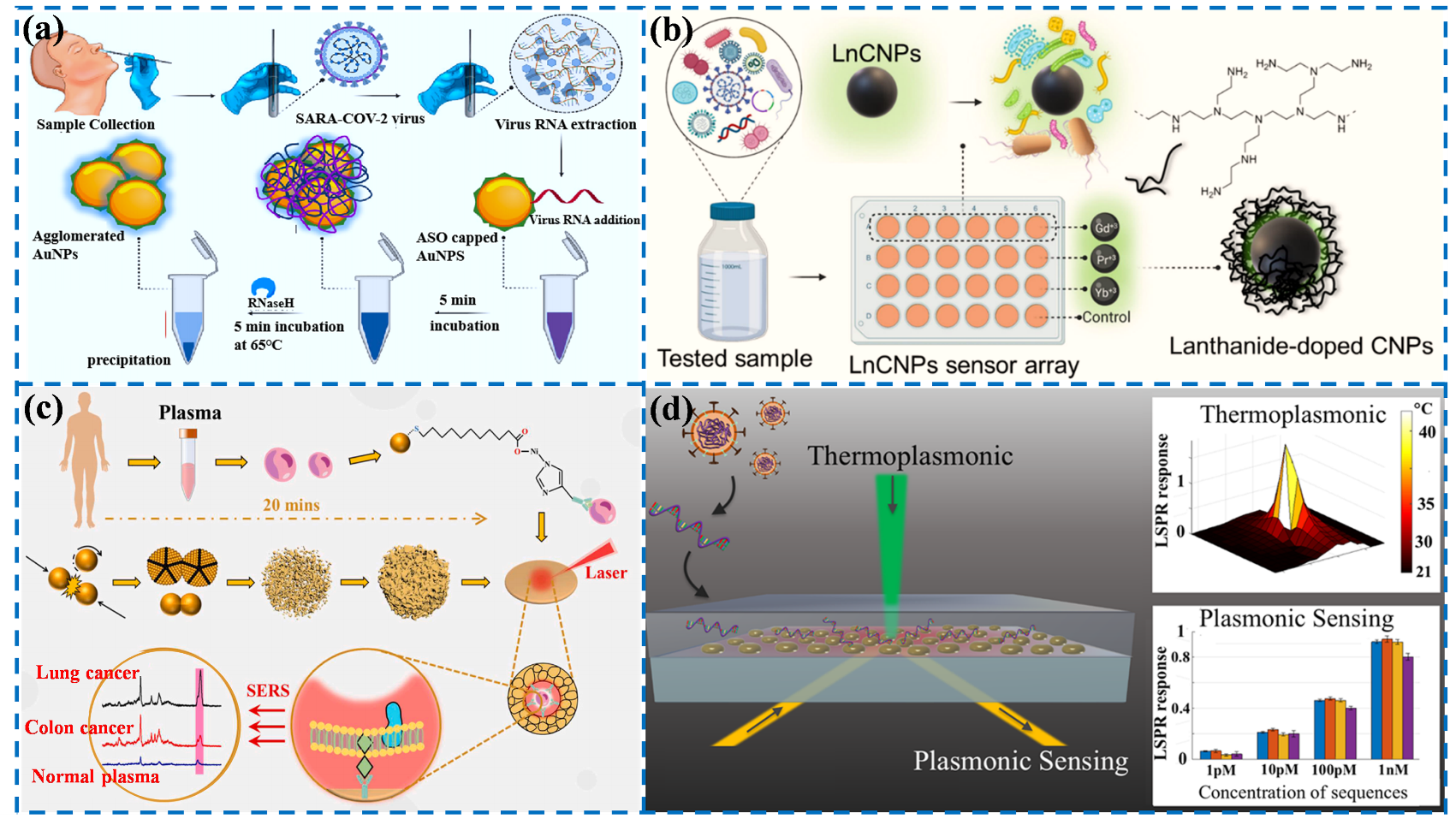
  26. Moitra, P.; Alafeef, M.; Dighe, K.; Frieman, M.B.; Pan, D. Selective naked-eye detection of SARS-CoV-2 mediated by N gene targeted antisense oligonucleotide capped plasmonic nanoparticles. ACS Nano 2020, 14, 7617–7627.
  27. Alafeef, M.; Dighe, K.; Moitra, P.; Pan, D. Monitoring the viral transmission of SARS-CoV-2 in still waterbodies using a lanthanide-doped carbon nanoparticle-based sensor array. ACS Sustain. Chem. Eng. 2022, 10, 245–258.
  28. Lin, C.; Liang, S.; Li, Y.; Peng, Y.; Huang, Z.; Li, Z.; Yang, Y.; Luo, X. Localized plasmonic sensor for direct identifying lung and colon cancer from the blood. Biosens. Bioelectron. 2022, 211, 114372.
  29. Qiu, G.; Gai, Z.; Tao, Y.; Schmitt, J.; Kullak-Ublick, G.A.; Wang, J. Dual-functional plasmonic photothermal biosensors for highly accurate severe acute respiratory syndrome coronavirus 2 detection. ACS Nano 2020, 14, 5268–5277.
  30. Pashchenko, O.; Shelby, T.; Banerjee, T.; Santra, S. A comparison of optical, electrochemical, magnetic, and colorimetric point-of-care biosensors for infectious disease diagnosis. ACS Infect. Dis. 2018, 4, 1162–1178.
  31. Huang, R.; He, L.; Li, S.; Liu, H.; Jin, L.; Chen, Z.; Zhao, Y.; Li, Z.; Deng, Y.; He, N. A simple fluorescence aptasensor for gastric cancer exosome detection based on branched rolling circle amplification. Nanoscale 2020, 12, 2445–2451.
  32. Li, B.; Yu, Q.; Duan, Y. Fluorescent labels in biosensors for pathogen detection. Crit. Rev. Biotechnol. 2015, 35, 82–93.
  33. Förster, T. Energy migration and fluorescence. J. Biomed. Opt. 2012, 17, 011002.
  34. Wu, B.Y.; Yan, X.P. Bioconjugated persistent luminescence nanoparticles for foster resonance energy transfer immunoassay of prostate specific antigen in serum and cell extracts without in situ excitation. Chem. Commu. 2015, 51, 3903–3906.
  35. Liao, J.; Madahar, V.; Dang, R.; Jiang, L. Quantitative FRET (qFRET) technology for the determination of protein-protein interaction affinity in solution. Molecules 2021, 26, 6339.
  36. Chen, T.; Shang, Y.; Zhu, Y.; Hao, S.; Yang, C. Activators confined upconversion nanoprobe with near-unity forster resonance energy transfer efficiency for ultrasensitive detection. ACS Appl. Mater. Inter. 2022, 14, 19826–19835.
  37. Teng, X.; Sun, X.; Pan, W.; Song, Z.; Wang, J. Carbon dots confined in silica nanoparticles for triplet-to-singlet föster resonance energy-transfer-induced delayed fluorescence. ACS. Appl. Nano Mater. 2022, 5, 5168–5175.
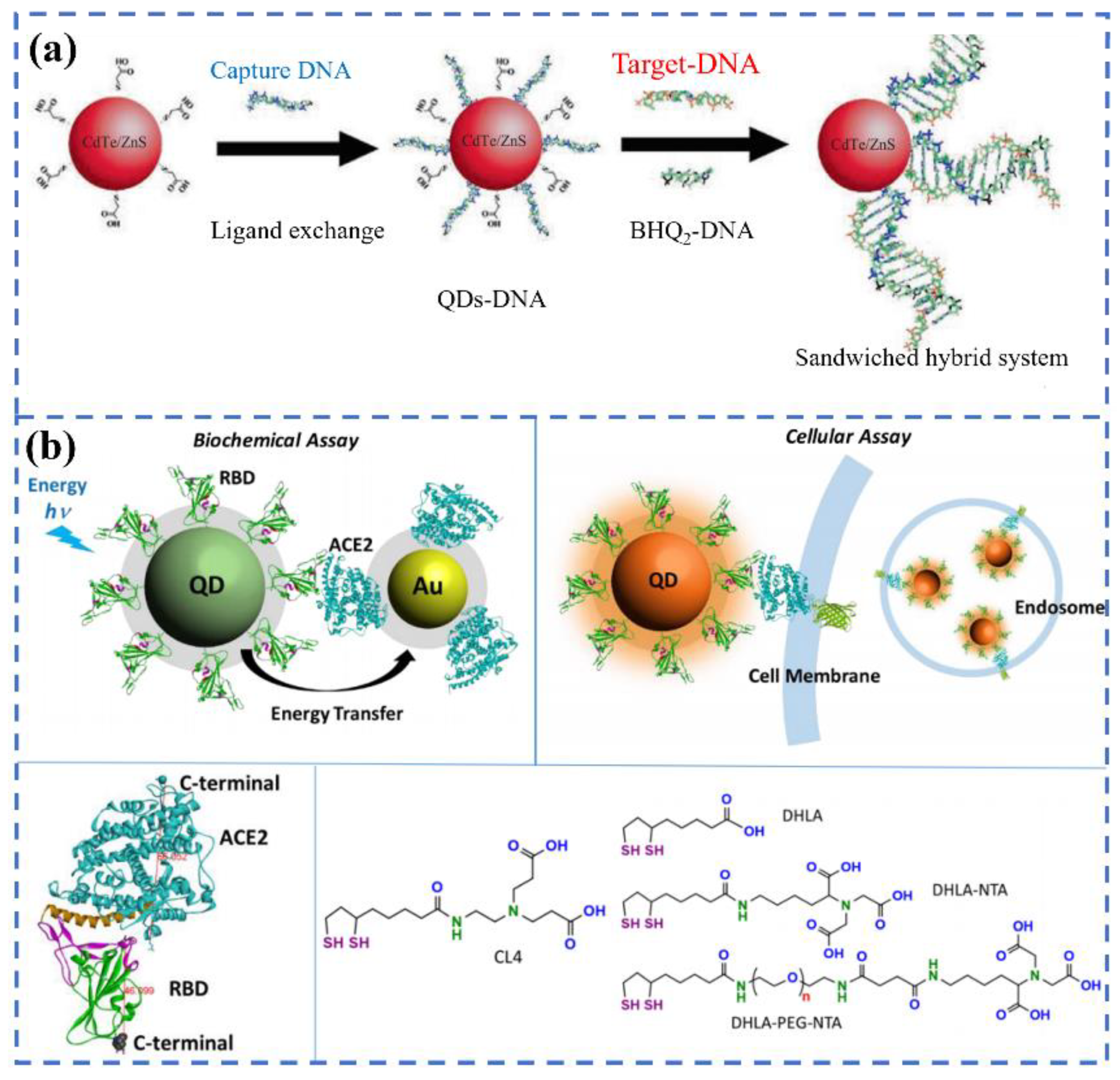
  38. Bardajee, G.R.; Zamani, M.; Mahmoodian, H.; Elmizadeh, H.; Yari, H.; Jouyandeh, L.; Shirkavand, R.; Sharifi, M. Capability of novel fluorescence DNA-conjugated CdTe/ZnS quantum dots nanoprobe for COVID-19 sensing. Spectrochim. Acta Part A Mol. Biomol. Spectrosc. 2022, 269, 120702.
  39. Gorshkov, K.; Susumu, K.; Chen, J.; Xu, M.; Pradhan, M.; Zhu, W.; Hu, X.; Breger, J.C.; Wolak, M.; Oh, E. Quantum dot-conjugated SARS-CoV-2 spike pseudo-virions enable tracking of angiotensin converting enzyme 2 binding and endocytosis. ACS Nano 2020, 14, 12234–12247.
  40. Bardajee, G.R.; Zamani, M.; Sharifi, M.; Rezanejad, H.; Motallebi, M. Rapid and highly sensitive detection of target DNA related to COVID-19 virus with a fluorescent bio-conjugated probe via a FRET mechanism. J. Fluoresc. 2022, 32, 1959–1967.
  41. Zhou, S.; Tu, D.; Liu, Y.; You, W.; Zhang, Y.; Zheng, W.; Chen, X. Ultrasensitive point-of-care test for tumor marker in human saliva based on luminescence-amplification strategy of lanthanide nanoprobes. Adv. Sci. 2021, 8, 2002657.
  42. Chen, R.; Ren, C.; Liu, M.; Ge, X.; Qu, M.; Zhou, X.; Liang, M.; Liu, Y.; Li, F. Early detection of SARS-CoV-2 seroconversion in humans with aggregation-induced near-infrared emission nanoparticle-labeled lateral flow immunoassay. ACS Nano 2021, 15, 8996–9004.
  43. Moitra, P.; Alafeef, M.; Dighe, K.; Sheffield, Z.; Dahal, D.; Pan, D. Synthesis and characterisation of N-gene targeted NIR-II fluorescent probe for selective localisation of SARS-CoV-2. Chem. Commun. 2021, 57, 6229–6232.
  44. Peters, J.A.; Djanashvili, K.; Geraldes, C.F.G.C.; Platas-Iglesias, C. The chemical consequences of the gradual decrease of the ionic radius along the Ln-series. Coord. Chem. Rev. 2020, 406, 213146.
  45. Zou, Q.; Huang, P.; Zheng, W.; You, W.; Li, R.; Tu, D.; Xu, J.; Chen, X. Cooperative and non-cooperative sensitization upconversion in lanthanide-doped LiYbF4 nanoparticles. Nanoscale 2017, 9, 6521–6528.
  46. Liu, Q.; Cheng, S.; Chen, R.; Ke, J.; Liu, Y.; Li, Y.; Feng, W.; Li, F. Near-infrared lanthanide-doped nanoparticles for a low interference lateral flow immunoassay test. ACS Appl. Mater. Inter. 2020, 12, 4358–4365.
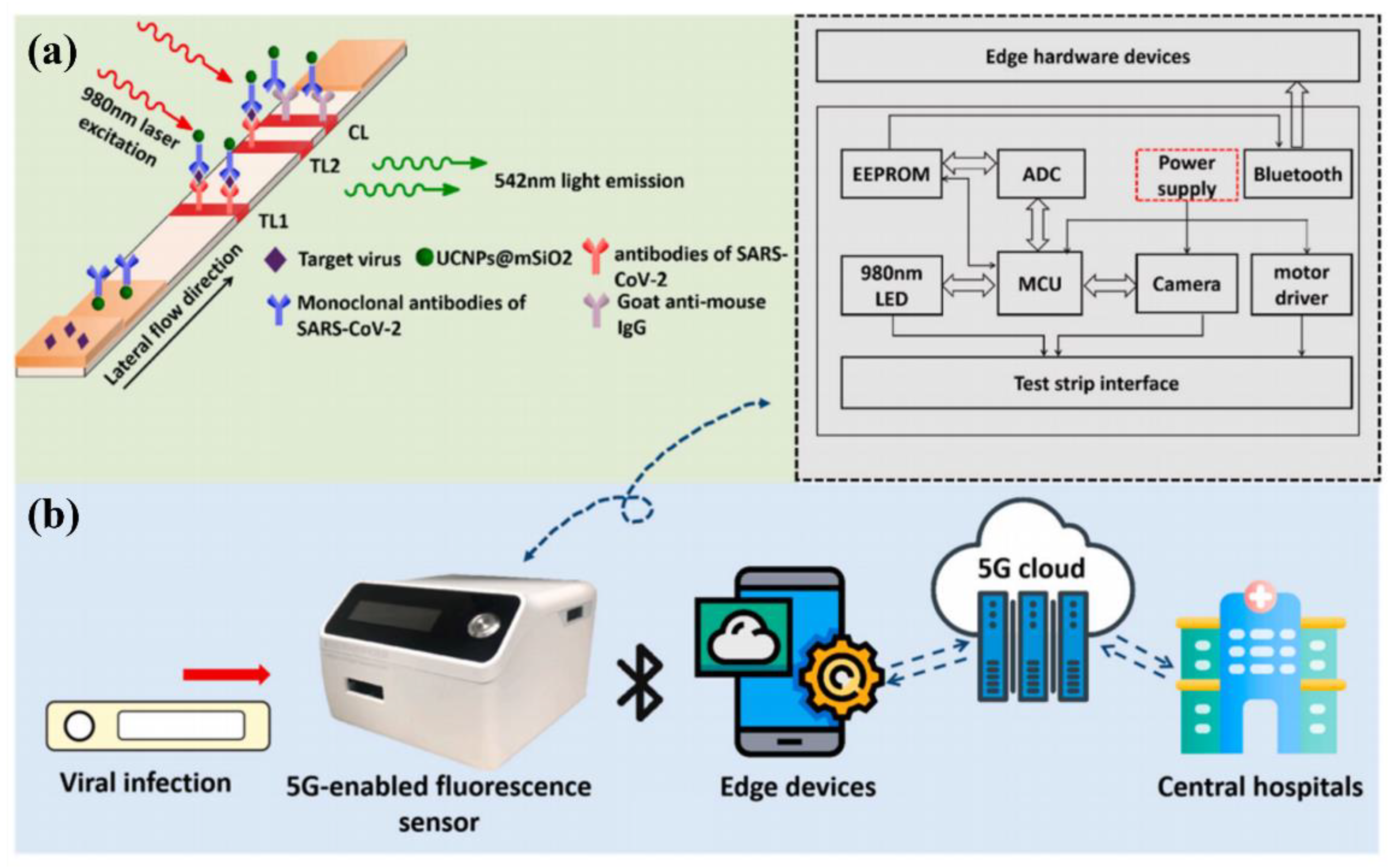
  47. Guo, J.; Chen, S.; Tian, S.; Liu, K.; Ni, J.; Zhao, M.; Kang, Y.; Ma, X.; Guo, J. 5G-enabled ultra-sensitive fluorescence sensor for proactive prognosis of COVID-19. Biosens. Bioelectron. 2021, 181, 113160.
  48. Feng, M.; Chen, J.; Xun, J.; Dai, R.; Zhao, W.; Lu, H.; Xu, J.; Chen, L.; Sui, G.; Cheng, X. Development of a sensitive immunochromatographic method using lanthanide fluorescent microsphere for rapid serodiagnosis of COVID-19. ACS Sens. 2020, 5, 2331–2337.
  49. Chen, Z.; Zhang, Z.; Zhai, X.; Li, Y.; Lin, L.; Zhao, H.; Bian, L.; Li, P.; Yu, L.; Wu, Y.; et al. Rapid and sensitive detection of anti-SARS-CoV-2 IgG, using lanthanide-doped nanoparticles-based lateral flow immunoassay. Anal. Chem. 2020, 92, 7226–7231.
  50. Hu, R.; Liao, T.; Ren, Y.; Liu, W.; Ma, R.; Wang, X.; Lin, Q.; Wang, G.; Liang, Y. Sensitively detecting antigen of SARS-CoV-2 by NIR-II fluorescent nanoparticles. Nano. Res. 2022, 15, 7313–7319.
  51. Serrano-Aroca, A.; Takayama, K.; Tunon-Molina, A.; Seyran, M.; Hassan, S.S.; Pal Choudhury, P.; Uversky, V.N.; Lundstrom, K.; Adadi, P.; Palu, G.; et al. Carbon-based nanomaterials: Promising antiviral agents to combat COVID-19 in the microbial-resistant era. ACS Nano 2021, 15, 8069–8086.
  52. Pinals, R.L.; Ledesma, F.; Yang, D.; Navarro, N.; Jeong, S.; Pak, J.E.; Kuo, L.; Chuang, Y.C.; Cheng, Y.W.; Sun, H.Y.; et al. Rapid SARS-CoV-2 spike protein detection by carbon nanotube-based near-infrared nanosensors. Nano. Lett. 2021, 21, 2272–2280.
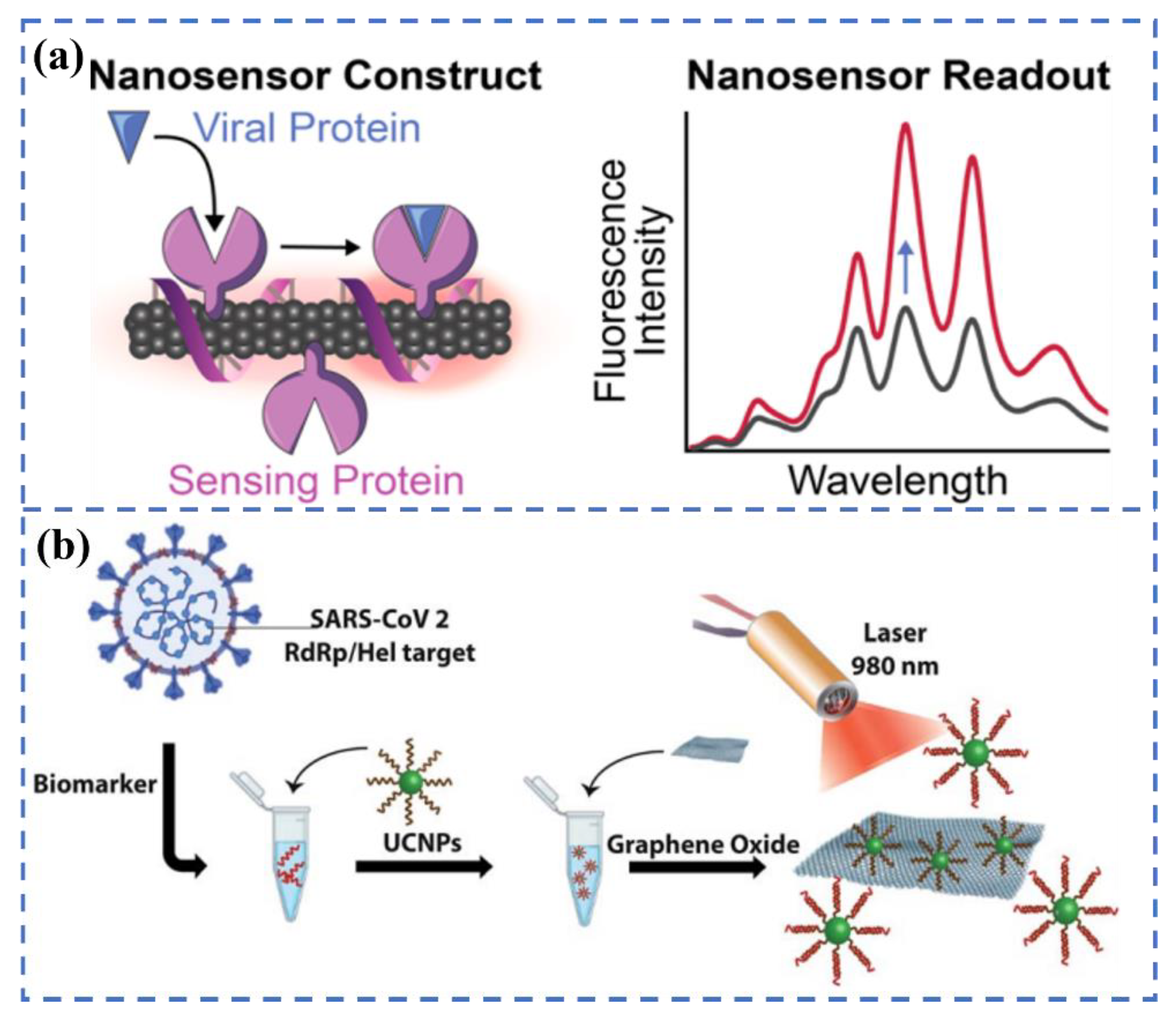
  53. Alexaki, K.; Kyriazi, M.E.; Greening, J.; Taemaitree, L.; El-Sagheer, A.H.; Brown, T.; Zhang, X.; Muskens, O.L.; Kanaras, A.G. A SARS-CoV-2 sensor based on upconversion nanoparticles and graphene oxide. RSC Adv. 2022, 12, 18445–18449.
  54. Masterson, A.N.; Liyanage, T.; Berman, C.; Kaimakliotis, H.; Johnson, M.; Sardar, R. A novel liquid biopsy-based approach for highly specific cancer diagnostics: Mitigating false responses in assaying patient plasma-derived circulating microRNAs through combined SERS and plasmon-enhanced fluorescence analyses. Analyst 2020, 145, 4173–4180.
  55. Song, C.; Li, J.; Sun, Y.; Jiang, X.; Zhang, J.; Dong, C.; Wang, L. Colorimetric/SERS dual-mode detection of mercury ion via SERS-Active peroxidase-like NPs. Sens. Actuators B Chem. 2020, 310, 127849.
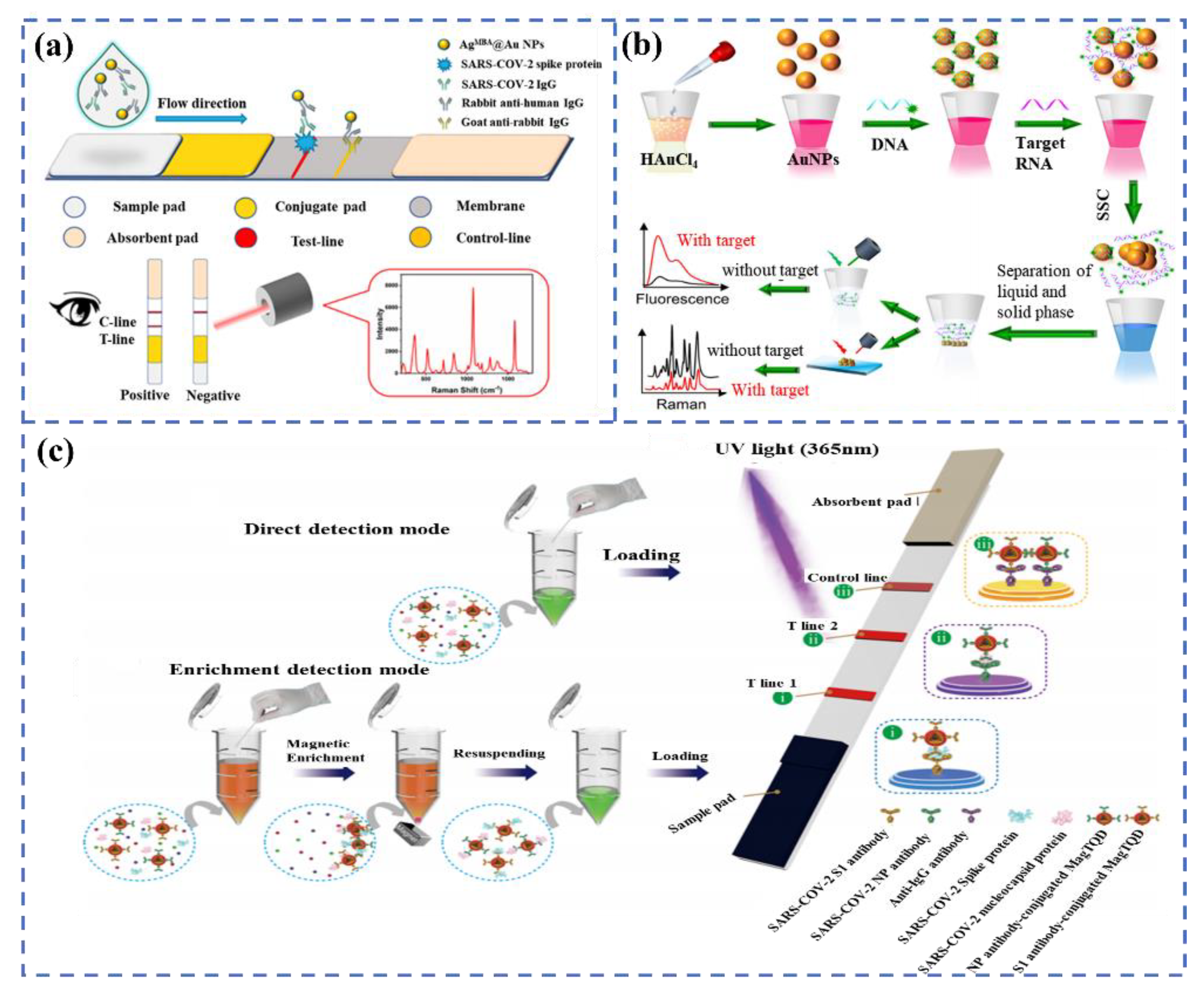
  56. Han, H.; Wang, C.; Yang, X.; Zheng, S.; Cheng, X.; Liu, Z.; Zhao, B.; Xiao, R. Rapid field determination of SARS-CoV-2 by a colorimetric and fluorescent dual-functional lateral flow immunoassay biosensor. Sens. Actuators B Chem. 2022, 351, 130897.
  57. Liang, P.; Guo, Q.; Zhao, T.; Wen, C.Y.; Tian, Z.; Shang, Y.; Xing, J.; Jiang, Y.; Zeng, J. Ag Nanoparticles with ultrathin au shell-based lateral flow immunoassay for colorimetric and SERS dual-mode detection of SARS-CoV-2 IgG. Anal. Chem. 2022, 94, 8466–8473.
  58. Gao, Y.; Han, Y.; Wang, C.; Qiang, L.; Gao, J.; Wang, Y.; Liu, H.; Han, L.; Zhang, Y. Rapid and sensitive triple-mode detection of causative SARS-CoV-2 virus specific genes through interaction between genes and nanoparticles. Anal. Chim. Acta. 2021, 1154, 338330.
  59. Wang, C.; Cheng, X.; Liu, L.; Zhang, X.; Yang, X.; Zheng, S.; Rong, Z.; Wang, S. Ultrasensitive and simultaneous detection of two specific SARS-CoV-2 antigens in human specimens using direct/enrichment dual-mode fluorescence lateral flow immunoassay. ACS Appl. Mater. Inter. 2021, 13, 40342–40353.
  60. Wang, C.; Yang, X.; Gu, B.; Liu, H.; Zhou, Z.; Shi, L.; Cheng, X.; Wang, S. Sensitive and simultaneous detection of SARS-CoV-2-specific IgM/IgG using lateral flow immunoassay based on dual-mode quantum dot nanobeads. Anal. Chem. 2020, 92, 15542–15549.
More
This entry is offline, you can click here to edit this entry!
Academic Video Service