Diffuse low-grade glioma (LGG), i.e., WHO grade II glioma [
1], is a rare brain cancer, whose ethiopathogeny is poorly understood, making difficult the prediction of its natural course, especially at an individual level [
2]. LGG spontaneously exhibits different stages in its evolution, namely (i) a pre-symptomatic period in which the tumor is usually slow-growing, as demonstrated in cases of incidental discovery [
3]; (ii) a symptomatic period in which the glioma induces clinical consequences, usually seizures and/or mild cognitive impairments visible on neuropsychological assessment [
4], while continuing to progress slowly but constantly (about 3–4 mm mean diameter per year) [
5]; and (iii) a period of malignant transformation (MT) with acceleration of the growth rate, resulting in more severe neurological deficits and ultimately death [
6]. However, LGG represents a heterogeneous group of tumors with various courses, which are difficult to predict at the individual level and at each stage of the disease. In addition, due to its unknown origin and diffuse features within the central nervous system, this is an incurable tumoral disease. Thus, the main goal of therapeutic management is to delay progression to a higher grade of malignancy while preserving quality of life (QoL) for as long as possible [
7]. To this end, early maximal safe surgical excision represents initial treatment, since reduction of the tumor volume decreases the risk of MT [
8] and thus prolongs overall survival (OS) [
9,
10,
11,
12,
13]. Moreover, because intratumoral heterogeneity is frequent in LGG, large resection increases the chances of detecting possible microfoci of high-grade glioma within the neoplasm, enabling better adaptation of the next strategy according to extensive histomolecular data [
14,
15,
16]. In other words, from an oncological perspective, variability in LGG is considerable not only at any given moment, then over months or years, but also from one patient to another one depending on the intrinsic glioma behavior (partly related to the genetic subtype), and depends also on the time of diagnosis in the natural history of the disease [
17].
In the same spirit, with regard to functional considerations, there are very high variations between brain organization across patients as well as in the same patient over time, due to physiological interindividual anatomo-functional variability, which is increased in the event of LGG [
18]. In fact, this type of slow-growing neoplasm which progresses over years or even decades [
19] may induce reactional processes of neural network reconfiguration that allow functional compensation, at least at the first stage of the disease [
20]. This explains why patients harboring LGG, mostly young adults, usually enjoy active familial and socio-professional lives, sometimes despite the presence of a voluminous tumor at diagnosis, possibly involving so-called “eloquent” areas. In practice, it is essential to better explore the mechanisms underpinning neuroplasticity at the individual level, since the patterns of cerebral reallocation may vary from one patient to another (for example, predominant compensatory recruitment of peritumoral structures versus predominant recruitment of contra-hemispheric homologous areas [
21]), as well as in a given patient over time—e.g., with a switch from a perilesional functional rearrangement to a more bilateral reshaping [
22]. Prediction of such potential for metaplasticity (plasticity of plasticity) is very complex, while the therapeutic strategy should be tailored according to these brain connectome dynamics for each individual glioma patient [
23]. This is particularly relevant for planning maximal surgical resection, which must be achieved up to the functional boundaries identified by intraoperative mapping in conscious patients, with the aim of preserving cortico-subcortical networks critical for neural functions at the individual level [
24]. Considering that the possibility of reoperation several months or years later will depend on the way the brain has reorganized (or not) since previous surgery, if optimization of the resection does not seem feasible because the limitations of neuroplasticity have been reached, medical adjuvant treatment represents a relevant alternative [
25].
2. Predicting Oncological Interindividual Variability and Its Changes over Time
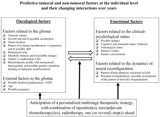
Distinct types of factors must be taken into consideration, including those related to the tumoral disease itself as well as external factors which may influence the course of glioma (Figure 1).
Figure 1. Predictive tumoral and non-tumoral factors at the individual level and their changing interactions over years.
2.1. Factors Related to the Glioma
2.1.1. Tumoral Volume
The volume of LGG at diagnosis is highly variable, from 0.39 to 386 cc according to recent studies [
4,
27]. Notably, it has long been considered that a larger size of LGG is an adverse prognostic factor; however, only one dimension of the tumor was classically measured, with a poorer prognosis when the size was ≥6 cm [
28,
29,
30]. More recently, it has been evidenced that a greater LGG volume (by definition, calculated in 3D) was significantly correlated with a higher risk of MT and with shorter OS [
9,
10,
26]. This may be explained by the fact that a more voluminous glioma reflects a more prolonged natural history of the disease, with a higher number of tumoral cells and therefore an increased risk of mutational accumulations [
31,
32]. Such a hypothesis is supported by a decreased risk of MT as well as a significant survival benefit in incidental LGG compared with symptomatic LGG discovered later [
3], although a higher volume at discovery of incidental LGG is also significantly associated with more progressive tumors [
33]. Consequently, a screening policy has been proposed to diagnose and treat LGG earlier, as well as to optimize the opportunity to better understand the origin of these tumors [
34]. In addition, during the follow-up of LGG patients already treated, it has also been suggested that further treatment(s) should be considered when the tumor volume re-increases over time, especially when it reaches a threshold of about 10–15 cc (even if the patient is asymptomatic at that time), in order to delay MT [
7].
2.1.2. Growth Rate
The velocity of a tumor’s spontaneous expansion, which can be plotted as a function of mean glioma diameter over time (computed from the volumes calculated by repeat MRIs) is predictive of long-term outcomes for LGG [
5]. Indeed, the slope of the mean tumor diameter growth curve is an independent prognostic factor for malignant progression-free survival and for OS as a continuous predictor—that is, showing a linear relationship between OS and growth rate [
35]. The relevant kinetics are very variable from one LGG to another at diagnosis, from less than 1 mm per year to 8 mm per year; over 8 mm/year, the LGG invokes a greater risk of MT [
35]. Interestingly, this marker is independent of the molecular profile [
35], in particular with regard to the IDH status [
36]. Thus, identifying rapidly growing LGG during the pretherapeutic period, i.e., tumors at higher risk of worsening evolution, can be helpful to decide when to start treatment(s) [
37], including for incidental LGG [
38]. This is also true during the surveillance of LGG patients already treated, in order to determine when to consider further therapy. Especially because the growth rate is similar before and after surgery [
39], acceleration of the velocity of the residual tumor in cases of incomplete resection may support earlier re-operation, even in asymptomatic patients [
25]. Furthermore, re-growth after a period of stabilization following chemotherapy and/or radiotherapy can prompt consideration of further treatment [
7]. The possibility of such changes favors deployment of a systematic control MRI every 3 to 6 months throughout life.
2.1.3. Pattern of Migration Versus Proliferation and Tumor Location
In addition to variable glioma volumes and kinetics, LGG may exhibit distinct patterns of progression within the brain, i.e., a more proliferative “bulky” pattern versus a more diffuse “migratory” one. Interindividual variability may be considerable, ranging from a very focal tumor to a gliomatosis with bi-hemispheric dissemination [
40]. Notably, in incidentally discovered focal LGG, the insular location was found to be a predictive factor of a more progressive tumor [
33]. In a study of 1097 cases, a tumor located in a nonfrontal area was an independent factor of poor prognosis [
10]. With its invasive profile, glioma has a high propension to migrate along the subcortical fibers, as shown radiologically [
41,
42] as well as pathologically: biopsy samples evidenced that the tumor cells followed the white matter tracts and were slightly more concentrated in the peripheral parts of those tracts [
43]. It seems that the myelin status could play a pivotal role not only in LGG invasion in adults but also possibly in its origin in teenagers [
44]. Biomathematical models have attempted to anticipate the profile of glioma evolution [
45], knowing nonetheless that this pattern might change over time, maybe at least partly in relation to the therapeutic effects; for example, a bulky LGG before surgery may switch towards a predominantly diffuse pattern after incomplete resection [
40]. Such a parameter is critical for treatment selection, because white matter connectivity represents the main limitation of neuroplasticity (see below). This could be a major problem for achieving massive surgical resection or for wide brain radiation therapy if the patient hopes to preserve an optimal QoL, particular in very diffuse LGG [
23].
2.1.4. The Peritumoral Zone
LGG is a heterogeneous and poorly circumscribed neoplasm with isolated tumor cells (ITC) that extend beyond the margins of the lesion depicted on MRI, as demonstrated by biopsy samples taken from within and beyond the “glioma core” (visible as a T2-FLAIR hypersignal on MRI); ITCs have been detected behind such signal abnormalities [
43,
46]. It is worth noting that the cycling tumor cell fraction was higher at the limits of the MRI-defined abnormalities than when closer to the center of the tumor, in 62.5% of patients [
47]. This could explain the high risk of glioma relapse at the periphery of the surgical cavity, even following large resection [
48]. Efforts to demarcate the glioma core from the surrounding healthy brain led to the definition of an intermediate region, the so-called peritumoral zone (PTZ) [
49]. An important interindividual variability exists regarding this PTZ, as demonstrated by samples which found ITC from 10 to 20 mm around the glioma core [
43,
46], in agreement with the fact that the tumor core might be more bulky or more diffuse (see above). Interestingly, recent investigations have indicated that this interface between the glioma core and the healthy brain represents a specific metabolic and cellular entity. Such characteristics of the PTZ that are being increasingly explored through radiomics and radiogenomics [
50,
51] may play a pivotal role for decision making in the management of diffuse LGG.
2.1.5. Metabolic Changes
While still a matter of debate from a radiological perspective, the occurrence of an enhancement during the course of LGG is usually associated with MT [
52,
53]. However, if the tumor has already become more aggressive when the (re)treatment is proposed, this means in essence that the opportunity for action has been missed [
38]. Therefore, since the main oncological goal is to prevent MT, additional non-invasive metabolic information may be useful in order to predict when the LGG has a higher risk of degeneration. First, an increase of perfusion or diffusion value(s) obtained through sequential and multimodal MRI could be a predictor of changes in glioma behavior [
54], possibly identifiable using new machine-learning classifiers [
55], and might prompt earlier (re)treatment. In the same spirit, recent advances in PET scanning using tracers easily accessible in routine practice (such as F-DOPA) have enabled an increase in sensitivity for the detection of foci of MT within the LGG, before the onset of enhancement [
56]. As mentioned, metabolic imaging could also be helpful to better investigate the PTZ [
49]. This additional information could be of utmost interest for deciding the best timings of new therapies during follow-up of LGG patients.
2.2. External Factors to the Glioma
2.2.1. Familial Predisposition
While the majority of gliomas are sporadic in origin, familial gliomas have been described, although these are exceptionally rare, especially in the form of LGG [
69,
70]. A potential heritable etiology for glioma families has been evoked; specifically, high-penetrance familial mutations and common low-penetrance susceptibility loci (e.g., single-nucleotide polymorphisms (SNPs)) may contribute to familial glioma risk [
70,
71]. Nonetheless, recent series have shown that familial gliomas, including LGG, showed similar genomic and molecular biomarker profiles to sporadic gliomas, consistent with the similarity in their clinical features [
72,
73]. However, identification of new susceptibility factors in familial LGG might help to elucidate the molecular pathogenesis of gliomas [
73]. In practice, to increase the chances of earlier diagnosis of possible LGG, screening can be offered to relatives of gliomas patients, and such an “intentional discovery” may lead to more rapid treatment in the first period of the disease [
74].
2.2.2. Age
Older age has been correlated with poorer oncological outcomes in LGG patients, even though the cut-off may vary, e.g., 40 years [
28,
29,
30] versus 55 years [
10]. Nevertheless, even if the prognosis seems to be directly linked to age per se, it cannot be rule out that glioma discovery in a younger patient may mean that the diagnosis was made at an earlier stage of the tumoral disease—therefore, with lesser volume and fewer mutational changes. Furthermore, LGG mainly affects young adults, explaining why screening policy design has suggested the application of MRI in a selected population before 40 years [
75]. In clinical routine, beyond the age at diagnosis and during the years (or even decades) of LGG management, practitioners be aware that the risk of MT is potentially increasing as the patient becomes older, justifying continuation of regular surveillance even in cases of long-term tumor stabilization [
40].
3. Predicting Neural Interindividual Variability and Its Changes over Time
Although linked, factors related to the clinical–psychological status of the patient and those related to the dynamics of neural network reconfiguration are here considered separately.
3.1. Factors Related to the Clinical–Psychological Status of the Patient
3.1.1. Epilepsy
Seizure is the first symptom in LGG, leading to diagnosis in the vast majority of patients [
104]. An epileptic symptomatology is linked to a better oncological outcome [
10]. Interestingly, computational models have evidenced that the onset of seizures corresponds with a time point which may already represent the overcompensation stage of cerebral plasticity, depending on the tumor growth rate and its pattern of progression, in particular in the event of massive invasion of the white matter tracts [
105]. Translation of these results into the clinical situation is another argument in favor of early surgery in asymptomatic patients, before the occurrence of epilepsy [
74]. Indeed, seizures can have a negative impact on daily life, in particular by preventing driving (and indirectly employment) for medico-legal reasons [
91,
106]. Furthermore, epilepsy is mainly elicited by the diffusion of the tumoral cells at the periphery of the LGG (and not by the glioma core itself) [
107]. This explains why larger surgical resection has a higher impact on epilepsy. Indeed, postoperative seizure control is more likely when EOR is ≥91% and/or residual tumor volume is ≤19 cc [
108]. This also supports the suggestion of supratotal resection (with removal of the PTZ) for functional reasons (in addition to the improvement of oncological outcomes), i.e., with an optimization of QoL as a result of freedom from epilepsy [
49].
Notably, important inter-individual variability has been observed, with about 15% of LGG patients experiencing intractable seizures, notably in temporal and/or paralimbic gliomas. In this situation, it has been proposed to remove the hippocampus, even if not invaded by the tumor according to preoperative MRI, since this can result in significant improvement of epilepsy control [
109]. During follow-up, the reappearance of seizures may be correlated with LGG relapse, and may prompt clinicians to propose reoperation prior to MT [
25]. When further resection is not possible due to diffusion within critical structures, for example within the Rolandic area which is very epileptogenic [
110], adjuvant medical treatment can have an impact on seizures [
111].
3.1.2. Cognitive and Emotional Status
Because LGG patients are generally young and enjoy active lives at diagnosis, presenting no or only slight deficit at so-called “standard neurological examination”, it has been claimed in the literature that these patients do not exhibit any significant functional disturbances [
6]. However, important variations are found across patients in terms of their neurocognitive status. In fact, a recent cohort including 157 LGG patients who benefited from extensive neuropsychological evaluation before treatment showed that 55.4% of them had already experienced cognitive decline, in particular with respect to language, verbal episodic memory, psychomotor speed, attention, and executive functions (phonological and categorical fluency) [
4]. Interestingly, neurocognitive impairments have also been found in patients with incidental LGG [
112].
3.1.3. Neurological Status
Due to earlier diagnosis of LGG, moderate or severe neurological deficits at clinical examination (e.g., hemiparesis or aphasia) are usually rare [
17]. Beyond possible episodes of transient worsening which might be elicited by repeat seizures, permanent impairments that arise are generally due to voluminous gliomas with mass effect and/or to MT, with acceleration of the neoplasm kinetics. In other words, with more “prophylactic management”, such major deteriorations should cease to occur before the last stage of the disease, thus giving the opportunity for LGG patients to enjoy active lives for many years or even decades [
7].
3.1.4. Patients’ Needs
Definition of QoL is eminently variable from one human being to another. Indeed, beyond the fact that patients do not want to experience hemiplegia or aphasia, especially following surgical resection, their expectations are very different according to their lifestyles: do they work? (if yes, what kind of employment they have, and do they want to resume their professional activities postoperatively and/or during medical treatment?); what are their hobbies? (e.g., do they practice sports, art, etc.?); what is their socio-cultural environment? (e.g., do they speak multiple languages?) [
127], do they need to drive? (if yes, what about possible medico-legal issues in case of visual field deficit and/or seizures?) [
106]. On the basis of these individual wishes, with the aim of enabling each patient to develop long-term projects (such as getting married, having a baby, buying a house, etc.), and also with due consideration given to the neurocognitive assessment at diagnosis (i.e., the presence or otherwise of some degrees of disturbance), it is possible to elaborate tailored management “à la carte”, beginning with a selection of optimal tasks to be performed during awake surgery [
128]. Moreover, therapeutic strategies should be re-adapted over time, not only according to the LGG course, but also taking into account possible changes in the patient’s priorities. An example of such an intra-individual variability could be when a patient would like to preserve executive functions during the first surgery because he or she was employed at the time, but sparing higher-order cognitive capacities is no longer absolutely mandatory a few years later because the patient has retired in the meantime. Therefore, a clear and extensive explanation of the principles of chronic tumoral disease should be provided to the patient and his or her relatives after the diagnosis. They should understand that management necessitates surveillance with constant anticipation of a specific lifelong therapeutic strategy, to give them the opportunity to make choices for their current and future life by thinking one step ahead—which increases the chance of finding a better psychological equilibrium [
7,
23].
3.2. Factors Related to the Dynamics of the Neural Network Reconfiguration
3.2.1. Patterns of Neuroplasticity
The structural anatomy of the brain is highly variable across healthy individuals, especially at the cortical level [
129], while variations are less pronounced at the level of the white matter tracts [
18]. Furthermore, advances in non-invasive functional imaging methods which permit the investigation of the functional connectivity have resulted in the development of a large database demonstrating between-subject variability in the distribution of neural networks [
130,
131]. Recent models of neurocognition employed in basic neuroscience have rejected the classical localizationist dogma (one cerebral site underpinning one specific function), and go beyond a simple network organization of the central nervous system (one cerebral circuit underpinning one specific function) by evidencing the critical role of dynamic interplay within and across neural networks which allows behaviors to be constantly adapted to the surrounding world [
132]. According to this meta-networking framework (based on a network of networks), complex cognitive abilities are made possible by the activation and coordination (combination or competition) of large-scale neural circuits involving domain-specific networks (e.g., movement or language circuits), and the activity of a multiple-demand system recruited during the performance of a wide range of cognitive-demanding activities with the aim of maintaining fluid intelligence [
132,
133].
Alongside this flexible and ever-changing physiological organization of the functional connectome, inter-individual anatomo-functional variability is significantly increased in brain-damaged patients, particularly in the event of slowly evolving lesions such as LGG [
134]. These mechanisms of neuroplastic functional reshaping permit neurological compensation during LGG growth (explaining why the vast majority of patients are active at diagnosis), at least to some extent, considering that over half of LGG patients already experience some degree of cognitive disturbance at the first neuropsychological evaluation, as previously mentioned [
4]. Maps of neuroplasticity have evidenced that potential for cortical reallocation is high (except for input such as the primary visual cortex and output such as the primary motor cortex), whereas axonal connectivity represents the main limit of functional reshaping [
135,
136,
137]. Thus, as in healthy subjects, variability across LGG patients is greater for cortical than subcortical reorganization [
17]. This implies that various dynamic processes of neural reconfiguration may be mobilized from one patient to another, such as peritumoral rearrangement, or recruitment of remote structures in the ipsilesional hemisphere and/or the contralateral side [
21,
22]. Inter-patient differences in such patterns of redistribution are strongly correlated with LGG characteristics, i.e., the volume of the tumor, the kinetics of the glioma (as plasticity is linked to the time course of the disease, with less compensation in more rapidly evolving lesions [
134]), and to the severity of brain invasion, with less plastic potential in more diffuse tumors which migrate more widely along the white matter pathways [
44,
138]. These connectomal considerations play a pivotal role when selecting the optimal therapeutic attitude, knowing that better prediction of the individual processes of neural reconfiguration may be valuable for anticipating the next treatment(s), thereby avoiding missing the opportunity for action from an oncological point of view.