Tonsilloliths, also known as tonsil stones, are soft aggregates of bacterial and cellular debris that form in the tonsillar crypts, the crevices of the tonsils. While they occur most commonly in the palatine tonsils, they may also occur in the lingual tonsils. Tonsil stones are common. Tonsilloliths have been recorded weighing from 0.3 g to 42 g. Protruding tonsilloliths may feel like foreign objects lodged in the tonsil crypt. They may be a nuisance and difficult to remove, but are usually not harmful. They are one of the causes of bad breath and always give off a putrid smell.
- tonsilloliths
- tonsil
- cellular
1. Signs and Symptoms
Tonsilloliths may produce no symptoms, or they may be associated with bad breath, or produce pain when swallowing.[1]
Tonsilloliths occur more frequently in teens than in adults or young children. Many small tonsil stones do not cause any noticeable symptoms. Even when they are large, some tonsil stones are only discovered incidentally on X-rays or CAT scans.
Other symptoms include a metallic taste, throat closing or tightening, coughing fits, and choking.
Larger tonsilloliths may cause multiple symptoms, including recurrent halitosis, which frequently accompanies a tonsil infection, sore throat, white debris, a bad taste in the back of the throat, difficulty swallowing, ear ache, and tonsil swelling.[2] A medical study conducted in 2007 found an association between tonsilloliths and bad breath in patients with a certain type of recurrent tonsillitis. Among those with bad breath, 75% of the subjects had tonsilloliths, while only 6% of subjects with normal halitometry values (normal breath) had tonsilloliths. A foreign body sensation may also exist in the back of the throat. The condition may also be an asymptomatic condition, with detection upon palpating a hard intratonsillar or submucosal mass.

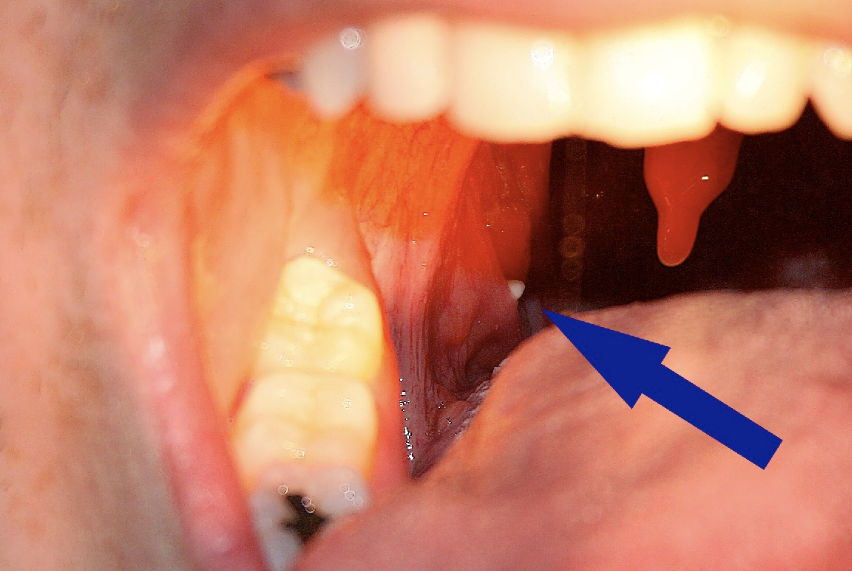
A tonsillolith protrudes from the tonsil

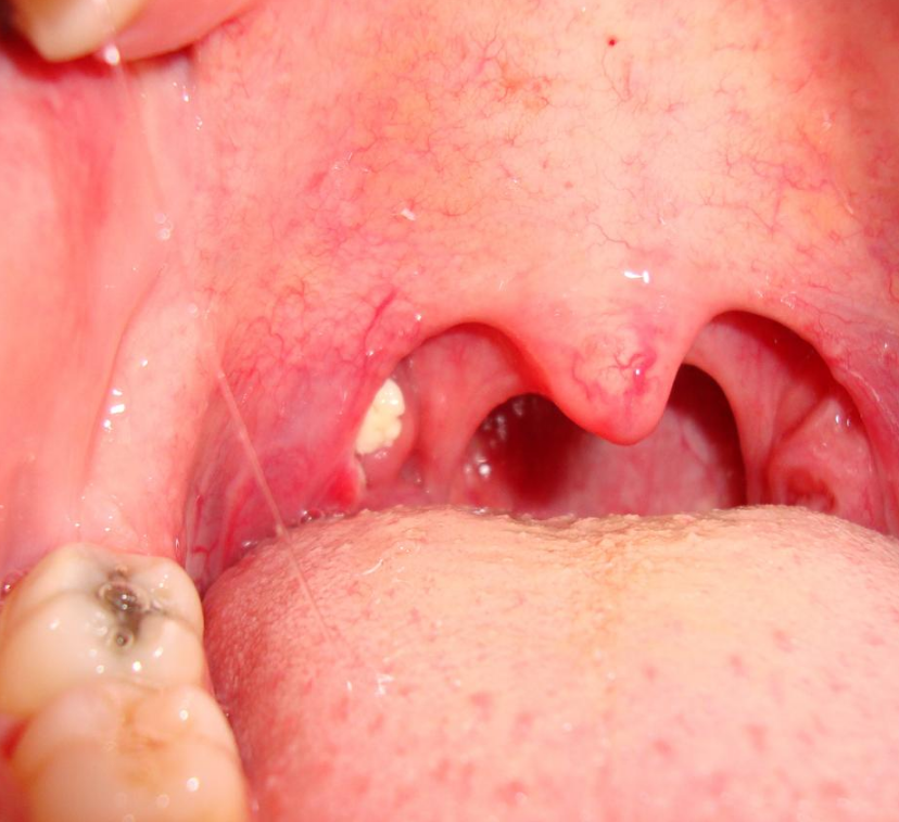
Large tonsillolith half exposed on tonsil

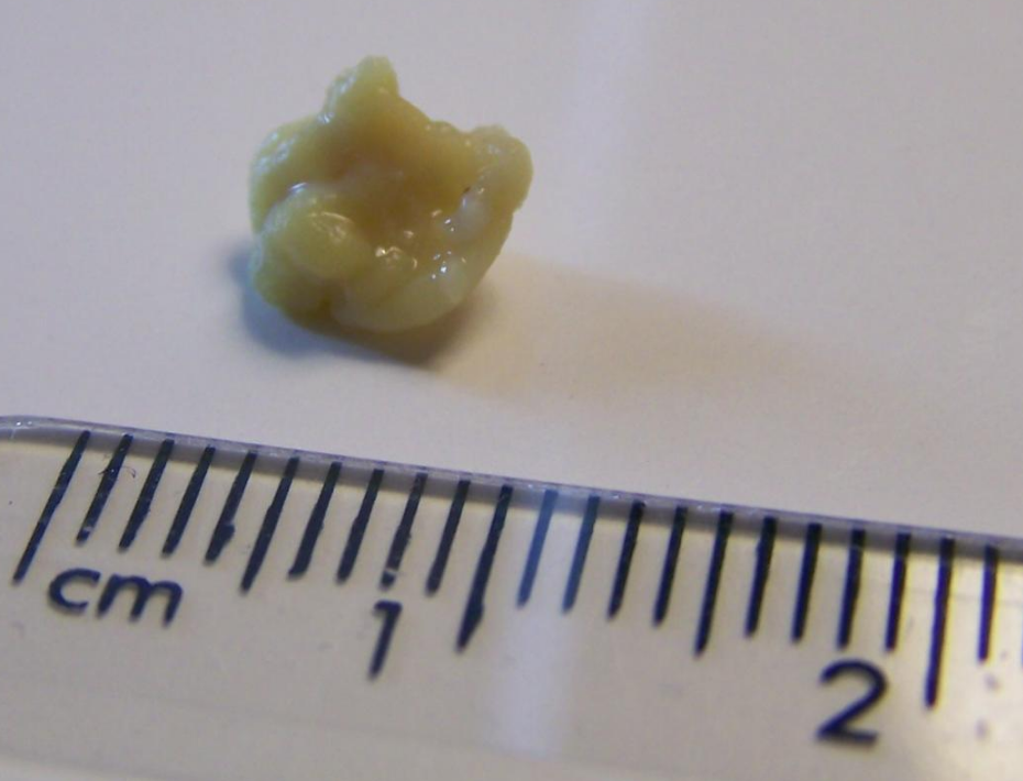
Closeup of a tonsillolith
2. Classification
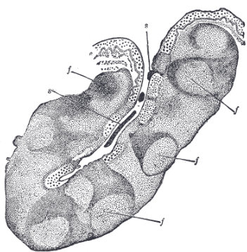
Tonsilloliths or tonsil stones are calcifications that form in the crypts of the palatal tonsils. They are also known to form in the throat and on the roof of the mouth. Tonsils are filled with crevices where bacteria and other materials, including dead cells and mucus, can become trapped. When this occurs, the debris can become concentrated in white formations that occur in the pockets.[2] Tonsilloliths are formed when this trapped debris accumulates and are expressed from the tonsil. They are generally soft, sometimes rubbery. This tends to occur most often in people who suffer from chronic inflammation in their tonsils or repeated bouts of tonsillitis.[2] They are often associated with post-nasal drip.
2.1. Giant
Much rarer than the typical tonsil stones are giant tonsilloliths. Giant tonsilloliths may often be mistaken for other oral maladies, including peritonsillar abscess, and tumours of the tonsil.[3]
3. Pathophysiology

The mechanism by which these calculi form is subject to debate,[4] though they appear to result from the accumulation of material retained within the crypts, along with the growth of bacteria and fungi – sometimes in association with persistent chronic purulent tonsillitis.
Recently, an association between biofilms and tonsilloliths was shown. Central to the biofilm concept is the assumption that bacteria form a three dimensional structure, dormant bacteria being in the center to serve as a constant nidus of infection. This impermeable structure renders the biofilm immune to antibiotic treatment. By use of confocal microscopy and microelectrodes, biofilms similar to dental biofilms were shown to be present in the tonsillolith, with oxygen respiration at the outer layer of tonsillolith, denitrification toward the middle, and acidification toward the bottom.[5]
4. Diagnosis
Diagnosis is usually made upon inspection. Differential diagnosis of tonsilloliths includes foreign body, calcified granuloma, malignancy, an enlarged temporal styloid process or rarely, isolated bone which is usually derived from embryonic rests originating from the branchial arches.[6]
Tonsilloliths are difficult to diagnose in the absence of clear manifestations, and often constitute casual findings of routine radiological studies.
Imaging diagnostic techniques can identify a radiopaque mass that may be mistaken for foreign bodies, displaced teeth or calcified blood vessels. Computed tomography (CT) may reveal nonspecific calcified images in the tonsillar zone. The differential diagnosis must be established with acute and chronic tonsillitis, tonsillar hypertrophy, peritonsillar abscesses, foreign bodies, phlebolites, ectopic bone or cartilage, lymph nodes, granulomatous lesions or calcification of the stylohyoid ligament in the context of Eagle syndrome (elongated styloid process).[7]
5. Treatment
Often, no treatment is needed, as few stones produce symptoms.[2]
Some people are able to remove tonsil stones using the tip of the tongue or finger. Oral irrigators are also simple yet effective and will thoroughly clean the tonsil crypts. Most electric oral irrigators are unsuitable for tonsil stone removal because they are too powerful and are likely to cause discomfort and rupture the tonsils, which could result in further complications such as infection. Irrigators that connect directly to the sink tap via a threaded attachment or otherwise are suitable for tonsil stone removal and everyday washing of the tonsils because they can jet water at low pressure levels that the user can adjust by simply turning the sink tap, allowing for a continuous range of pressures to suit each user's specific requirements.[2]
More simply still, gargling with warm, salty water may help alleviate the discomfort of tonsillitis, which often accompanies tonsil stones. Vigorous gargling each morning can also keep the tonsil crypts clear of all but the most persistent tonsilloliths.[2]
5.1. Curettage
Larger tonsil stones may require removal by curettage (scooping) or otherwise, although thorough irrigation will still be required afterwards to effectively wash out smaller pieces. Larger lesions may require local excision, although these treatments may not completely help the bad breath issues that are often associated with this condition.
5.2. Laser
Another option is to decrease the surface area (crypts, crevices, etc.) of the tonsils via laser resurfacing. The procedure is called laser cryptolysis. It can be performed using a local anesthetic. A scanned carbon dioxide laser selectively vaporizes and smooths the surface of the tonsils. This technique flattens the edges of the crypts and crevices that collect the debris, preventing trapped material from forming stones.
5.3. Surgery
Tonsillectomy may be indicated if bad breath due to tonsillar stones persists despite other measures.[8]
5.4. Home Remedies
This may include gargling with salt water or attempts to remove with a cotton swab.[2]
6. Epidemiology
Tonsilloliths or tonsillar concretions occur in up to 10% of the population, frequently due to episodes of tonsillitis.[9] While small concretions in the tonsils are common, true tonsilloliths or stones are rare.[4] They commonly occur in young adults and are rare in children.[4]
The content is sourced from: https://handwiki.org/wiki/Medicine:Tonsillolith
References
- "An unusual tonsillolithiasis in a patient with chronic obstructive sialoadenitis". Dentomaxillofac Radiol 34 (4): 247–50. July 2005. doi:10.1259/dmfr/19689789. PMID 15961601. https://dx.doi.org/10.1259%2Fdmfr%2F19689789
- "Tonsil Stones (Tonsilloliths)". http://www.webmd.com/oral-health/tonsil-stones-tonsilloliths-treatment-and-prevention. Retrieved 6 March 2016.
- "Giant tonsillolith simulating tumour of the tonsil – a case report". Indian J Cancer 21 (2): 90–1. May–Jun 1984. PMID 6530236. http://www.ncbi.nlm.nih.gov/pubmed/6530236
- "Pseudo bilateral tonsilloliths: a case report and review of the literature". Oral Surg Oral Med Oral Pathol Oral Radiol Endod 98 (1): 110–4. July 2004. doi:10.1016/j.tripleo.2003.11.015. PMID 15243480. https://dx.doi.org/10.1016%2Fj.tripleo.2003.11.015
- Stoodley, P; Debeer, D; Longwell, M; Nistico, L; Hall-Stoodley, L; Wenig, B; Krespi, YP (September 2009). "Tonsillolith: not just a stone but a living biofilm.". Otolaryngology–Head and Neck Surgery 141 (3): 316–21. doi:10.1016/j.otohns.2009.05.019. PMID 19716006. https://dx.doi.org/10.1016%2Fj.otohns.2009.05.019
- Images http://www.ijri.org/getarticleimages.asp?a=IndianJRadiolImaging_2001_11_1_31_28307
- "Giant tonsillolith: report of a case" (PDF). Medicina oral, patología oral y cirugía bucal 10 (3): 239–42. 2005. PMID 15876967. http://www.medicinaoral.com/medoralfree01/v10i3/medoralv10i3p239.pdf.
- "Indications for tonsillectomy and adenoidectomy". Laryngoscope 112 (8 Pt 2 Suppl 100): 6–10. August 2002. doi:10.1002/lary.5541121404. PMID 12172229. https://dx.doi.org/10.1002%2Flary.5541121404
- S. G. Nour; Mafee, Mahmood F.; Valvassori, Galdino E.; Galdino E. Valbasson; Minerva Becker (2005). Imaging of the head and neck. Stuttgart: Thieme. p. 716. ISBN 1-58890-009-6.
