Thirty years after the first encounter, there is revolutionary progress in the field of CNTs. Currently, a variety of synthetic techniques are being employed with modified approaches and tweaks to produce CNTs with some exceptional features due to the recent revelation of CNT application in the pharmaceutical division.
Electric-arc discharge, laser ablation, and chemical vapor deposition (CVD) are commonly used to produce several types of CNTs.
Arc discharge and laser ablation are both energy-intensive processes, therefore comparisons between them revealed some noteworthy commonalities. Such a situation is exceedingly uneconomical for performance at an industrial level. Both techniques have extremely rigorous purification protocols and huge graphite requirements as a target material, which restricts their use in large-scale industrial manufacturing.
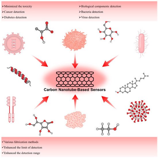
3. Applications of Carbon Nanotubes as Biosensors
3.1. Carbon Nanotube-Based Sensors for Detection of Cancer
3.1.1. CD44 Expressing Cancer Cell
Recently, cancer stem cells (CSCs) were identified as rare tumor-initiating cell populations which show self-renewal, pluripotent, and highly tumorigenic which makes them more resistant to breast cancer treatment. These cells are mainly responsible for breast cancer recurrence since even though most of the cells were killed by therapy, still few CSCs can regenerate tumors. Notably, CSCs are isolated from various cancer types including breast, brain, lung, colon, and skin cancer or melanoma. In particular, the case of breast cancer CSCs is identified by the presence of characteristic biomarkers namely CD44 and CD24 as well as one of the enzyme activities (ALDH1). This literature suggests that it is very important to detect and target the CSCs and their daughter cells responsible for cancer regeneration to achieve double remission.
3.1.2. EpCAM Expressing Cancer Cell
Epithelial cell adhesion/activating molecule abbreviated as EpCAM is the first tumor-associated antigen and currently it is considered as the most intensely and frequently expressed tumor-associated antigen. It is found to be expressed in a great variety of cancer types and it can be utilized as a biosensor application. Circulating tumor cells (CTCs) can also express EpCAM antigen and a nanoprobe or sensor can be developed to study the presence of these biomarkers on cancer cells by using anti-EpCAM antibody. Neoh et al. developed a CNT chip containing promising microfluidic technology for the effective capture and release of the CTCs. This technique allowed to perform downstream analysis of CTCs such as molecular and functional analyses. Researchers successfully developed a chip platform with the ability to not only capture the CTSs but also release them in a pH-responsive manner with higher sensitivity. This platform was tested for the clinical samples for the optimization of a device in order to maximize the cell capture and release efficiency, viability as well as application of this technology for single-cell molecular profiling and in vitro culture. Since EpCAM is a widely expressed antigen by various cancer cells, this platform could be generalized for different types of CTCs capture and detailed analysis.
3.1.3. CA19-9 Expressing Cancer Cell
Carbohydrate antigen 19-9 (CA 19-9) is a cell protein glycoprotein also known as Sialyl Lewis-a produced by ductal cells in the pancreas, salivary gland, biliary system, and epithelial cells in the stomach and colon. It is the most used biomarker for the diagnosis and management of prognosticating pancreatic ductal adenocarcinoma (PDAC) [
136]. Its widespread expression in several tumor cells makes it useful for the diagnosis of other tumor types apart from its historical use in the case of PDAC. Considering its diagnostic potential Thapa et al. developed a highly sensitive biosensor to detect the pancreatic cancer biomarker CA19-9. This developed biosensor based on nanomaterials promises cheaper, faster, and more efficient early diagnosis of pancreatic cancer as compared to traditional bulky devices. To fabricate the device, MWCNTs with functionalized anti-CA19-9 antibody were utilized.
3.1.4. VEGF Expressing Cancer Cell
Vascular endothelial growth factor (VEGF) is considered a main angiogenic factor in the case of many malignant tumors. VEGF acts by specific effects by stimulating cell growth and migration as well as increasing vascular permeability. It is a promising biomarker, especially in the case of the prognosis of cancer cells [
138]. Electrochemical aptasensors are promising agents due to their advantages of being cheaper and the possibility to have quantitative analysis.
3.2. Carbon Nanotube-Based Sensors for Detection of Diabetes
Two primary approaches are used while incorporating nanotechnology for glucose sensing applications. In the first approach, sensors can be designed by using micro or macroscopic components while incorporating nanomaterials in the sensing device. These nanomaterials in the sensor design offer several advantages such as higher surface area and enhanced catalytic activity. In the case of the second approach, nanofabrication can generate nanoscale sensors for glucose sensing. These sensors have some advantages such as offering continuous monitoring and avoiding foreign body responses of the immune system resulting in a longer life as compared to traditional sensors. In the case of diabetes, CNTs incorporation is heavily investigated as enzymatic electrode detection of glucose due to the electron transfer ability of the CNT and their surface areas [
146]. CNT-based electrochemical biosensors immensely helped with glucose sensing. Both single-walled CNTs as well as multi-walled CNTs have been explored as a nanomaterial for the detection of glucose. Functionalization of MWCNTs is less complex as compared to the SWCNTs since GOx could be directly adsorbed on the surface of MWCNTs as compared to the SWCNTs where a covalent linkage is required. It is possible to fabricate the best-performing glucose sensing devices when they are combined with the other nanomaterials [
147].
Enzymatic sensors are based on the use of enzymes for the conversion of an electro-inactive substrate into an electro-active product such as the use of glucose oxidase enzyme on a platinum electrode. On the other hand, non-enzymatic glucose sensors are based on the direct electrochemical oxidation of glucose. Most of the researchers focused on the development of enzymatic electrochemical sensors using glucose oxidase, but recently, non-enzymatic sensors using direct electrochemistry of glucose on noble metals are coming forward as next-generation glucose sensing technology [
148]. Here are examples of enzymatic and non-enzymatic sensors fabricated to enhance the ultrasensitive detection of glucose. Researchers tried to use carbon nanotubes to either modify the sensitivity of enzyme-based sensors that are prone to temperature-based degradation or used an alternative non-enzymatic sensing strategy by combining the carbon nanotubes in the device fabrications. Additionally, we have presented a tabular form of CNT-based enzymatic and non-enzymatic biosensors (
Table 2).
Table 2. Summarization of CNT-based enzymatic and non-enzymatic biosensors.
| Methods |
Analytes |
Limit of Detection |
Detection Range |
Ref. |
| Enzymatic |
Lactate |
Not reported |
5–20 nM |
[149] |
| Uric acid |
9.91 μM |
50 to 650 μM |
[150] |
| Glucose |
0.58 μM |
0.8 to 250 μM |
[151] |
| Glucose |
3 × 10⁻⁴ M |
(1–15) × 10−3 M |
[152] |
| Glucose |
5 × 10⁻⁵ M |
(0–5) × 10−3 M |
[153] |
| Glucose |
2.99 × 10⁻⁶ M |
(3–14) × 10−3 M |
[154] |
| Ethanol |
1 × 10⁻⁵ M |
(1–5) × 10−4 M |
[155] |
| Urease |
67 μM |
1.0–25.0 mM |
[156] |
| Alcohol dehydrogenase |
10 μM |
0.1 to 0.5 μM |
[152] |
| Choline |
0.6 μM |
3–120 μM |
[157] |
| Non-enzymatic |
Pyruvic acid |
0.048 μM |
0.1–200 μM |
[158] |
| Human epidermal growth factor receptor 2 |
7400 pg/mL |
10–110 ng mL−1 |
[159] |
| Cholesterol |
0.5 nM |
0.001–3 μM |
[160] |
| glucose |
500 nM |
2–19,600 μM |
[161] |
| Zearalenone |
0.15 pg mL−1 |
0.001–0.1 |
[162] |
| Long non-coding RNAs |
42.8 fM |
10−14–10−7 M |
[163] |
| MicroRNA 21 |
0.01 fM |
10−17–10−6 M |
[164] |
| Thrombin |
0.08 pM |
0.001–4 nM |
[165] |
| Human epidermal growth factor receptor 2 |
50 fg mL−1 |
0.1 pg mL−1–1 ng mL−1 |
[166] |
| Cardiac troponin T |
0.04 pg mL−1 |
0.1–8 pg mL−1 |
[167] |
| Urea |
4.7 nM |
0.066–20,600 µM |
[168] |
| Ascorbic acid |
0.85 nM |
0.001–8000 µM |
[169] |
| Glucose |
645 nM |
20–10,500 µM |
[170] |
| Glucose |
0.33 nM |
10–2000 µM |
[171] |
| Dopamine |
9.5 nM |
0.033–1 µM |
[172] |
| Potassium ions |
Not reported |
1000–32,000 µM |
[173] |
| Hydrogen peroxide |
Not Reported |
5 × 10⁻⁶–5 × 10⁻³ M |
[174] |
| MicroRNA 155 |
3.34 × 10⁻¹⁴ M |
1 × 10⁻¹³–1 × 10⁻⁹ M |
[175] |
| Digoxin |
7.95 × 10⁻¹² M |
2.65 × 10⁻¹¹–6.8 × 10⁻¹⁰ M |
[176] |
| Sequence specific to chronic myelogenous leukemia |
1 fM |
10−15–10−6 M |
[177] |
| Myeloperoxidase |
327 ng mL−1 |
Not reported |
[178] |
| SARS-CoV-2 spike protein |
35 mg L−1 |
Not reported |
[179] |
| SARS-CoV-2 spike protein |
0.55 fg mL−1 |
0.0055–5.5 pg mL−1 |
[180] |
| Ascorbic acid |
76.5 pM |
100 pM to 1 mM |
[181] |
3.3. Carbon Nanotube for Biological Components Detection
The detection of biological components is very important in biology, clinical science, and in hospitals facing real patients. In particular, it can make an early diagnosis of the disease, thereby allowing the patient to receive a faster and more correct response to the disease [
185]. This is closely related to the quality of life of patients. However, even if the patient has a specific disease, if the biological component cannot be detected or not, the patient’s life will be very difficult and painful, the treatment cost will increase significantly, and a cure cannot be guaranteed. In fact, many patients around the world suffer from misdiagnosis and late diagnosis [
186]. In order to fundamentally solve these problems, it is urgent and very important to develop materials, devices, and equipment that can detect biological components with high performance and high sensitivity.
CNTs have also been studied for use in the detection of biological components. CNTs have many advantages in the detection of various components due to their unique physical properties, such as large surface area [
187], tubular three-dimensional structure [
188], and the possibility of multiple modifications [
189]. Here, we introduce studies of CNTs for the detection of biological components. Recently, much research has been conducted to develop a sensor that detects glucose using CNTs. The detection of glucose in the serum is very important to mankind and has been performed for a long time. Measurement of blood glucose levels, especially in diabetic patients, is indispensable that must be performed daily and frequently. Scientific, patient-friendly, and modern blood glucose level measurement began with Clinistix developed by Kohn in 1957 [
190], and Dextrostix developed by Ernie Adams in 1965 [
191]. In a recent glucose detection study, a biosensor using ZnFe
2O
4, CNT, and glucose oxidase was developed [
151]. Briefly, ZnFe
2O
4 was conjugated with CNT by a one-step solvothermal approach using acid-treated CNT as a precursor. Glucose oxidase (GOD) was linked to ZnFe
2O
4-conjugated CNT by coupling reaction between the amine group and carboxyl group (ZnFe
2O
4-CNT-GOD). When glucose is added to ZnFe
2O
4-CNT-GOD, glucose is oxidized by GOD. In this process, the intermediate product, hydrogen peroxide, oxidizes the 3,3′,5,5′-tetramethylbenzidine (TMB) substrate and is eventually visualized in blue. In this process, ZnFe
2O
4 acts as a peroxidase and not only accelerates the overall reaction but also increases the intensity of the detected signal.
ZnFe
2O
4-CNT-GOD has a glucose detection range of 0.8 to 250 μM with a detection limit of 0.58 μM. ZnFe
2O
4-CNT-GOD did not react with lactose, maltose, fructose, sucrose, uric acid, dopamine, cystine, albumin, and ascorbic acid, so there was no component detection, but only a glucose-specific reaction occurred. In addition, the ZnFe
2O
4-CNT-GOD showed only a negligible change in the detection sensitivity of glucose even in the presence of 100 μM of copper, zinc, potassium, calcium, and iron ions. The authors report that the fabrication method of ZnFe
2O
4-CNT-GOD is simple, maintains the sensing activity for at least 20 days, and can be reused at least five times. Wang C et al. reported enzyme-functionalized CNTs and their application in glucose and Fe
2+ detection [
192]. Briefly, CNT was modified with carboxylation for functionalization. Later, carboxylated CNTs were covalently conjugated with GOD and/or horseradish peroxidase (HRP) (CNT-HRP-GOD). CNT-HRP-GOD detects glucose through the chain reaction between glucose, GOD, and HRP. In this process, the intermediate product, hydrogen peroxide, oxidizes the 3,3′,5,5′-tetramethylbenzidine (TMB) substrate and results in the production of colorimetric products (blue). Fe
2+ reacts with hydrogen peroxide and leads to a lower concentration of hydrogen peroxide, which in turn decreases the oxidation state of TMB and eventually causes a lower colorimetric absorbance of the solution. CNT-HRP-GOD has a glucose and Fe
2+ detection range of 1 to 100 μM with a detection limit of glucose and Fe
2+ of 0.3 and 0.22 μM, respectively. CNT-HRP-GOD did not react with other sugars except glucose and did not react with albumin and ascorbic acid, so it was verified as a biosensor through a glucose-specific reaction.
Alcohol detection can be applied to a variety of fields, from the quality analysis of bio-alcohol, which has been actively researched and manufactured recently, to checking the blood alcohol concentration and analyzing the quality of alcoholic beverages. Wilson, T et al. reported a CNT-based alcohol biosensor [
155]. They modified CNTs using polytyramine (PT) and glassy carbon (GC). PT has been electro-deposited onto MWCNT-modified GC electrodes via oxidation of tyramine (GC/MWCNT/PT). ADH immobilization for alcohol detection was improved by the PT layer. The polymeric film was formed on the electrode surface of MWCNT and it was confirmed using SEM and XPS. In order to detect alcohol with GC/MWCNT/PT, GC/MWCNT/PT was immersed in 0.05 M of sodium phosphate buffer (PBS) which contained 1 mg mL
−1 of alcohol dehydrogenase (ADH). The EDC/NHS coupling reaction was performed for one hour for the conjugation of the two substances (GC/MWCNT/PT/ADH). This alcohol biosensor showed a sensitivity of 4.28 ± 0.06 μA mM
−1 cm
−2, a regression coefficient of 0.9993, and a response time of 5 s. Furthermore, it had a 10 μM limit of detection and it almost accurately detected the alcohol content of commercial alcoholic beverages at a level of recovery of 97.4–102.1%.
Ascorbic acid is one of the water-soluble vitamins, and since L-gulono-γ-lactone oxidase is not present in the body, ascorbic acid must be consumed as a food or nutritional supplement [
193]. A deficiency of this causes scurvy [
194]. Ascorbic acid acts as an antioxidant in the body, protecting normal cells from various reactive oxygen species [
195] and helping to maintain the immune system [
196]. In addition, it suppresses various inflammatory reactions [
197] and aging [
198], and helps the elderly to maintain cognitive ability and memory, thereby helping to prevent Alzheimer’s disease [
199]. Zhao, Y et al. reported an ultra-sensitive biosensor for the voltammetric determination of ascorbic acid (AA) [
200]. For the fabrication of CNT-based high-sensitivity ascorbic acid sensors, MWCNTs were surface modified with glassy carbon electrodes (GCEs), graphene oxide (GO), and gold nanorods (AuNRs). Since the aggregation of MWCNTs reduces the detection sensitivity of biological components, prevention of aggregation improves the sensitivity. In this study, they used GO to prevent the aggregation of MWCNTs. Furthermore, overpotential was reduced and the peak current of AA oxidation was increased by positively charged AuNRs. The electrochemical properties of biosensors were investigated by cyclic voltammetry (CV). Finally, this ascorbic acid biosensor sensitively detected ascorbic acid with low working potential (0.036 V), low detection limit (0.85 nM), and high sensitivity (7.61 μA μM
−1 cm
−2).
CNTs can be applied as biosensors for the detection of uric acid and it has already been proven in several studies [
201,
202,
203]. Some chronically ill patients have hyperuricemia, in which there is too much uric acid in the body [
204]. It accumulates in the cartilages and produces tophi and uric acid crystals, which cause great pain to the patient. In addition, the accumulation of tophi and uric acid induces an inflammatory response in the cartilages and causes lasting damage to cartilage and bones [
205]. This phenomenon also entails great pain for the patient. Tophi and uric acid accumulate in the kidneys as well as in the cartilages, and just as they accumulate in the cartilages, they cause very severe organ irritation and pain [
206]. For the detection of uric acid, Huang B et al. reported a standing electrochemical sensor based on CNT for the determination of uric acid [
207]. They developed a free-standing electrochemical biosensor. CNT was modified with 3D graphene foam (GF) and gold nanoparticles (GNPs) for the fabrication of biosensors (GF/CNTs/GNPs). Cyclic voltammetry (CV) and differential pulse voltammetry (DPV) were used for the investigation of the electrochemical properties of GF/CNTs/GNPs. GF/CNTs/GNPs show outstanding electrocatalytic activity toward dopamine and uric acid. Detection of uric acid with GF/CNTs/GNPs shows remarkable sensitivity of 3.36 μA μM
−1 cm
−2, the low detection limits of 33.03 nM (S/N = 3), with a wide linear range of 0.50–60 μM. Furthermore, GF/CNTs/GNPs evaluated the quantification of uric acid with human urine. GF/CNTs/GNPs show good agreement in the concentration of uric acid (μM), total found (μM), and recovery (%) with high-performance liquid chromatography.
CNT-based biosensors can detect not only single molecules but also protein-based components. Here, we introduce the detection of biological components for the diagnosis of Alzheimer’s disease. Amyloid-β accumulates in the brains of people with Alzheimer’s disease and this phenomenon is a pathological mechanism of Alzheimer’s disease [
208]. Amyloid precursor protein is one of the proteins that plays a very important role in regulating the homeostasis of the neuronal system, such as neuronal development and signal transfer between neurons [
209]. However, various precursor protein cleavage products produced by the cleavage of amyloid precursor protein are very closely related to Alzheimer’s disease and induce dysfunction of the neuronal system [
210]. Oh, J et al. reported a carbon nanotube (CNT) film-based biosensor with a metal-semiconductor field effect transistor structure (MESFET) for amyloid-β detection in human serum [
211]. Briefly, for the fabrication of CNT-MESFET, the top gate was modified by depositing Au (10 nm) only in the middle of the semiconducting CNT channel. These immobilized antibodies on CNT-MESFETs were controlled by the antibody-binding proteins. In order to detect HRP used as the model analyte, anti-HRP antibodies were immobilized on the Au top gate with protein G or auto-displayed Zdomains of protein A as the antibody-binding protein. CNT-MESFET exhibited a higher sensitivity than the antibodies immobilized biosensor using a chemical linker. CNT-MESFET could detect the HRP at levels as low as 1 fg mL
−1 in serum. Finally, they applied the CNT-MESFET to the detection of amyloid-β in human serum. This CNT-MESFET could detect the amyloid-β at the level of 1 pg mL
−1 in human serum. It can be applied as a CNT-based biological material detection sensor with very high sensitivity, and further research is needed to see if it can detect other substances besides amyloid-β.
Thrombin is a protein closely related to blood clotting [
212]. During bleeding, platelets are destroyed, and thromboplastin is released into the plasma, which is activated in the presence of calcium ions in the blood and becomes thrombin. It catalyzes the reaction of hydrolysis of soluble fibrinogen in the blood, which is the essence of blood coagulation, into insoluble fibrin [
213]. Therefore, quantitative detection of thrombin in bleeding patients or patients undergoing surgery can prevent accidents caused by bleeding and judge the patient’s condition for bleeding more clearly. Su, Z et al. reported an amperometric thrombin aptamer sensor (aptasensor) as a thrombin biosensor [
165]. For the fabrication of the aptasensor, polyaniline-coated MWCNT was placed on the glassy carbon electrode (GCE). Later thiolated thrombin-specific aptamers were conjugated with polyaniline by the thiol-ene reaction. The surface of the aptasensor was coated with bovine serum albumin to prevent non-specific binding. The modified GCE shows a pair of well-defined redox peaks (at 50/−25 mV) and the tethered TTA–thrombin interaction shows a decreased electrochemical signal. Thrombin in spiked human serum (0.2 to 4 nM) was accurately detected by the aaptasensor and it shows recoveries that ranged from 95 to 102%.
Accurately detecting COVID-19 is very important in the situation of the pandemic. Early detection and accurate diagnosis of the virus can prevent the spread of coronavirus infection. In addition, diagnosis of severe acute respiratory syndrome coronavirus 2 (SARS-CoV-2) is crucial for tracking the route of transmission and suitable treatment for patients in the event of a pandemic [
214,
215]. Pinals, R et al. introduced an SWCNT-based optical sensing approach toward this end [
179]. SARS-CoV-2 enters the host cell through binding to the ACE2 receptor [
216]. They used ACE2 to fabricate the noncovalently functionalized SWCNT as a virus sensor since ACE2 has a high binding affinity to the SARS-CoV-2 spike protein. Biosensor fluorescence was increased (2-fold) in the presence of the SARS-CoV-2 spike protein. They evaluated biosensor stability and confirmed preserving sensing responses in saliva and virus delivery media. In addition, it was demonstrated that the biosensor had a 73% fluorescence-on response within 5 s of exposure to 35 mg L
−1 SARS-CoV-2 virus-like particles. The biosensor shows a 100% turn-on response in fluorescence upon the addition of 1 μM CoV-2 S spike protein receptor-binding domain (S RBD).
Zamzami, M et al. developed a fast (2–3 min), easy-to-use, low-cost, and quantitative electrochemical biosensor based on a CNT field-effect transistor (CNT-FET) that allows digital detection of the SARS-CoV-2 S1 [
28]. It can quickly and accurately detect SARS-CoV-2 S1 antigens in saliva samples. The anti-SARS-CoV-2 S1 was immobilized on a Si/SiO2 surface by CNT printing for the fabrication of a CNT-FET biosensor. The CNT-FET biosensor effectively detected the SARS-CoV-2 S1 antigen in 10 mM ammonium acetate buffer at concentrations from 0.1 fg mL
−1 to 5.0 pg mL
−1. The limit of detection (LOD) of the CNT-FET biosensor was 4.12 fg mL
−1. In order to confirm whether the biosensor can specifically detect only the target antigen, selectivity tests were performed using target SARS-CoV-2 S1 and non-target SARS-CoV-1 S1 and MERS-CoV S1 antigens. The biosensor has good detection sensitivity with SARS-CoV-2 S1 antigen. However, it shows no detection response to SARS-CoV-1 S1 and MERS-CoV S1 antigen. The developed CNT-FET biosensor was verified to be capable of sensitive, fast, and accurate detection of SARS-CoV-2 S1 in human saliva.
3.4. Carbon Nanotube for Bacteria and Virus Detection
As mentioned before, CNTs and their derived structures have excellent physical properties including electrical conductivity, SERS, FRET, and so on. They can be utilized as sensing channels to detect bacteria, viruses, virus DNA, etc. [
217,
218]. Especially, the need for high-performance virus sensing platforms has increased for well-being and better human life, so many CNT-based sensing systems have been introduced. For example, Lee and his co-workers introduced virus DNA detection via an electrical biosensing platform which is composed of magnetically aligned NPs decorated CNT on the IDE [
219]. In this study, firstly gold and magnetic nanoparticles were modified on the surface of CNT (Au/MNP-CNT) and they were laid on the Pt-IDE via an external magnetic field. After that, the thiol-modified probe DNA was immobilized on the Au NP. In this case, Au and CNT played a role as electrical sensing channels and MNP was the moiety for alignment. Due to the synergic properties between the three nanomaterials, this sensing platform showed high sensitivity with a LOD of 8.4 pM for influenza virus DNA and 8.8 pM for norovirus DNA. Furthermore, it showed high selectivity against mismatched DNA strains. Therefore, this sensing platform could show excellent sensing performance.
On the other hand, metallic nanoparticles (NPs)-decorated CNT was also used as a plasmonic substrate for a plasmonic resonance energy transfer (PRET)-based FL immune sensing system. In this case, metallic NPs such as gold or silver NPs and CNT possess plasmonic properties; thus, their hybrid structure-based plasmonic material has synergic properties. For instance, the plasmonic property of gold NP-decorated CNT (gold-CNT) assisted in the detection of influenza viruses [
220]. In this study, gold-CNT was modified with influenza virus Ab, so it could capture the target virus and subsequently, fluorescent quantum dots (QDs)-Ab were added into the mixture and bound with virus-Ab-gold CNTs, and finally, sandwich structures were induced. As a result, depending on the concentrations of the target influenza virus, the FL of QD was changed linearly. According to the results of detection performance, the limit of detection of viruses was estimated at around 0.1 pg mL
−1. Furthermore, the influenza virus from a clinical sample was also monitored with excellent sensitivity was 50 PFU mL
−1 in the range of 50–10,000 PFU mL
−1. It meant that a metal NP-CNT structure-based fluoro-immuno sensing system could be potentially applied for virus detection.
In another study, CNT could be utilized as a sensing channel to detect the dengue virus (DENV) via an electrochemical approach. Wasik et al. fabricated a heparin-SWCNT hybrid structure on the electrode to monitor the dengue virus and the resistance difference was measured depending on the concentration of the virus [
221]. The limit of detection of this system showed 8 DENV/chip and this system showed excellent selectivity against the influenza virus. Therefore, a CNT-based electrochemical sensing platform also could be developed for high-performance biosensors.
On the other hand, some bacteria have caused critical diseases, and they have threatened human life. So, a highly sensitive and selective bacteria sensing system was required, and several bacteria were also successfully detected by using a CNT-based sensing platform. For instance, Zhang et al. reported a gold-CNT-based sensing system that could detect
Escherichia coli (
E. coli) through an electrochemical approach [
222]. Firstly, they fabricated the gold-CNT/GCE for the sensing channel. Then, the captured Ab was immobilized on the surface of gold-CNT to monitor the
E. coli. The sensing behavior of the gold-CNT-based sensing system was characterized depending on the amounts of bacteria from 2.0 × 10
2 to 2.0 × 10
6 CFU mL
−1. This system showed linear response depending on the amount of
E. coli and good sensitivity and selectivity was also proven. Especially,
E. coli in sludge was also successfully detected, thus this system exhibited excellent sensing performance.
In another study, the detection of
E. coli O157:H7 based on an MWCNT electrical sensing system was introduced by Li and co-workers [
223]. In this study, MWCNT was covered by polystyrene sulfonate (PSS) and the authors produced a PSS-MWCNT-based layer-by-layer (LBL) structure using poly(ethyleneimine) (PEI) to apply for the electrical sensing channel. O157:H7 Abs were modified on the surface of MWCNT to capture the target bacteria. The electrical signal was monitored depending on the concentration of O157:H7 and a linear response was shown. Interestingly, the authors collected the bacteria that were captured (O157:H7) by the sensing channel and isolated specific DNA from the collected O157:H7. Subsequently, the concentration of O157:H7 DNA was estimated by loop-mediated isothermal amplification (LAMP). Based on the sensing performance results, the LOD of this system was around 1 PFU mL
−1. Therefore, they developed highly sensitive bacteria detection systems using MWCNT.
E. coli in dairy products were also detected by using CNT and gold NP mixture [
224]. In this case, the surface of CNT was modified with target Ab by an EDC/NHS coupling reaction and horseradish peroxidase (HRP) enzyme at the same time. On the other hand, gold NPs were coated by poly(amidoamine) dendrimer and specific Ab for
E. coli was attached to the surface of NPs. In this environment, if target bacteria existed in the sample, these two nanomaterials could be formed as a sandwich structure with bacteria and its electrochemical sensing signal might be changed. The sensing performance of this system was tested under a range of 1.0 × 10
2 to 1.0 × 10
6 CFU mL
−1 and a LOD of around 50 CFU mL
−1 was estimated. Therefore, a CNT-based sensing system could be applied to the bacteria screening platform.
Liu et al. have reported that molecular imprinted TiO
2-coated multiwalled carbon nanotubes (MI-TiO
2@CNTs) were fabricated to detect microcystin-LR (MC-LR), a type of cyanobacterial toxin in water by the photoelectrochemical method. The molecular imprinted TiO
2 showed enhanced detection in comparison to traditional TiO
2 and non-imprinted TiO
2. This sensor resulted in a wide linear range from 1.0 pM to 3.0 nM for the detection of MC-LR. MI-TiO
2@CNTs achieved magnificent selectivity towards MC-LR. Moreover, this promising sensor showed high sensitivity for the detection of MC-LR which could be a potential candidate for water purification [
29].
He and co-workers developed an enzyme-free and dual-signal readout immunosensor that was used to detect MC-LR while an enzyme-based biosensor with great obstacles such as instability, sensitivity, temperature, and pH should be considered. Initially, gold nanoparticle-decorated CNT (AgNP-CNTs) was fabricated for the detection of MC-LR and secondly, silver nanorods were coated over AgNP-CNTs to detect via dual-signal mode. These sensors showed the determination of MC-LR in a linear range from 0.005 μg L
−1 to 20 μg L
−1 with a LOD of 2.8 ng L
−1. In terms of reproducibility, high selectivity, and sensitivity, these sensors indicated their promising application in environment monitoring [
225].
In another study, Han et al. proposed an MWCNT-based electrochemical biosensor that was demonstrated to monitor the MC-LR in drinking water supplies. This biosensor was fabricated in well-aligned and millimeter-long MWCNT arrays by water-assisted CVD. In addition, monoclonal antibodies were decorated to specify MC-LR toxin detection. A linear range from 0.05 to 20 μg L
−1 was observed for the detection of MC-LR with a LOD of 1 μg L
−1 in drinking water [
226]. To reduce the burden of cost-effectiveness and increase the rapid detection of MC-LR in environmental water, Queiros and co-workers proposed label-free potentiometric sensors composed of MWCNTs. These sensors were synthesized by imprinted polymer and polyvinyl chloride membranes. This method was applied successfully to detect MC-LR with great selectivity and sensitivity. Moreover, this method benefited with easy production and cost-effectiveness [
227].
Cholera is another devastating disease that has taken uncountable lives over the past few decades. The detection of cholera toxin (CT) was highly required to eradicate cholera from our lives. Viswanathan et al. proposed a sensitive method to detect CT by using an electrochemical immunosensor. This immunosensor was composed of potassium ferrocyanide, ganglioside (GM1)-functionalized liposomes, and monoclonal antibodies on the surface of Nafion-supported multi-walled carbon nanotubes. The detection mechanism was proposed by a sandwich-type assay, where the toxin was first coupled with an anti-CT antibody and followed by a GM1-functionalized liposome. This sandwich method resulted in the detection of CT in ultra-trace levels. The detection of CT showed a linear range of 10
−14−10
−7 g mL
−1 with a LOD of 10
−16 g of CT [
228].
In another study, Palomar et al. proposed an impedimetric immunosensor based on CNTs to improve sensing performances by increasing electroactive surface areas on CNTs. These systems were modified with polypyrrole-nitrilotriacetic acid (poly(pyrrole-NTA)) and Cu (II) complex to produce sensor devices. With great sensitivity and easy reproducibility, the cholera sensor showed a promising linear detection range from 10
−13–10
−5 g mL
−1 with a LOD of 10
−13 g mL
−1, which could be a potential sensing platform to detect cholera in the environment [
229].
4. Conclusions
Over the years, researchers and scientists have used a diverse range of nanomaterials such as metal nanoparticles (NPs), metal oxide NPs, nanofibers [
273], quantum dots (QDs), and carbon nanomaterials such as carbon quantum dots [
274], graphene, and carbon nanotubes (CNTs) to fabricate high-performance and sensitive biosensors. CNTs and their derivatives have gained great attention in the field of advanced functional materials today. It has been explored in diverse fields from defense to electronics. The field of biomedical applications has investigated CNTs and their derivatives extensively as potential candidates. Although the physical and chemical properties are not completely understood, it has been exploited by the electronics industry over the years. CNTs showed excellent properties in device fabrications as well as sensing behaviors. CNTs and their derivatives have been utilized for bio and chemical sensing due to having similar sizes to the analytes and bio-species. Due to their small size and high aspect ratio, CNTs exhibit unusual optical, mechanical, electrical, and chemical properties due to their small diameter and high aspect ratio. Utilizing them, a wide class of sensors is fabricated. It has been shown that CNTs have improved cell penetration properties and stability, as well as chirality and diameter-based physicochemical properties. On the account of synthesis, the materials that are necessary for CNT production are profuse, and they can be crafted with only a modest amount of raw materials. Further functionalization without damaging the covalent backbone extends the desired application of CNTs. Although one major drawback of CNT production is reproducibility, structurally and chemically reproducible batch production with minimal impurities is an immediate concern. Another two important properties that distinguish them from other nanomaterials are temperature stability (2800 °C in vacuum and ~750 °C in air) and hydrophilicity. While considering the mechanical properties, extremely high Young’s modulus values (1–1.8 TPa range) allow them to act as an excellent candidate for probe tips for scanning microscopy. Although some other disadvantages are always associated with CNTs, namely cellular toxicity, incompatibility with biological mediums, agglomeration, accumulation, and long-term persistence which require a strong action for mitigation. Numerous studies on CNTs and their derivatives have reported their interactions with analytes and their toxicology profiles. However, to allow their commercialization, there are limits in terms of cost-effectiveness, purity, and high density of perfect alignment during industrialization. A huge number of studies are conducted on biosensors to enable their commercialization. Interestingly, CNTs have been investigated as biosensors as in vivo devices, while many efforts have been made to minimize their toxicity profile.