Obesity, Body Composition, and Nutrition in COVID-19 Pandemia: History
Please note this is an old version of this entry, which may differ significantly from the current revision.
Contributor: , , , , , ,

The coronavirus disease 2019 (COVID-19) pandemic has spread worldwide, infecting nearly 500 million people, with more than 6 million deaths recorded globally. Obesity leads people to be more vulnerable, developing worse outcomes that can require hospitalization in intensive care units (ICU). Most studies showed that not only body fat quantity but also its distribution seems to play a crucial role in COVID-19 severity. Compared to the body mass index (BMI), visceral adipose tissue and intrathoracic fat are better predictors of COVID-19 severity and indicate the need for hospitalization in ICU and invasive mechanical ventilation. High volumes of epicardial adipose tissue and its thickness can cause an infection located in the myocardial tissue, thereby enhancing severe COVID-related myocardial damage with impairments in coronary flow reserve and thromboembolism. 

  • SARS-CoV-2
  • COVID-19
  • obesity
  • body mass index
  • intensive care units

1. Introduction

The coronavirus disease 2019 (COVID-19) pandemic has spread around the entire globe, infecting nearly 500 million people since the end of 2019, with more than 6 million deaths recorded globally [1]. A major proportion of these infected people have recovered with irrelevant clinical complications. However, a subgroup of individuals affected by COVID-19 developed worse outcomes leading to hospitalization in semi-intensive or intensive care units (ICU) and have shown a high rate of mortality [1].
Obesity is defined as an excessive amount of fat deposition in adipose tissue, according to the World Health Organization (WHO) [2][3]. Several studies have reported a higher prevalence of obesity in patients experiencing a severe COVID-19 clinical course, with serious complications requiring hospitalization and admission to ICU as well as a higher rate of mortality.
A meta-analysis conducted by Popkin et al. in June 2020 showed that, compared to non-obese patients, individuals with obesity have an increased risk of COVID-19 infection (over 46%), a higher risk of hospitalization (113%), a higher need for ICU admission (74%), and a risk of mortality (>48%) [4]. Obesity alone is responsible for 30% of all COVID-19 hospitalizations [5], and the rate of the latter may grow when obesity is also associated with impaired metabolic health (i.e., type 2 diabetes and hypertension) [6] even at a younger age [7][8]. Systematic reviews and meta-analyses confirmed that obesity was significantly associated with more severe forms of the disease and mortality in patients with COVID-19 [5]. These findings were also confirmed in the ICU and showed that a higher BMI is associated with higher inflammation levels, muscle damage, and in-hospital mortality during the first 28 days [9]. Moreover, being overweight in critically ill COVID-19 patients requiring invasive mechanical ventilation significantly increases their risk of death [10][11].
Even though the underlying mechanisms are still not fully understood, behind the severe prognosis of COVID-19 in individuals with obesity, increasing evidence suggests several hypotheses. Firstly, the presence of uncontrolled weight-related comorbidities (e.g., type 2 diabetes, cardiovascular, pulmonary, and renal diseases) make this population more vulnerable [12]. Pulmonary complications particularly in this population may present primary fertile soil for respiratory tract infection [13], especially lung fat embolism [14]. Secondly, due to the abnormal fat deposition, immune system alterations may facilitate a systemic diffusion of infection, and make the condition difficult to treat [15][16]. Thirdly, an increased risk of nosocomial infections [17], and the lack of full knowledge about optimum antimicrobial doses suitable for patients with obesity that fit their body weight [18] may also lead to difficulties in treating these patients in time, with possible life-threatening consequences [19].
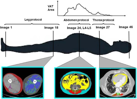
In the field of obesity research in the last decades, body composition has gained importance as a risk factor for unfavorable health-related outcomes. Computer tomography (CT) and magnetic resonance imaging (MRI) have been established as the standard reference techniques for studies that evaluate body fat distribution (Figure 1). CT scans were considered the reference method in performing diagnoses with suspected pneumonia in symptomatic hospitalized COVID-19 patients and, as a consequence, in the last two years, many studies have investigated the relationship between thoracic and high abdominal fat distribution and several important health-related outcomes in this population. Growing available evidence on this topic should be evaluated in order to improve clinical management and better address future research.
Figure 1. Computer tomography imaging protocol for both whole-body and regional (i.e., left to right, leg, abdomen, thorax, etc.) measures of adipose tissue and lean tissue.

2. The Role of Adipose Tissue Distribution in Patients with Severe COVID-19

2.1. Visceral Adipose Tissue

Epidemiological studies show that visceral adipose tissue (VAT, Figure 1, abdomen protocol, in yellow) may be a better predictor of COVID-19 severity than BMI [20][21]. Favre et al. show that for low VAT content, the linearity of the relationship is lost, at least in part due to a maldistribution of VAT values in the sample studied [21]. Interestingly, the authors showed that the correlation lost statistical significance after adjustment for sex and age, suggesting that VAT could explain the relation between sex, age, and the severity of COVID-19, which is greater in men and older populations. VAT is recognized as a risk factor for the severity and mortality of COVID-19, and multiple pathogenetic mechanisms have been proposed [22]. A previous meta-analysis identified VAT as a risk factor for hospitalization, ICU admission, and invasive mechanical ventilation [20]. Therefore, not only body fat quantity but also its distribution can play a crucial role in COVID-19 severity even though the underlying mechanisms linking central adiposity to severe COVID-19 are not completely understood. Through the possible mechanisms, inconsistent glycemic control strongly related to VAT depot [23] is associated with significantly increased mortality in COVID-19 [24]. Furthermore, during an infection, uncontrolled glycemia can lead to an immunity dysfunction that worsens patient outcomes. Individuals with obesity could undergo an additional worsening of glycemic control when dexamethasone, effective and frequently used in COVID-19 severe illness treatment, is introduced, as steroids can alter glycemic homeostasis [25].
VAT and intrathoracic fat can alter respiratory physiology by decreasing respiratory compliance. In these patients, airway resistance and respiratory work are increased. Another negative consequence is that patients with obesity use a larger fraction of oxygen uptake to support respiratory work, resulting in a decreased functional reserve. Central fat distribution has a mechanical effect, determining a reduction in lung elastic recoil, peripheral airway size, and chest wall compliance, affecting pulmonary volumes [26]. All this evidence can support the idea that visceral obesity can lead to an increased risk of respiratory failure in critically ill COVID-19 subjects.
The use of the ACE2 receptor by SARS-CoV-2 could also lead to increased neutrophil recruitment, capillary permeability, and pulmonary edema, as found by previous studies on the SARS-CoV-1 virus, which uses the same receptor to enter cells [27]. In particular, VAT presents high angiotensin converting enzyme (ACE) 2 expression. For this reason, it has been hypothesized that, as with other diseases (i.e., influenza A, HIV, cytomegalovirus), SARS-CoV-2 could use adipose tissue as a reservoir.

3. The Role of Skeletal Muscle Mass and Function in SARS-CoV-2 Infection

Skeletal muscle mass accounts for approximately 40% of body weight and is the major regulator of glucose homeostasis [28]. Moreover, this specialized tissue is involved in several essential activities, such as breathing, transmitting strength to the bones, maintaining posture, gait, and global locomotion [28]. Together with muscle mass, muscle strength has also emerged as a predictor of a long lifespan [29]. Higher muscle strength is associated with healthy aging and a lower risk of developing acute illness or chronic diseases, disability, hospitalization, and long-term mortality [30][31][32].
With advancing age, older adults may frequently experience sarcopenia, defined as the decline in muscle mass, muscle strength, and physical performance [33]. With sarcopenia, two different conditions, associated with a concomitant increase in frailty and muscle weakness, might occur: (i) Weight loss caused by a reduced appetite, characterized by a lower amount of protein intake, leading to malnutrition [34]; or (ii) weight gain caused by a disproportionate caloric intake due to the combination of poor physical activity and overeating, leading to sarcopenic obesity [34].
The relation between sarcopenia and COVID-19 is controversial. Some studies [35] have highlighted that the presence of sarcopenia can increase the vulnerability to COVID-19 and vice versa. The presence of sarcopenia can markedly increase infection rates, since sarcopenic patients have a poor immune response and have a predisposition to lipotoxicity, metabolic dysregulation, and inflammation [36]. Furthermore, sarcopenia is a risk factor for aspiration pneumonia caused by a loss in swallowing function, frequently observed in the oldest populations [37]. Finally, failures in respiratory muscle strength and function observed in patients with sarcopenia hamper the treatment for severe pneumonia and acute respiratory distress syndrome [36]. Hence, patients with sarcopenia are more likely to develop a severe form of COVID-19 associated with important unfavorable health outcomes.
On the other hand, severe and prolonged COVID-19 can lead to sarcopenia onset [38], since several studies [34][39] have shown skeletal muscle loss and damage in hospitalized patients. During the acute stage, the symptoms reported by patients are muscle soreness, fatigue, weaknesses, and deficits in lower extremity muscle contraction. In this scenario, older individuals with negative health conditions such as frailty, obesity, metabolic, and cardiovascular disorders are more prone to experiencing muscle impairments [35]. Aging is characterized by low-grade chronic inflammation, so-called inflammaging (71, 76). However, COVID-19-associated “cytokine storm” (i.e., an increase in IL-6, tumor necrosis factor-α, and c-reactive protein level), which enhances the inflammatory state, coupled with oxidative stress (that intensifies reactive oxygen species generation), worsens sarcopenia through a “catabolic crisis” with rapid protein degradation [40]. It is worth noting that the duration of immobility and bed rest associated with lower levels of routine physical activity could facilitate sarcopenia onset/deterioration after COVID-19 infection [37].
Prolonged immobilization reduces mechanical overloading, which acts as a stimulus for bone and muscle health homeostasis [41]. Regarding muscle mass, in a recent study, Narici and colleagues [42] summarized the impact of COVID-19-related sedentarism, reporting that even over short periods (i.e., 5 days) of bed rest, a significant reduction in quadriceps muscle mass can be observed. Muscle atrophy further deteriorated over time, with a loss of 10% and 15%, after 30 and 60 days, respectively [42], which, in physiological conditions, corresponds to a drop observed in more than 10 years [37]. After 5 days of bed rest, the loss of quadriceps strength is greater (i.e., 9%) compared to muscle mass [42].

4. Nutrition in SARS-CoV-2 Prevention and Treatment

Through the modulation of immune function, nutritional status has the potential of influencing the course of viral infections, with implications for the duration, severity, and overall outcome of the disease [43][44]. It is also increasingly evident that the relationship between nutritional status and SARS-CoV-2 infection is bidirectional. Indeed, if poor nutritional status increases the risk of infection and negatively affects its course [45][46], on the other hand, the infection itself is a risk factor for worsening nutritional status [47]. Malnutrition is widely prevalent among SARS-CoV-2 patients at the time of hospitalization, indicating an increased risk of infection in malnourished patients [48]; moreover, poor nutritional status (in both under and overnutrition patients) has a negative impact on the course of the SARS-CoV-2 disease, with a growing body of evidence of increased severity, need for invasive treatment, and mortality [48][49][50][51]. The evaluation of nutritional status and the implementation of nutritional interventions are gaining a leading role in the approach to patients with SARS-CoV-2 infection, both in general healthcare and ICU settings.

4.1. Nutritional Prevention

Regarding prevention, although it is known that nutritional interventions can act as immunostimulators helping to prevent viral infections, it should be emphasized that data from randomized clinical trials are still lacking [52]. During the pandemic, the WHO confirmed the indications for proper nutrition, based on the guidelines already known, which recommend a Mediterranean diet, with a prevalent consumption of fresh and unprocessed foods, along with vegetables, in which the use of sugars and saturated fats and an excessive amount of salt are not recommended [53]. Furthermore, part of the general nutritional approach for the prevention of viral infections is the supplementation or at least adequate intake of vitamins and micronutrients, to maximize natural antiviral defenses; micronutrients such as vitamin A, vitamin E, cyanocobalamin, vitamin B 6, zinc, and selenium have different roles in supporting the functions of mucosal immunity and the integrity of the epithelial barrier and enhancing adaptive and innate immune functions [54]. As with other respiratory tract infections, a link between vitamin D deficiency and SARS-CoV-2 infection is emerging, as observational studies reported an association between low levels of 25-OH vitamin D and susceptibility to lower respiratory tract infections [55][56][57]. As pointed out by Mechanick et al., several fundamental gaps in the evidence remain [52], as to whether specific foods, macronutrients, or micronutrients can reduce the overall risk for SARS-CoV-2 infection or severity. As suggested by international guidelines, as a prevention strategy in the overall population and in those at risk of severe disease (e.g., the elderly and patients with altered nutritional status), it is reasonable to optimize the nutritional status, ensuring adequate caloric, protein, and micronutrients intake [58].

4.2. Nutritional Risk Assessment

It is increasingly apparent that nutritional care, including the identification of nutritional risk and the use of nutrition support, should be a fundamental part of management in SARS-CoV-2 inpatients. These subjects present a high nutritional risk for many reasons, including a high prevalence of comorbidities, as well as an increase in energy-protein requirements, hypercatabolism, the possible reduction in intake in the presence of gastrointestinal disorders and reduced appetite, dysphagia, dyspnea, or the need for support with invasive or non-invasive ventilation. In patients with SARS-CoV-2 infection, poor nutritional status is associated with poorer clinical outcomes [49][50][51]. Thus, the rapid assessment, identification, and treatment of poor nutritional status are essential for improving clinical outcomes in severely and critically ill SARS-CoV-2 patients. Based on the available evidence, the European Society for Parenteral and Enteral Nutrition (ESPEN) and the American Society for Parenteral and Enteral Nutrition (ASPEN) formulated recommendations regarding nutritional intervention in hospitalized patients with SARS-CoV-2 infection [58][59]. Most of the literature agrees on the need for nutritional screening with validated tools such as the Malnutrition Universal Screening Tool (MUST), Mini Nutritional Assessment (MNA), and Nutritional Risk Screening-2002 (NRS-2002) [60]. Moreover, regardless of nutritional status, it is important to identify a reduction in food intake early on, and to introduce nutritional treatment as soon as the caloric intake is less than 70% of the daily requirement. In line with previous recommendations for critically ill patients requiring the ICU who do not reach energy and protein targets through oral feeding, the importance of giving priority to enteral nutrition is emphasized, with the proposal of a parenteral route in case of specific limitations or even enteral nutrition if it is not possible to reach the target. In ventilated patients subjected to prono-supination cycles, the preferential recommendation for enteral nutrition remains valid, as the prone position per se does not represent a contraindication [61].

This entry is adapted from the peer-reviewed paper 10.3390/nu14173493

References

  1. Chen, J. Novel statistics predict the COVID-19 pandemic could terminate in 2022. J. Med. Virol. 2022, 94, 2845–2848.
  2. El Ghoch, M.; Fakhoury, R. Challenges and New Directions in Obesity Management: Lifestyle Modification Programmes, Pharmacotherapy and Bariatric Surgery. J. Popul. Ther. Clin. Pharmacol. 2019, 26, e1–e4.
  3. Apovian, C.M. Obesity: Definition, Comorbidities, Causes, and Burden. Am. J. Manag. Care 2016, 22, s176–s185.
  4. Popkin, B.M.; Du, S.; Green, W.D.; Beck, M.A.; Algaith, T.; Herbst, C.H.; Alsukait, R.F.; Alluhidan, M.; Alazemi, N.; Shekar, M. Individuals with obesity and COVID-19: A global perspective on the epidemiology and biological relationships. Obes. Rev. 2020, 21, e13128.
  5. Ho, J.S.; Fernando, D.I.; Chan, M.Y.; Sia, C.-H. Obesity in COVID-19: A Systematic Review and Meta-analysis. Ann. Acad. Med. Singap. 2020, 49, 996–1008.
  6. O’Hearn, M.; Liu, J.; Cudhea, F.; Micha, R.; Mozaffarian, D. Coronavirus Disease 2019 Hospitalizations Attributable to Cardiometabolic Conditions in the United States: A Comparative Risk Assessment Analysis. J. Am. Heart Assoc. 2021, 10, e019259.
  7. Williamson, E.J.; Walker, A.J.; Bhaskaran, K.; Bacon, S.; Bates, C.; Morton, C.E.; Curtis, H.J.; Mehrkar, A.; Evans, D.; Inglesby, P.; et al. Factors associated with COVID-19-related death using OpenSAFELY. Nature 2020, 584, 430–436.
  8. Onder, G.; Palmieri, L.; Vanacore, N.; Giuliano, M.; Brusaferro, S.; The Italian National Institute of Health COVID-19 mortality group; Agazio, E.; Andrianou, X.; Barbariol, P.; Bella, A.; et al. Nonrespiratory Complications and Obesity in Patients Dying with COVID-19 in Italy. Obesity 2020, 29, 20–23.
  9. Rossi, A.P.; Gottin, L.; Donadello, K.; Schweiger, V.; Nocini, R.; Taiana, M.; Zamboni, M.; Polati, E. Obesity as a risk factor for unfavourable outcomes in critically ill patients affected by COVID-19. Nutr. Metab. Cardiovasc. Dis. 2021, 31, 762–768.
  10. Shabanpur, M.; Pourmahmoudi, A.; Nicolau, J.; Veronese, N.; Roustaei, N.; Jahromi, A.J.; Hosseinikia, M. The importance of nutritional status on clinical outcomes among both ICU and Non-ICU patients with COVID-19. Clin. Nutr. ESPEN 2022, 49, 225–231.
  11. Czapla, M.; Juárez-Vela, R.; Gea-Caballero, V.; Zieliński, S.; Zielińska, M. The Association between Nutritional Status and In-Hospital Mortality of COVID-19 in Critically-Ill Patients in the ICU. Nutrients 2021, 13, 3302.
  12. Khaodhiar, L.; McCowen, K.C.; Blackburn, G.L. Obesity and its comorbid conditions. Clin. Cornerstone 1999, 2, 17–31.
  13. Cortes-Telles, A.; Ortiz-Farias, D.L.; Pou-Aguilar, Y.; Almeida-De-La-Cruz, L.; Perez-Padilla, J.R. Clinical impact of obesity on respiratory diseases: A real-life study. Lung India 2021, 38, 321–325.
  14. Colleluori, G.; Graciotti, L.; Pesaresi, M.; Di Vincenzo, A.; Perugini, J.; Di Mercurio, E.; Caucci, S.; Bagnarelli, P.; Zingaretti, C.M.; Nisoli, E.; et al. Visceral fat inflammation and fat embolism are associated with lung’s lipidic hyaline membranes in subjects with COVID-19. Int. J. Obes. 2022, 46, 1009–1017.
  15. Francisco, V.; Pino, J.; Campos-Cabaleiro, V.; Ruiz-Fernández, C.; Mera, A.; Gonzalez-Gay, M.A.; Gómez, R.; Gualillo, O. Obesity, Fat Mass and Immune System: Role for Leptin. Front. Physiol. 2018, 9, 640.
  16. Bähr, I.; Spielmann, J.; Quandt, D.; Kielstein, H. Obesity-Associated Alterations of Natural Killer Cells and Immunosurveillance of Cancer. Front. Immunol. 2020, 11, 245.
  17. Huttunen, R.; Karppelin, M.; Syrjänen, J. Obesity and nosocomial infections. J. Hosp. Infect. 2013, 85, 8–16.
  18. Falagas, M.E.; Karageorgopoulos, D.E. Adjustment of dosing of antimicrobial agents for bodyweight in adults. Lancet 2009, 375, 248–251.
  19. Miles, J.; Anderson, D.P.; Engelke, M.; Kirkpatrick, M.K.; Pories, M.L.; Waters, W.G.; Watkins, F.R.; Pokorny, M.E.; Rose, M.A. Barriers to transition of obese patients from hospital to community. Am. J. Manag. Care 2012, 18, e234–e237.
  20. Huang, Y.; Lu, Y.; Huang, Y.-M.; Wang, M.; Ling, W.; Sui, Y.; Zhao, H.-L. Obesity in patients with COVID-19: A systematic review and meta-analysis. Metabolism 2020, 113, 154378.
  21. Favre, G.; Legueult, K.; Pradier, C.; Raffaelli, C.; Ichai, C.; Iannelli, A.; Redheuil, A.; Lucidarme, O.; Esnault, V. Visceral fat is associated to the severity of COVID-19. Metabolism 2020, 115, 154440.
  22. Petersen, A.; Bressem, K.; Albrecht, J.; Thieß, H.-M.; Vahldiek, J.; Hamm, B.; Makowski, M.R.; Niehues, A.; Niehues, S.M.; Adams, L.C. The role of visceral adiposity in the severity of COVID-19: Highlights from a unicenter cross-sectional pilot study in Germany. Metabolism 2020, 110, 154317.
  23. Engin, A.B.; Engin, A. Obesity and Lipotoxicity; Springer: Berlin/Heidelberg, Germany, 2017; Volume 960, ISBN 3-319-48382-X.
  24. Zhu, L.; She, Z.-G.; Cheng, X.; Qin, J.-J.; Zhang, X.-J.; Cai, J.; Lei, F.; Wang, H.; Xie, J.; Wang, W.; et al. Association of Blood Glucose Control and Outcomes in Patients with COVID-19 and Pre-existing Type 2 Diabetes. Cell Metab. 2020, 31, 1068–1077.e3.
  25. Gounarides, J.S.; Korach-André, M.; Killary, K.; Argentieri, G.; Turner, O.; Laurent, D. Effect of Dexamethasone on Glucose Tolerance and Fat Metabolism in a Diet-Induced Obesity Mouse Model. Endocrinology 2007, 149, 758–766.
  26. Rossi, A.P.; Watson, N.L.; Newman, A.B.; Harris, T.B.; Kritchevsky, S.B.; Bauer, D.C.; Satterfield, S.; Goodpaster, B.H.; Zamboni, M. Effects of Body Composition and Adipose Tissue Distribution on Respiratory Function in Elderly Men and Women: The Health, Aging, and Body Composition Study. J. Gerontol. Ser. A 2011, 66, 801–808.
  27. Kuba, K.; Imai, Y.; Rao, S.; Gao, H.; Guo, F.; Guan, B.; Huan, Y.; Yang, P.; Zhang, Y.; Deng, W.; et al. A crucial role of angiotensin converting enzyme 2 (ACE2) in SARS coronavirus–induced lung injury. Nat. Med. 2005, 11, 875–879.
  28. Riuzzi, F.; Sorci, G.; Sagheddu, R.; Chiappalupi, S.; Salvadori, L.; Donato, R. RAGE in the pathophysiology of skeletal muscle. J. Cachexia Sarcopenia Muscle 2018, 9, 1213–1234.
  29. McLeod, M.; Breen, L.; Hamilton, D.; Philp, A. Live strong and prosper: The importance of skeletal muscle strength for healthy ageing. Biogerontology 2016, 17, 497–510.
  30. Rantanen, T. Muscle strength, disability and mortality: Strengths and disablement. Scand. J. Med. Sci. Sports 2003, 13, 3–8.
  31. Gariballa, S.; Alessa, A. Impact of poor muscle strength on clinical and service outcomes of older people during both acute illness and after recovery. BMC Geriatr. 2017, 17, 123.
  32. Guadalupe-Grau, A.; Carnicero, J.A.; Gómez-Cabello, A.; Avila, G.G.; Humanes, S.; Alegre, L.M.; Castro, M.; Rodríguez-Mañas, L.; García-García, F.J. Association of regional muscle strength with mortality and hospitalisation in older people. Age Ageing 2015, 44, 790–795.
  33. Cruz-Jentoft, A.J.; Bahat, G.; Bauer, J.; Boirie, Y.; Bruyère, O.; Cederholm, T.; Cooper, C.; Landi, F.; Rolland, Y.; Sayer, A.A.; et al. Sarcopenia: Revised European consensus on definition and diagnosis. Age Ageing 2019, 48, 16–31.
  34. Ali, A.M.; Kunugi, H. Skeletal Muscle Damage in COVID-19: A Call for Action. Medicina 2021, 57, 372.
  35. Ali, A.; Kunugi, H. Physical Frailty/Sarcopenia as a Key Predisposing Factor to Coronavirus Disease 2019 (COVID-19) and Its Complications in Older Adults. BioMed 2021, 1, 11–40.
  36. Wang, P.-Y.; Li, Y.; Wang, Q. Sarcopenia: An underlying treatment target during the COVID-19 pandemic. Nutrition 2020, 84, 111104.
  37. Beaudart, C.; Veronese, N.; Sabico, S. Sarcopenia: Research and Clinical Implications; Springer: Berlin/Heidelberg, Germany, 2021.
  38. Menozzi, R.; Valoriani, F.; Prampolini, F.; Banchelli, F.; Boldrini, E.; Martelli, F.; Galetti, S.; Fari’, R.; Gabriele, S.; Palumbo, P.; et al. Impact of sarcopenia in SARS-CoV-2 patients during two different epidemic waves. Clin. Nutr. ESPEN 2021, 47, 252–259.
  39. Silva, R.N.; Goulart, C.D.L.; Oliveira, M.R.; Tacao, G.Y.; Back, G.D.; Severin, R.; Faghy, M.A.; Arena, R.; Borghi-Silva, A. Cardiorespiratory and skeletal muscle damage due to COVID-19: Making the urgent case for rehabilitation. Expert Rev. Respir. Med. 2021, 15, 1107–1120.
  40. Kirwan, R.; McCullough, D.; Butler, T.; de Heredia, F.P.; Davies, I.G.; Stewart, C. Sarcopenia during COVID-19 lockdown restrictions: Long-term health effects of short-term muscle loss. GeroScience 2020, 42, 1547–1578.
  41. Bettis, T.; Kim, B.-J.; Hamrick, M.W. Impact of muscle atrophy on bone metabolism and bone strength: Implications for muscle-bone crosstalk with aging and disuse. Osteoporos. Int. 2018, 29, 1713–1720.
  42. Narici, M.; De Vito, G.; Franchi, M.; Paoli, A.; Moro, T.; Marcolin, G.; Grassi, B.; Baldassarre, G.; Zuccarelli, L.; Biolo, G.; et al. Impact of sedentarism due to the COVID-19 home confinement on neuromuscular, cardiovascular and metabolic health: Physiological and pathophysiological implications and recommendations for physical and nutritional countermeasures. Eur. J. Sport Sci. 2021, 21, 614–635.
  43. Calder, P.C.; Carr, A.C.; Gombart, A.F.; Eggersdorfer, M. Optimal Nutritional Status for a Well-Functioning Immune System Is an Important Factor to Protect against Viral Infections. Nutrients 2020, 12, 1181.
  44. Keusch, G.T. The History of Nutrition: Malnutrition, Infection and Immunity. J. Nutr. 2003, 133, 336S–340S.
  45. Li, T.; Zhang, Y.; Gong, C.; Wang, J.; Liu, B.; Shi, L.; Duan, J. Prevalence of malnutrition and analysis of related factors in elderly patients with COVID-19 in Wuhan, China. Eur. J. Clin. Nutr. 2020, 74, 871–875.
  46. Li, G.; Zhou, C.-L.; Ba, Y.-M.; Wang, Y.-M.; Song, B.; Cheng, X.-B.; Dong, Q.-F.; Wang, L.-L.; You, S.-S. Nutritional risk and therapy for severe and critical COVID-19 patients: A multicenter retrospective observational study. Clin. Nutr. 2020, 40, 2154–2161.
  47. Di Filippo, L.; De Lorenzo, R.; D’Amico, M.; Sofia, V.; Roveri, L.; Mele, R.; Saibene, A.; Rovere-Querini, P.; Conte, C. COVID-19 Is Associated with Clinically Significant Weight Loss and Risk of Malnutrition, Independent of Hospitalisation: A Post-Hoc Analysis of a Prospective Cohort Study. Clin. Nutr. 2021, 40, 2420–2426.
  48. Allard, L.; Ouedraogo, E.; Molleville, J.; Bihan, H.; Giroux-Leprieur, B.; Sutton, A.; Baudry, C.; Josse, C.; Didier, M.; Deutsch, D.; et al. Malnutrition: Percentage and Association with Prognosis in Patients Hospitalized for Coronavirus Disease 2019. Nutrients 2020, 12, 3679.
  49. Yu, Y.; Ye, J.; Chen, M.; Jiang, C.; Lin, W.; Lu, Y.; Ye, H.; Li, Y.; Wang, Y.; Liao, Q.; et al. Malnutrition Prolongs the Hospitalization of Patients with COVID-19 Infection: A Clinical Epidemiological Analysis. J. Nutr. Health Aging 2020, 25, 369–373.
  50. Földi, M.; Farkas, N.; Kiss, S.; Dembrovszky, F.; Szakács, Z.; Balaskó, M.; Erőss, B.; Hegyi, P.; Szentesi, A. Visceral Adiposity Elevates the Risk of Critical Condition in COVID-19: A Systematic Review and Meta-Analysis. Obesity 2020, 29, 521–528.
  51. Simonnet, A.; Chetboun, M.; Poissy, J.; Raverdy, V.; Noulette, J.; Duhamel, A.; Labreuche, J.; Mathieu, D.; Pattou, F.; Jourdain, M. High Prevalence of Obesity in Severe Acute Respiratory Syndrome Coronavirus-2 (SARS-CoV-2) Requiring Invasive Mechanical Ventilation. Obesity 2020, 28, 1195–1199.
  52. Mechanick, J.I.; Carbone, S.; Dickerson, R.N.; Hernandez, B.J.; Hurt, R.T.; Irving, S.Y.; Li, D.; McCarthy, M.S.; Mogensen, K.M.; Gautier, J.B.O. Clinical Nutrition Research and the COVID-19 Pandemic: A Scoping Review of the ASPEN COVID-19 Task Force on Nutrition Research. J. Parenter. Enter. Nutr. 2021, 45, 13–31.
  53. Zampelas, A. Nutritional Habits and Recommendations in the COVID-19 Era. Nutrients 2022, 14, 693.
  54. James, P.T.; Ali, Z.; Armitage, A.E.; Bonell, A.; Cerami, C.; Drakesmith, H.; Jobe, M.; Jones, K.S.; Liew, Z.; Moore, S.E.; et al. The Role of Nutrition in COVID-19 Susceptibility and Severity of Disease: A Systematic Review. J. Nutr. 2021, 151, 1854–1878.
  55. Chiodini, I.; Gatti, D.; Soranna, D.; Merlotti, D.; Mingiano, C.; Fassio, A.; Adami, G.; Falchetti, A.; Eller Vainicher, C.; Rossini, M. Vitamin D Status and SARS-CoV-2 Infection and COVID-19 Clinical Outcomes. Front. Public Health 2021, 9, 1968.
  56. Grant, W.B.; Lahore, H.; McDonnell, S.L.; Baggerly, C.A.; French, C.B.; Aliano, J.L.; Bhattoa, H.P. Evidence that Vitamin D Supplementation Could Reduce Risk of Influenza and COVID-19 Infections and Deaths. Nutrients 2020, 12, 988.
  57. Liu, N.; Sun, J.; Wang, X.; Zhang, T.; Zhao, M.; Li, H. Low vitamin D status is associated with coronavirus disease 2019 outcomes: A systematic review and meta-analysis. Int. J. Infect. Dis. 2021, 104, 58–64.
  58. Barazzoni, R.; Bischoff, S.C.; Breda, J.; Wickramasinghe, K.; Krznaric, Z.; Nitzan, D.; Pirlich, M.; Singer, P. ESPEN expert statements and practical guidance for nutritional management of individuals with SARS-CoV-2 infection. Clin. Nutr. 2020, 39, 1631–1638.
  59. Martindale, R.; Patel, J.J.; Taylor, B.; Arabi, Y.M.; Warren, M.; McClave, S.A. Nutrition Therapy in Critically Ill Patients with Coronavirus Disease 2019. JPEN J. Parenter. Enter. Nutr. 2020, 44, 1174–1184.
  60. Schueren, M.A.v.B.-d.v.d.; Guaitoli, P.R.; Jansma, E.P.; de Vet, H.C. Nutrition screening tools: Does one size fit all? A systematic review of screening tools for the hospital setting. Clin. Nutr. 2014, 33, 39–58.
  61. Reignier, J.; Thenoz-Jost, N.; Fiancette, M.; Legendre, E.; Lebert, C.; Bontemps, F.; Clementi, E.; Martin-Lefevre, L. Early enteral nutrition in mechanically ventilated patients in the prone position. Crit. Care Med. 2004, 32, 94–99.
More
This entry is offline, you can click here to edit this entry!