Your browser does not fully support modern features. Please upgrade for a smoother experience.
Please note this is an old version of this entry, which may differ significantly from the current revision.
Intracranial hemorrhaging is considered a type of disease that affects the brain and is very dangerous, with high-mortality cases if there is no rapid diagnosis and prompt treatment. CT images are one of the most important methods of diagnosing intracranial hemorrhages. CT images contain huge amounts of information, requiring a lot of experience and taking a long time for proper analysis and diagnosis. Thus, artificial intelligence techniques provide an automatic mechanism for evaluating CT images to make a diagnosis with high accuracy and help radiologists make their diagnostic decisions.
- CNN models
- hybrid method
- ANN
- SVM
- LBP
- GLCM
- PCA
1. Introduction
An intracranial hemorrhage (ICH) involves bleeding that occurs in an intracranial vault [1]. This disease represents a high-risk health problem that requires prompt diagnosis and treatment. ICHs account for about 10–30% of strokes, but it is the most dangerous type of stroke, accounting for 35–52% of deaths compared with other strokes. They are also the fifth leading cause of death in the United States of America [2]. The most critical factors of infection of an ICH are high blood pressure, weak blood vessels, external trauma to the skull, drug abuse and leakage into the veins associated with the ICH. The major types of ICHs are epidural hemorrhages, subdural hemorrhages, subarachnoid hemorrhages and intraventricular and intraparenchymal hemorrhages [3]. According to the American Stroke Association, early diagnosis and prompt and timely treatment of the patient is necessary, because these cases can kill the patient after they occur [4]. For medical treatment of disorders that cause cerebral hemorrhaging, determining the type and location of bleeding is essential for the patient’s treatment. Therefore, to examine disorders, doctors rely on many imaging techniques of the brain’s internal structures, such as angiography (CTA), magnetic resonance imaging and computer tomography (CT) of the head. CT is considered one of the best medical techniques used to diagnose cerebral hemorrhages because of its advantages, such as its high sensitivity to blood and the short scanning time, showing the bleeding area with a high density [5]. Thus, CT provides accurate and reliable information that the radiologist can diagnose an ICH with, as well as the amount of bleeding and its location in the patient. Strokes caused by reduced blood flow to the brain have clinical characteristics similar to ICHs, and therefore treatment differs significantly between strokes and cerebral hemorrhages. Therefore, prompt and accurate diagnosis by doctors and radiologists is critical to dealing with a cerebral hemorrhage and its location [6], though there is a lack of highly experienced radiologists, especially in developing countries. Additionally, analyzing all the data in CT images requires highly experienced specialists, as well as great effort and a lot of time. Due to the increase of big data in CT, patients have the risk of misdiagnosis. Although highly experienced radiologists are limited in analyzing massive CT image data [7], it is not easy to obtain correct diagnostic results in a short time [8]. Thus, artificial intelligence techniques helped experts, radiologists and patients with their high speeds to analyze all CT image data with high accuracy and determine the type and location of a cerebral hemorrhage. Artificial intelligence algorithms have achieved effective performance and high speeds in recent years. Deep learning models have shown superior generalizability for solving complex medical problems such as analyzing and forming diagnoses from medical images [9], discovering diseases [10] and detecting internal organs [11]. CNN models are the most efficient and effective because they have many convolutional layers to extract more complex deep feature maps that cannot be detected manually. Several CNN models have been developed, such as GoogLeNet, AlexNet, ResNet-xx, MobileNet and VGG [12]. All of these models were extensively trained on a dataset called ImageNet, which contains more than a million images for classifying into more than a thousand classes [13]. However, ImageNet does not contain many medical images, though these models can be used with the transfer learning method. The transfer learning method builds on the experience gained by CNN models to perform new tasks of classifying medical datasets [14,15,16]. In this study, several proposed systems, materials and methods are applied to diagnose the CT images of a hemorrhage dataset. The first proposed system is pretrained CNN models to perform new tasks on a new dataset. The second system uses hybrid techniques between CNN models and machine learning algorithms. The third system extracts features in a hybrid method between CNN models and GLCM and LBP algorithms and then feeds them to ANN neural networks to classify them.
2. ICH and IVH
ICH and IVH were split at the same time by CT scans using minimally invasive surgery, alteplase and clot lysis [43]. A probabilistic technique based on deep Gaussian processes was developed for training by multi-instance learning and predicting cerebral intracranial hemorrhages. The technique takes the relationships between features using multiple Gaussian layers that outperform single-layer Gaussian operations [44]. Numerous patients with neurological problems received head CT scans. The link between hematoma and clinical factors was demonstrated using a number of univariate and multivariate techniques [45]. The ResNet-18 model was used to discriminate between normal CT and ICH images. In addition to using a Grad-weighted class activation mapping method to detect ResNet-18 decisions [46]. Based on radiology records, a pretrained deep learning network may identify brain haemorrhages. It has been observed that the effectiveness of fine-tuned deep learning models improves the classification results [47]. Using a three-dimensional deep learning network, the subtypes of haemorrhages are segmented in [48]. The CT images were segmented using the area growth method and then optimized.
In this study, three proposed methods have been discussed for evaluating CT images to make a diagnosis for rapid detection of an intracranial hemorrhage. Each proposed system contains more than one model with various methodologies and materials. The study aimed to find an automatic method that helps doctors and radiologists in the rapid diagnosis of an intracranial hemorrhage and determine its location for the patient to receive appropriate treatments. All CT images were improved, all artifacts were removed, and the edges of the hemorrhagic region appeared with the same filters for all the proposed methods. The data augmentation method was used to avoid the overfitting problem during the training phase.
Due to the dataset’s limited subject availability and the usage of artificial intelligence techniques in the developed systems, the classification was concentrated at the level of the slide (image) rather than the level of the subject (patient). Additionally, the dataset comprised various numbers of slides for every subject. It is sufficient to diagnose the patient as having hemorrhagic disease if a hemorrhage appears on one of the many patient slides. The dataset was divided into the training, validation and testing stages according to the patient. In contrast to obtaining results at the patient level, results were acquired based on each image, which was more precise. To be more detailed, for the hemorrhagic class, four patients were chosen during the testing phase. Each patient had the following number of slides: 196, 195, 23 and 123. In addition, five patients from the non-hemorrhagic class were chosen. Each patient had 131, 217, 134, 123 and 129 slides. In Figure 21, for instance, we see four images of non-hemorrhagic classes that were incorrectly classified as hemorrhagic and six images of hemorrhagic classes that were wrongly classified as non-hemorrhagic when looking at the confusion matrix. Additionally, due to the small number of images relative to the number of images for each patient, these incorrectly classified images did not accurately represent the patient. Therefore, artificial intelligence systems are classifying images one after the other. As a result, this study diagnosed the CT images of all the proposed systems at the slide level.
The proposed methods in this study are the following. The first proposed method for evaluating CT images to make a diagnosis of the hemorrhagic dataset by CNNs involves pretrained GoogLeNet, ResNet-50 and AlexNet models. The second proposed method is hybrid technology between the CNN models (GoogLeNet, ResNet-50 and AlexNet). The third proposed method is to diagnose the hemorrhagic dataset by an ANN network based on extracting the hybrid features between the CNN models and the GLCM and LBP algorithms and fusing them together.
The first proposed method was the GoogLeNet, ResNet-50 and AlexNet models, where the parameters and training options were tuned to extract deep feature maps and classify them. The GoogLeNet, ResNet-50 and AlexNet models achieved overall accuracies of 94%, 91.7% and 91.5%, respectively. The second proposed method represents a hybrid technique consisting of two blocks: the first block is CNN models to extract the feature maps, and the second block is the SVM algorithm for classifying the feature maps. The GoogLeNet + SVM, ResNet-50 + SVM and AlexNet + SVM networks achieved overall accuracies of 97.4%, 97.2% and 95.7%, respectively. The third proposed method is to diagnose the hemorrhage dataset with an ANN based on the hybrid features extracted by the GoogLeNet, ResNet-50 and AlexNet models and apply the PCA algorithm to reduce the dimensions of the feature maps and combine them with the hybrid features extracted using the GLCM and LBP algorithms. The ANN network based on combining GoogLeNet feature maps with the features of the GLCM and LBP algorithms achieved 98.9% overall accuracy. When the ANN network was based on the combination of ResNet-50 feature maps with the features of the GLCM and LBP algorithms, the overall accuracy was 99.1%. In comparison, the ANN network reached a 99.3% overall accuracy based on the combination of AlexNet feature maps with the features of the GLCM and LBP algorithms.
Table 7 summarizes the evaluative performance of all proposed methods for CT imaging for the rapid detection of intracranial hemorrhages. First, all the proposed methods reached superior results for the hemorrhage class. The ANN algorithm based on the hybrid features between the CNN models and GLCM and LBP features achieved the best diagnosis of the hemorrhage dataset. It is noted that the best diagnosis of hemorrhage images was accomplished by an ANN classifier based on the features of ResNet-50 + (GLCM and LBP), which reached an accuracy of 99.8%, while the best diagnosis of the hemorrhage images was by the ANN classifier based on the features of GoogLeNet + features (GLCM and LBP), which reached an accuracy of 99.2%.
Table 7. Performance of all proposed methods for CT imaging diagnostics for early detection of hemorrhage disease.
| System | Diseases | Hemorrhage | Non-Hemorrhage | Accuracy (%) |
|---|---|---|---|---|
| GoogLeNet | 92 | 95.3 | 94 | |
| Deep Learning | ResNet-50 | 93.1 | 90.8 | 91.7 |
| AlexNet | 79.1 | 99.1 | 91.5 | |
| GoogLeNet + SVM | 95.9 | 98.3 | 97.4 | |
| Hybrid | ResNet-50 + SVM | 96.8 | 97.4 | 97.2 |
| AlexNet + SVM | 94.2 | 96.5 | 95.7 | |
| GoogLeNet + (GLCM and LBP) | 98.5 | 99.2 | 98.9 | |
| Hybrid Features | ResNet-50 + (GLCM and LBP) | 99.8 | 98.1 | 99.1 |
| ANN | AlexNet + (GLCM and LBP) | 98.9 | 99.5 | 99.3 |
Figure 23 shows the performance of all the proposed methods for CT diagnostics for the fast detection of hemorrhage disease in graph form.
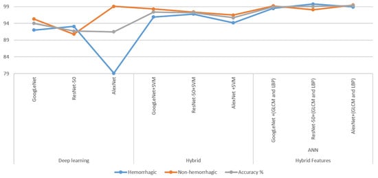
Figure 23. Performance of all proposed methods for fast detection of hemorrhage disease in graph form.
This entry is adapted from the peer-reviewed paper 10.3390/electronics11152460
This entry is offline, you can click here to edit this entry!