Apoptosis and (in) Pain—Potential Clinical Implications: History
Please note this is an old version of this entry, which may differ significantly from the current revision.

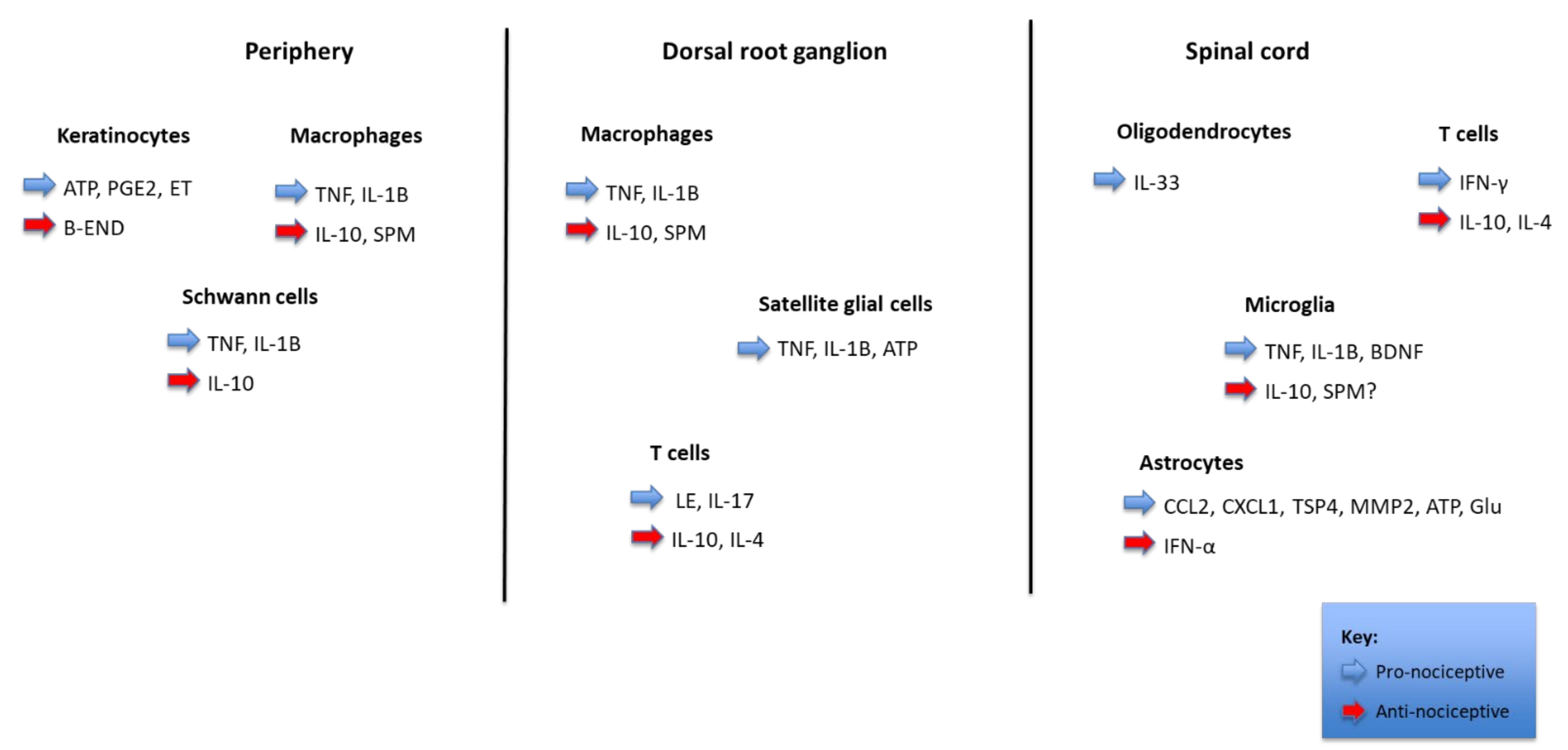
The deregulation of apoptosis is involved in the development of several pathologies, and recent evidence suggests that apoptosis may be involved in chronic pain, namely in neuropathic pain. Neuropathic pain is a chronic pain state caused by primary damage or dysfunction of the nervous system. Recently, it was found that nerve endings contain transient receptor potential (TRP) channels that sense and detect signals released by injured tissues and respond to these damage signals. TRP channels are similar to the voltage-gated potassium channels or nucleotide-gated channels that participate in calcium and magnesium homeostasis. TRP channels allowing calcium to penetrate into nerve terminals can activate apoptosis, leading to nerve terminal destruction. Further, some TRPs are activated by acid and reactive oxygen species (ROS). ROS are mainly produced in the mitochondrial respiratory chain, and an increase in ROS production and/or a decrease in the antioxidant network may induce oxidative stress (OS). Depending on the OS levels, they can promote cellular proliferation and/or cell degeneration or death. Previous studies have indicated that proinflammatory cytokines, such as tumor necrosis factor-α (TNF-α), play an important role in the peripheral mediation of neuropathic pain. 

  • apoptosis
  • pain
  • cell signaling
  • oxidative stress
  • inflammation

1. Definition and Mechanisms of Pain

The IASP conducted a review about the definition of pain in 2020, and it is now considered as an “unpleasant sensory and emotional experience associated with, or resembling that associated with, actual or potential tissue damage” [1].
It was Charles Sherrington, winner of the Nobel Prize in Medicine in 1932, who divided for the first time the mechanisms of propioception, exteroception, and interoception [2], with pain, temperature, and pressure also part of exteroception [3].
Nociception refers to the processing of a noxious stimulus resulting in the perception of pain by the brain. Various neurotransmitters, nociceptors, and cellular components are involved in the mechanisms of transduction, perception, transmission, and modulation of pain [4], as shown in Figure 1.
Figure 1. Pain regulation by non-neuronal cells and inflammation. The figure shows the interactions between distinct parts of a nociceptor (periphery, dorsal root ganglion, and spinal cord) with different types of non-neurons cells (keratinocytes, Schwann cells, satellite glial cells, oligodendrocytes, and astrocytes), immune cells (macrophages, T cells, and microglia), cancer cells, and bone marrow stem cells. These cells produce pronociceptive (highlighted in blue) and antinociceptive (highlighted in red) mediators, which modulate the nociceptor sensitivity and excitability through binding to their respective receptors. In the spinal cord dorsal horn, the central terminal of the nociceptor forms a nociceptive synapse with a postsynaptic neuron to mediate pain transmission in the Central Nervous System (CNS).
Transduction is the conversion of a noxious stimulus (mechanical, chemical, or thermal) into electrical energy by a peripheral nociceptor (free afferent nerve ending). Transmission describes the propagation through the peripheral nervous system via first-order neurons. 
Pain depends on a multifactorial system with multiple pathways for numerous cortex areas, the cortical body matrix [5], which explains why it is a multidimensional experience that can significantly impair an individual’s quality of life [6].
The most abundant pain sensors are in the skin and are called the transient receptor potential cation channels (TRP) [7]. The TRP family contains many members, such as the vanilloid receptors (TRPV), the canonical receptors (TRPC), the ankyrin repeat receptors (TRPA), the polycystin recaptors (TRPP), the melastatin receptors (TRPM), and the mucolipin receptors (TRPML) [7]. TRP channels are activated by histamine, bradykinin, fatty acid metabolites, endocannabinoids, cannabinoids, adenosine triphosphate (ATP), acid, heat, cold, prostaglandins, cytokines, capsaicin, monoterpenoids, among other stimuli. The initial sensation can be heat, cold, pain, or itching that can be several minutes in duration. Usually, prolonged activation of the TRP channels (for more than 20–30 min or so) results in long-term deactivation and pain relief [7]. Recently, nine TRP channels (TRPA1, TRPC5, TRPM2, TRPM4, TRPM7, TRPV1, TRPV2, TRPV3, and TRPV4) have been demonstrated to be activated by oxidative stress [8] and in neurons related to nociception (as in dorsal root ganglion (DRG) and trigeminal ganglia neurons), the expression levels of four TRP channels (TRPA1, TRPM2, TRPV1, and TRPV4) are high [9]. From [10][11], TRPV1 and TRPA1 have been demonstrated in neuropathic pain associated with diabetes or the administration of chemotherapeutics, probably mediated by the synthesis of reactive oxygen and nitrogen species [12][13], which are well-known TRPA1 activators [14][15]. Further, TRP channels have been associated with pain in neurodegenerative disorders (NDD) through oxidative stress-induced cell death, particularly related to inflammation and calcium homeostasis deregulation [15]. All these processes are connected with microglial activation, a proposed mechanism inductor of inflammation and neuropathic pain [16], which leads to neuronal degeneration and cell death in NDD, and simultaneously to neuropathic and inflammatory pain [17][18].
Mitochondria play important roles in a myriad of cellular processes, and mitochondrial dysfunction has been implicated in multiple neurological disorders [19]. The five major mitochondrial functions (the mitochondrial energy generating system, ROS generation, mitochondrial permeability transition pore, apoptotic pathways, and intracellular calcium mobilization) may play critical roles in neuropathic and inflammatory pain [19].
Oxidative stress is a central mediator of apoptosis, neuroinflammation, metabolic disturbances, and bioenergetic failure in neurons [20]. Oxidative stress and apoptosis, as observed in the DRG neurons, play an essential role in inducing and developing pain hypersensitivity [21].
It should also be noted that apoptosis is also regulated by proinflammatory cytokines, such as Interferon gamma (IFN-g), tumor necrosis factor-α (TNF-α), interleukina-1 beta (IL-1 b), and IL-6 [22]. The roles of IL-1 and TNF- α in apoptosis in neurodegenerative diseases such as Alzheimer’s disease were previously described [22][23]. More importantly, it is known that these proinflammatory cytokines have crucial roles in the modulation of neuropathic pain [22][24].

2. Apoptosis Pathways as Mediators of Pain Formation

2.1. Apoptosis Cell Signaling Pathways

The homeostasis of a multicellular organism depends on cell turnover, as it is related to the capacity to maintain the equilibrium between cell proliferation and death [25][26].
Physiological cell death occurs primarily through an evolutionary and conserved form of cellular suicide, called apoptosis (from the Greek word for decay, as with leaf fall, for example), in response to a series of intrinsic and extrinsic stimuli [25][26]. It was discovered in 1952. However, in 1972, the Australian pathologist John FR Kerr and the Scots Andrew H. Wylie and Alistair R. Currie showed the importance of this cell death process in developing an adult organism [25]. Unlike the other type of death, cell necrosis, in which the cell is a passive victim, cell apoptosis is an active form of death, in which the cell expends energy to carry out this genetically programmed cellular suicide process. For this reason, it is often called programmed cell death [25][27].
The induction of apoptosis can be divided into three steps: the inducing agent’s interaction with the cell, the biochemical transduction of the death signal, and the execution by the apoptotic machinery. Different extracellular signals, such as an increase in ROS production [28] and/or a decrease in antioxidants resulting in oxidative stress, inflammatory mediators such as TNF- α and IL1-β [29], moderate hyperactivity stimulation of the N-methyl-D-aspartate (NMDA) receptors [30], and the endoplasmic reticulum stress [31] could activate signal transduction pathways that converge on a final common pathway leading to the execution phase of cellular apoptosis. At least two pathways are involved, the mitochondria-dependent pathway (intrinsic or mitochondrial pathway), which is mediated by mitochondria and the B-cell lymphoma-2 (BCL-2) family proteins, and the extrinsic or membrane pathway involving external signaling via membrane receptors from the tumor necrosis factor (TNF) family, such as TNF-R1, FAS, TNF-apoptotic inducing ligand receptors 1 and 2 (TRAIL-R1 and 2, or death receptors DR4 and 5 [32], and the low-affinity nerve growth factor (NGF) receptor, p75-NTR [13][33]. Moreover, the p38MAPK pathway causes the activation of apoptosis in neuronal cells, inducing pain (such as bone cancer pain-related hyperalgesia) [34].
However, all the pathways culminate in the activation of a series of cysteine proteases, called caspases. The effector caspases, such as caspase 3, cleave a wide variety of protein targets that are responsible for the characteristic changes in apoptotic cells. These include cell shrinkage, nuclear condensation, membrane blebbing, fragmentation into apoptotic bodies, and membrane changes that can lead to phagocytosis of the affected cell [11][20]. An alternative, proinflammatory form of apoptosis [27] in macrophages infected with Salmonella or Shigella, termed “pyroptosis” [35]. Since its discovery, pyroptosis has been observed in the central nervous system [36] and the cardiovascular system, suggesting that this form of cell death is biologically significant. [34][35].
The knowledge of this active physiologic mode of cell death has profound implications on our understanding of several diseases. In fact, the deregulation of apoptosis is involved in the development of several diseases, such as cancer [37], neurodegenerative diseases as Alzheimer’s and Parkinson’s, and autoimmune or rheumatoid arthritis. Recent evidence suggests that apoptosis may be involved in pain, namely neuropathic pain [30].
The involvement of apoptosis in pain and the clinical relevance of this knowledge in the discovery of new potential biomarkers and therapeutic targets may be useful for the development of more effective and targeted drugs to treat pain.

2.2. Ubiquitin Proteasome Pathway and Apoptosis

Regulatory proteins involved in normal cell proliferation and differentiation are degraded by a proteolytic pathway, which involves lysosomal enzymes, and a non-lysosomal pathway, known as the ubiquitin–proteasome pathway (UPP) [38].
The UPP encompasses several enzymes, namely the ubiquitin-activating enzyme (E1), the ubiquitin-conjugating enzyme (E2), and a ligase (E3) that catalyzes the binding of ubiquitin to the target protein, the deubiquitinating enzyme (DUB), and proteasomes (namely the 26S proteasome) [39].
The degradation of proteins by the UPP involves two successive and distinct steps: first, several ubiquitin molecules bind to the target protein to be subsequently degraded in the proteasomes. Some of the regulatory proteins with levels are controlled by this degradative pathway include proteins involved in cell death by apoptosis, namely the BCL2 family proteins such as BAX protein [38], and the C-terminal fragment of the BID protein the BIDt [40], and the IAP family and the inhibitor of the NF-kB, IkB [41][42].
The UPP is relevant in neural development and brain structure and also in maintenance of their functions, and is implicated in synaptic plasticity, formation, and maintenance of memory. Recently, it was found that the dysfunction of this proteasomal degradation pathway leads to changes in cell death by apoptosis in diverse cell types and may be implicated in the pathogenesis of several diseases, either through hypofunction.
One of the most important enzymes is the ubiquitin-conjugating enzyme E2B (Ube2b), also known as RAD6B. Ube2b is a member of the ubiquitin-conjugating enzyme family that has been identified to be vital in neural DNA double-strand DNA breaks (DSBs). Further, the Ube2b modulates the DNMT3a (DNA methyltransferase 3 alpha) ubiquitination degradation and CaMKK1 (calcium/calmodulin-dependent protein kinase) gene promoter demethylation increasing CaMKK1 level.
Moreover, the nuclear factor-κB (NF-κB) is a key regulator of molecules and pathways important for inflammation and pain [43][44][45]. NF-κB can exist as homo or heterodimers composed of the Rel family proteins (the p65/p50 subunits in humans) and is bound to the κB inhibitor (IκBα) in the cytoplasm, when in an inactive state. Upon activation by proinflammatory cytokines and growth factors, among others, the IκB kinase (IKK) phosphorylates IκBα, targeting its degradation in the UPP. As a result, IκBα releases the p65/p50 complex, which subsequently translocates to the nucleus and initiates the transcription of several genes, including inflammatory genes such as TNFα, IL-1β, and cyclooxogenase-2 (COX-2) that directly or indirectly influence pain [43][44][45]. Thus, targeting the NF-kB pathway, namely by inhibiting the IkB degradation in the UPP could be a new therapeutic approach to treating pain [31][43][44].

2.3. Endoplasmic Reticulum Stress Signalling and Apoptosis

Endoplasmic reticulum (ER) stress causes the activation of caspase signaling pathway-dependent apoptosis in neuronal cells and induces bone cancer pain-related hyperalgesia.
The ER is the cellular organelle in which protein folding, lipid biosynthesis, and calcium storage take place. Many factors can cause an imbalance in the homeostasis of ER function, resulting in ER stress. Such ER stress initiates an evolutionarily conserved signaling cascade called the unfolded protein response (UPR), which is a self-protective signaling pathway. The accumulation of unfolded proteins in the ER can activate the UPR to restore ER function. Besides the increase in ER stress as a self-protective signal transcription pathway after mild injury, the failure of this system to relieve prolonged or excessive ER stress may cause cell apoptosis, and plays an important role in neuropathic pain, specifically in rat models of bone cancer pain [31].

2.4. Oxidative Stress

When the synthesis of oxidative species is superior to the capacity of the cells to counteract them, there is the occurrence of oxidative stress. The generation of reactive oxygen species (ROS) in mitochondria is a consequence of oxidative phosphorylation related to the respiratory chain [46]. ROS acts physiologically as signaling molecules, but when produced in abundance has deleterious consequences to DNA, protein, lipids, initiation of inflammatory events, excitotoxicity, and apoptosis [47].
Mitochondrial dysfunction, the increase in ROS and reactive nitrogen species (RNS), is involved in the pathogenesis of chronic neuropathic pain [48], leading to the degeneration of primary afferents [49], and ROS accumulation originates a feed-forward mechanism of nitroxidative lesion that triggers proapoptotic factors [50][51].
In fact, mitochondrial dysfunction is observed in various neuropathic pain phenotypes, such as chemotherapy-induced neuropathy, diabetic neuropathy, HIV-associated neuropathy, Charcot–Marie–Tooth neuropathy, and trauma-induced painful mononeuropathy [51].
More recently, it was shown that damage to mitochondria and aberrant mitochondrial transport in peripheral neurons are common features of peripheral neuropathy [52]. The complex and highly energy-consuming processes of neurotransmission by peripheral neurons are critically dependent on mitochondrial metabolic functions [53].

2.5. Inflammation

For five decades, apoptosis has been recognized as fundamental for almost all the critical biological processes that occur in multicellular organisms, since embryogenesis and throughout life, to maintain the homeostatic balance of tissues and organs as well as the progression and final resolution of inflammation [54][55][56].
However, this physiological form of cell death was very early on associated with the absence of an inflammatory reaction by the surrounding tissues. This was initially explained by the preservation of membrane integrity in such a way that cellular content does not come into contact with neighbouring cells [54][57][58][59][60][61].
Inside, they contain well-preserved organelles and nuclear fragments, which will be prompt digested and removed from tissues by professional phagocytes or non-professional neighboring cells that specifically recognize apoptotic cells [57][59].
Although initially it was considered that the maintenance of the integrity of the cellular membrane was the only reason why apoptosis did not cause inflammation, it is now known and consensual that the mechanisms involved in the recognition and subsequent phagocytosis and clearance of apoptotic cells will determine whether apoptosis is immunologically “silent” or anti-inflammatory [60][61][62][63][64][65].
It seems clear that during apoptosis, the inflammatory response could be blocked [65] Regarding biochemical explanation, the fact that the inflammatory response is absent during apoptosis was initially explained by the exposure of phagocytes to phosphatidylserine after a process of translocation from the inner to the outer leaflet of the cell membrane ([66] This “eat me” signal that attracts and allows the recognition of apoptotic cells and their subsequent elimination plays a central role in phagocytosis and is one of the hallmarks of apoptosis [64][67][68].
Caspase activation, changes in mitochondrial membrane potential, and DNA cleavage are also players in apoptosis, as they contribute to disrupt cellular functions and mark cells for a “silent” phagocytic clearance [69][70].
Due to activation of caspases that cleave membrane channel proteins results in the release of adenosine monophosphate (AMP) [70].
Then, through the 5′-nucleotidase a phosphate group is removed from the AMP to produce adenosine that binds to the A2a receptors on the surface of phagocytes to generate the anti-inflammatory response [70].
Currently, other molecules, such as lysophosphatidylcholine, ATP and uridine 5’-triphosphate (UTP), sphingosine-1-phosphate, CX3CL1/fractalkine, thrombospondin-1, monocyte chemoattractant protein-1 (MCP-1), lactoferrin, are also known as effective in the “silent” clearance of apoptotic cells [71] These molecules act by sending chemotactic signals that attract phagocytes that recognize apoptotic cell-associated molecular patterns (ACAMPs) on the surface of apoptotic cells, therefore contributing to the anti-inflammatory characteristic of apoptosis.

2.6. The p38MAPK Pathway and Apoptosis

Numerous studies have shown that activation of Mitogen-activated protein kinases (MAPK), particularly c-Jun N-terminal kinase (JNK) and p38, contributes to neuropathic pain pathology [31][72][73][74].
The activation of the p38MAPK signaling pathway plays an essential role in the generation and maintenance of neuropathic pain by regulating transcription, protein synthesis, receptor expression, and inducing apoptosis. The protein kinase A (PKA) is a key protein involved in neuropathic pain signaling, which by activating the p38MAPK intervenes in the apoptosis of spinal cord cell [34]. Studies have shown that PKA is closely related to inflammatory pain and bone cancer pain [74] but it is not clear its involvement in neuropathic pain caused by nerve damage. Scholars hypothesized that PKA is involved in neuropathic pain by mediating spinal cord cell apoptosis through p38MAPK pathway activation, which culminates in a decrease in the antiapoptotic protein BCL2 and an increase in the proapoptotic proteins, TNF-alfa, IL1-beta, BAX, Caspase3 and 9 (Figure 1) [34]. Further, it has been suggested that the inhibition of either JNK or p38 may represent a potent clinical target for neuropathic pain management [34][72].

3. Apoptosis and Clinical Implications

3.1. Apoptosis and Neuropathic Pain

Nerve damage due to oxidative stress and mitochondrial dysfunction is a key pathogenic mechanism involved in chemotherapy-induced peripheral neuropathy (CIPN) [8][10][12][13][14][15][75]. On the other hand, TRP channels allow calcium to penetrate into nerve terminals. This can activate apoptosis mechanisms that result in nerve terminal destruction [20] in the skin which can cause long-term pain relief [20]. Further, cisplatin, oxaliplatin, and paclitaxel-induced mitochondrial oxidative stress, inflammation, cold allodynia, and hyperalgesia, through an increase in TRPA1 and TRPV4, leading to Ca2+ influx through direct channel activation or excessive production of oxidative stress and induction of apoptosis. The pain resulting from this Ca2+ overload is mediated through substance P (SP) and excitatory amino acid production. This chemotherapy-induced oxidative stress in DRG neurons that contribute to peripheral pain may be prevented with TRPA1 and TRPM8 antagonists such as reduced glutathione (GSH) and selenium [9][10][14][76][77][78].

2.2. Biomarkers and Circulating Mediators in Pain

Oxidative damage to peripheral neurons can cause damage to the myelin sheath, mitochondrial proteins, and other antioxidant enzymes [20]. Identification of levels of malondialdehyde, glutathione (GSH), superoxide dismutase (SOD), and activities of mitochondrial enzymes such as citrate synthase and ATP synthase can help monitor the course of peripheral neuropathy and response of neuropathy to the treatment [20].

2.3. Pain Therapy through Modulating Apoptosis Activities

Only one-third of patients receive pain relief from current analgesics, such as opiates, nonsteroidal anti-inflammatory drugs, local anesthetics, tricyclic antidepressants, and anticonvulsants, including carbamazepine and gabapentin. Therefore, it seems notable that recent developments in understanding the mechanisms that produce pain, either individually or collectively, have disclosed new potential therapeutic targets for developing more effective drugs [30].
Isoflurane, a general inhalation anesthetic used for the induction and maintenance of general anesthesia, promotes PI3K/AKT activation, upregulates B-cell lymphoma 2 (Bcl-2)-associated X protein Bcl-2 expression levels, and reduces the expression levels of caspase 3 and caspase 8 in myocardial cells. Isoflurane is beneficial for pain attenuation and inhibits apoptosis in myocardial cells via the PI3K/AKT signaling pathway in mice during cardiac surgery [79].
Paeoniflorin can not only significantly inhibit the activation of ASK1 and simulate the analgesic effect of ASK1 inhibitors, but also significantly inhibit the response of glial cells and neuroinflammation induced by CCI [80]. Acting in the same pathway, administration of a p38 inhibitor and a JNK inhibitor ameliorates neuropathic pain symptoms in rodent models [81][82].
Spare nerve injury (SNI) induces a significant increase in PKA expression in the spinal cord, and PKA is involved in neuropathic pain by activating the p38MAPK pathway to mediate spinal cord cell apoptosis [34].
Tetramethylpyrazine (TMP) attenuated neuropathic pain-associated hyperalgesia and neuronal apoptosis in the spinal dorsal horn, which was demonstrated by a decreased number of TUNEL-positive cells, upregulation in BCL-2 expression, and downregulation in caspase-3 expression in the spinal dorsal horn. These results suggest that TMP is beneficial for treating neuropathic pain by inhibiting apoptosis via the modulation of BCL-2 and caspase-3 proteins [83].
The crucial role of NF-kB in several pathologies that accompany pain provides evidence that targeting the NF-kB signaling cascade, including UPP, might have beneficial antinociceptive effects [43][44][45]. Several proteasome inhibitors are already approved to treat hematological neoplasias, such as multiple myeloma [84]. The generation of new proteasome inhibitors may represent a new pharmacotherapy for inflammatory pain [39]. Several studies show that UPP inhibitors can prevent inflammatory pain following injury and infection. The proteasome inhibitor MG132 can inhibit the activation of NF-κB, reversing the inflammatory pain [85].

4. Conclusions

Neuropathic pain is a chronic pain condition caused by primary damage or dysfunction of the nervous system, but the fine molecular mechanisms have not yet been fully elucidated.
Previous studies have indicated that NF-kB, proinflammatory cytokines such as TNF-α, and oxidative stress play an important role in peripheral mediation of neuropathic pain through apoptosis modulation. Further, the degree of DRG neuronal apoptosis has recently been associated with spinal nerve injury via caspase signaling and/or PKA activation through the p38MAPK pathway, generating and maintaining neuropathic pain.
The adoption of measures to prevent and protect mitochondrial function may constitute a promising therapeutic strategy to alleviate or prevent chronic pain conditions [86] since mitochondria are a primary source of cellular ROS [75]. Pharmacological interventions aimed at maintaining normal mitochondrial function may be an alternative therapeutic approach to the direct free radical scavengers for the treatment of CIPN [75]
Medications that can modulate proinflammatory cytokine expression, the NF-kB pathway, and apoptosis, such as TNF-alpha modulators, G-CSF, proteasome inhibitors, and flavonoids, among others, have the potential to treat neuropathic pain [22]. However, the timing of administration, dose, and indications for neuropathic pain treatment still need to be validated by clinical studies [22].
Further, the inhibition of apoptosis via the modulation of BCL-2 and caspase-3 is beneficial for the treatment of neuropathic pain [83].
The involvement of apoptosis in pain and the clinical relevance of this knowledge in the potential discovery of new biomarkers and therapeutic targets can result in the development of more effective and targeted drugs to treat chronic pain, particularly neuropathic pain.

This entry is adapted from the peer-reviewed paper 10.3390/biomedicines10061255

References

  1. Raja, S.N.; Carr, D.B.; Cohen, M.; Finnerup, N.B.; Flor, H.; Gibson, S.; Keefe, F.J.; Mogil, J.S.; Ringkamp, M.; Sluka, K.A.; et al. The revised international association for the study of pain definition of pain: Concepts, challenges, and compromises. Pain 2020, 161, 1976–1982.
  2. Nobelstiftelsen. Nobel Lectures in Physiology or Medicine; Elsevier for the Nobel Foundation: Amsterdam, The Netherlands; London, UK, 1965; pp. 1922–1941.
  3. Valenzuela-Moguillansky, C.; Reyes-Reyes, A.; Gaete, M.I. Exteroceptive and interoceptive body-self awareness in fibromyalgia patients. Front. Hum. Neurosci. 2017, 11, 117.
  4. Ji, R.-R.; Chamessian, A.; Zhang, Y.-Q. Pain regulation by non-neuronal cells and inflammation. Science 2016, 354, 572–577.
  5. Garcia-Larrea, L.; Peyron, R. Pain matrices and neuropathic pain matrices: A review. Pain 2013, 154, S29–S43.
  6. Orr, P.M.; Shank, B.C.; Black, A.C. The role of pain classification systems in pain management. Crit. Care Nurs. Clin. N. Am. 2017, 29, 407–418.
  7. Adams, J.D. Editorial: Apoptosis is critical to pain control. Open J. Apoptosis 2013, 2, 23–24.
  8. Ogawa, N.; Kurokawa, T.; Mori, Y. Sensing of redox status by TRP channels. Cell Calcium 2016, 60, 115–122.
  9. Carrasco, C.; Naziroǧlu, M.; Rodríguez, A.B.; Pariente, J.A. Neuropathic pain: Delving into the oxidative origin and the possible implication of transient receptor potential channels. Front. Physiol. 2018, 9, 95.
  10. Nazıroğlu, M.; Braidy, N. Thermo-sensitive TRP channels: Novel targets for treating chemotherapy-induced peripheral pain. Front. Physiol. 2017, 8, 1040.
  11. Jardín, I.; López, J.J.; Diez, R.; Sánchez-Collado, J.; Cantonero, C.; Albarrán, L.; Woodard, G.E.; Redondo, P.C.; Salido, G.M.; Smani, T.; et al. TRPs in pain sensation. Front. Physiol. 2017, 8, 392.
  12. Yowtak, J.; Wang, J.; Kim, H.Y.; Lu, Y.; Chung, K.; Chung, J.M. Effect of antioxidant treatment on spinal GABA neurons in a neuropathic pain model in the mouse. Pain 2013, 154, 2469–2476.
  13. Mai, L.; Huang, F.; Zhu, X.; He, H.; Fan, W. Role of nerve growth factor in orofacial pain. J. Pain Res. 2020, 13, 1875–1882.
  14. Braidy, N.; Smani, T.; Naziroglu, M. Editorial: Involvements of TRP channels, oxidative stress and apoptosis in neurodegenerative diseases. Front. Physiol. 2021, 12, 649230.
  15. Duitama, M.; Vargas-López, V.; Casas, Z.; Albarracin, S.L.; Sutachan, J.-J.; Torres, Y.P. TRP channels role in pain associated with neurodegenerative diseases. Front. Neurosci. 2020, 14, 782.
  16. Haraguchi, K.; Kawamoto, A.; Isami, K.; Maeda, S.; Kusano, A.; Asakura, K.; Shirakawa, H.; Mori, Y.; Nakagawa, T.; Kaneko, S. TRPM2 Contributes to inflammatory and neuropathic pain through the aggravation of pronociceptive inflammatory responses in mice. J. Neurosci. 2012, 32, 3931–3941.
  17. Carniglia, L.; Ramírez, D.; Durand, D.; Saba, J.; Turati, J.; Caruso, C.; Scimonelli, T.N.; Lasaga, M. Neuropeptides and microglial activation in inflammation, pain, and neurodegenerative diseases. Mediat. Inflamm. 2017, 2017, 5048616.
  18. Suter, M.R.; Wen, Y.-R.; Decosterd, I.; Ji, R.-R. Do glial cells control pain? Neuron Glia Biol. 2007, 3, 255–268.
  19. Sui, B.; Xu, T.-Q.; Liu, J.-W.; Wei, W.; Zheng, C.-X.; Guo, B.-L.; Wang, Y.-Y.; Yang, Y.-L. Understanding the role of mitochondria in the pathogenesis of chronic pain. Postgrad. Med. J. 2013, 89, 709–714.
  20. Areti, A.; Yerra, V.G.; Naidu, V.; Kumar, A. Oxidative stress and nerve damage: Role in chemotherapy induced peripheral neuropathy. Redox Biol. 2014, 2, 289–295.
  21. Ding, R.; Sun, B.; Liu, Z.; Yao, X.; Wang, H.; Shen, X.; Jiang, H.; Chen, J. Advanced oxidative protein products cause pain hypersensitivity in rats by inducing dorsal root ganglion neurons apoptosis via NADPH Oxidase 4/c-Jun N-terminal kinase pathways. Front. Mol. Neurosci. 2017, 10, 195.
  22. Liao, M.-F.; Lu, K.-T.; Hsu, J.-L.; Lee, C.-H.; Cheng, M.-Y.; Ro, L.-S. The role of autophagy and apoptosis in neuropathic pain formation. Int. J. Mol. Sci. 2022, 23, 2685.
  23. Yuan, J.; Yankner, B.A. Apoptosis in the nervous system. Nature 2000, 407, 802–809.
  24. Grace, P.M.; Hutchinson, M.R.; Maier, S.F.; Watkins, L.R. Pathological pain and the neuroimmune interface. Nat. Rev. Immunol. 2014, 14, 217–231.
  25. Duke, R.C.; Ojcius, D.M.; Young, J.D.-E. Cell suicide in health and disease. Sci. Am. 1996, 275, 80–87.
  26. Thompson, C.B. Apoptosis in the pathogenesis and treatment of disease. Science 1995, 267, 1456–1462.
  27. D’Arcy, M.S. Cell death: A review of the major forms of apoptosis, necrosis and autophagy. Cell Biol. Int. 2019, 43, 582–592.
  28. Redza-Dutordoir, M.; Averill-Bates, D.A. Activation of apoptosis signalling pathways by reactive oxygen species. Biochim. Biophys. Acta (BBA)—Mol. Cell Res. 2016, 1863, 2977–2992.
  29. Sekiguchi, M.; Sekiguchi, Y.; Konno, S.-I.; Kobayashi, H.; Homma, Y.; Kikuchi, S.-I. Comparison of neuropathic pain and neuronal apoptosis following nerve root or spinal nerve compression. Eur. Spine J. 2009, 18, 1978–1985.
  30. Corasaniti, M.T.; Amantea, D.; Russo, R.; Bagetta, G. The crucial role of neuronal plasticity in pain and cell death. Cell Death Differ. 2006, 13, 534–536.
  31. He, Q.; Wang, T.; Ni, H.; Liu, Q.; An, K.; Tao, J.; Chen, Y.; Xu, L.S.; Zhu, C.; Yao, M.; et al. Endoplasmic reticulum stress promoting caspase signaling pathway-dependent apoptosis contributes to bone cancer pain in the spinal dorsal horn. Mol. Pain 2019, 15, 1744806919876150.
  32. Peter, M.E.; Kischkel, F.C.; Hellbardt, S.; Chinnaiyan, A.M.; Krammer, P.H.; Dixit, V.M. CD95 (APO-1/Faz)—associating signalling proteins. Cell Death Differ. 1996, 3, 161–170.
  33. Lewin, G.R.; Nykjaer, A. Pro-neurotrophins, sortilin, and nociception. Eur. J. Neurosci. 2014, 39, 363–374.
  34. Deng, Y.; Yang, L.; Xie, Q.; Yang, F.; Li, G.; Zhang, G.; Li, S.; Wu, Z.; Wang, J.; Kang, X. Protein kinase a is involved in neuropathic pain by activating the p38MAPK pathway to mediate spinal cord cell apoptosis. Mediat. Inflamm. 2020, 2020, 6420425-17.
  35. Fink, S.L.; Cookson, B.T. Apoptosis, pyroptosis, and necrosis: Mechanistic description of dead and dying eukaryotic cells. Infect. Immun. 2005, 73, 1907–1916.
  36. Liu, X.-H.; Kwon, D.; Schielke, G.P.; Yang, G.-Y.; Silverstein, F.S.; Barks, J.D.E. Mice deficient in interleukin-1 converting enzyme are resistant to neonatal hypoxic-ischemic brain damage. J. Cereb. Blood Flow Metab. 1999, 19, 1099–1108.
  37. Sarmento-Ribeiro, A.B.; Dourado, M.; Paiva, A.; Freitas, A.; Silva, T.; Regateiro, F.; Oliveira, C.R. Apoptosis deregulation influences chemoresistance to azaguanine in human leukemic cell lines. Cancer Investig. 2012, 30, 331–342.
  38. Li, B.; Dou, Q.P. Bax degradation by the ubiquitin/proteasome-dependent pathway: Involvement in tumor survival and progression. Proc. Natl. Acad. Sci. USA 2000, 97, 3850–3855.
  39. Cheng, J.; Deng, Y.; Zhou, J. Role of the ubiquitin system in chronic pain. Front. Mol. Neurosci. 2021, 14, 674914.
  40. Breitschopf, K.; Zeiher, A.M.; Dimmeler, S. Ubiquitin-mediated degradation of the proapoptotic active form of bid. J. Biol. Chem. 2000, 275, 21648–21652.
  41. Zhang, H.-G.; Wang, J.; Yang, X.; Hsu, H.-C.; Mountz, J.D. Regulation of apoptosis proteins in cancer cells by ubiquitin. Oncogene 2004, 23, 2009–2015.
  42. Qiu, J.H.; Asai, A.; Chi, S.; Saito, N.; Hamada, H.; Kirino, T. Proteasome inhibitors induce cytochrome c–caspase-3-like protease-mediated apoptosis in cultured cortical neurons. J. Neurosci. 2000, 20, 259–265.
  43. Hartung, J.E.; Eskew, O.; Wong, T.; Tchivileva, I.E.; Oladosu, F.A.; O’Buckley, S.C.; Nackley, A.G. Nuclear factor-kappa B regulates pain and COMT expression in a rodent model of inflammation. Brain Behav. Immun. 2015, 50, 196–202.
  44. Niederberger, E.; Geisslinger, G. The IKK-NF-κB pathway: A source for novel molecular drug targets in pain therapy? FASEB J. 2008, 22, 3432–3442.
  45. Fakhri, S.; Abbaszadeh, F.; Jorjani, M. On the therapeutic targets and pharmacological treatments for pain relief following spinal cord injury: A mechanistic review. Biomed. Pharmacother. 2021, 139, 111563.
  46. Matés, J.M.; Segura, J.A.; Alonso, F.J.; Márquez, J. Oxidative stress in apoptosis and cancer: An update. Arch. Toxicol. 2012, 86, 1649–1665.
  47. Wu, Y.; Chen, M.; Jiang, J. Mitochondrial dysfunction in neurodegenerative diseases and drug targets via apoptotic signaling. Mitochondrion 2019, 49, 35–45.
  48. Flatters, S.J. The contribution of mitochondria to sensory processing and pain. Prog. Mol. Biol. Transl. Sci. 2015, 131, 119–146.
  49. Bennett, G.J.; Doyle, T.; Salvemini, D. Mitotoxicity in distal symmetrical sensory peripheral neuropathies. Nat. Rev. Neurol. 2014, 10, 326–336.
  50. Sinha, K.; Das, J.; Pal, P.B.; Sil, P.C. Oxidative stress: The mitochondria-dependent and mitochondria-independent pathways of apoptosis. Arch. Toxicol. 2013, 87, 1157–1180.
  51. Lim, T.; Rone, M.B.; Lee, S.; Antel, J.P.; Zhang, J. Mitochondrial and bioenergetic dysfunction in trauma-induced painful peripheral neuropathy. Mol. Pain 2015, 11, 58.
  52. English, K.; Barton, M.C. HDAC6: A key link between mitochondria and development of peripheral neuropathy. Front. Mol. Neurosci. 2021, 14, 175.
  53. Todorova, V.; Blokland, A. Mitochondria and synaptic plasticity in the mature and aging nervous system. Curr. Neuropharmacol. 2016, 15, 166–173.
  54. Haanen, C.; Vermes, I. Apoptosis and inflammation. Mediat. Inflamm. 1995, 4, 5–15.
  55. Wyllie, A.; Kerr, J.; Currie, A. Cell death: The significance of apoptosis. Int. Rev. Cytol. 1980, 68, 251–306.
  56. Wallach, D.; Kovalenko, A. Keeping inflammation at bay. eLife 2014, 3, e02583.
  57. Kerr, J.F.R.; Wyllie, A.H.; Currie, A.R. Apoptosis: A basic biological phenomenon with wideranging implications in tissue kinetics. Br. J. Cancer 1972, 26, 239–257.
  58. Haslett, C.; Savill, J.S.; Whyte, M.K.B.; Stern, M.; Dransfield, I.; Meagher, L.C.; Nagata, S. Granulocyte apoptosis and the control of inflammation. Philos. Trans. R. Soc. B: Biol. Sci. 1994, 345, 327–333.
  59. Nagata, S. Apoptosis and clearance of apoptotic cells. Annu. Rev. Immunol. 2018, 36, 489–517.
  60. Tsuchiya, K. Switching from apoptosis to pyroptosis: Gasdermin-elicited inflammation and antitumor immunity. Int. J. Mol. Sci. 2021, 22, 426.
  61. Hart, S.P.; Dransfield, I.; Rossi, A.G. Phagocytosis of apoptotic cells. Methods 2008, 44, 280–285.
  62. Ellis, R.E.; Yuan, J.; Horvitz, H.R. Mechanisms and functions of cell death. Annu. Rev. Cell Biol. 1991, 7, 663–698.
  63. Cohen, J.J. Apoptosis. Immunol. Today 1993, 14, 126–130.
  64. Arienti, S.; Barth, N.; Dorward, D.A.; Rossi, A.G.; Dransfield, I. Regulation of apoptotic cell clearance during resolution of inflammation. Front. Pharmacol. 2019, 10, 891.
  65. Voll, R.E.; Herrmann, M.; Roth, E.A.; Stach, C.; Kalden, J.R.; Girkontaite, I. Immunosuppressive effects of apoptotic cells. Nature 1997, 390, 350–351.
  66. Martin, S.; Reutelingsperger, C.E.M.; McGahon, A.J.; Rader, J.; Van Schie, R.C.A.A.; LaFace, D.M.; Green, D. Early redistribution of plasma membrane phosphatidylserine is a general feature of apoptosis regardless of the initiating stimulus: Inhibition by overexpression of Bcl-2 and Abl. J. Exp. Med. 1995, 182, 1545–1556.
  67. Huynh, M.-L.N.; Fadok, V.A.; Henson, P.M. Phosphatidylserine-dependent ingestion of apoptotic cells promotes TGF-β1 secretion and the resolution of inflammation. J. Clin. Investig. 2002, 109, 41–50.
  68. Segawa, K.; Nagata, S. an apoptotic ‘eat me’ signal: Phosphatidylserine exposure. Trends Cell Biol. 2015, 25, 639–650.
  69. Ravichandran, K.S.; Lorenz, U. Engulfment of apoptotic cells: Signals for a good meal. Nat. Rev. Immunol. 2007, 7, 964–974.
  70. Yamaguchi, H.; Maruyama, T.; Urade, Y.; Nagata, S. Immunosuppression via adenosine receptor activation by adenosine monophosphate released from apoptotic cells. eLife 2014, 3, e02172.
  71. Szondy, Z.; Sarang, Z.; Kiss, B.; Garabuczi, É.; Köröskényi, K. Anti-inflammatory mechanisms triggered by apoptotic cells during their clearance. Front. Immunol. 2017, 8, 909.
  72. Shin, J.; Yin, Y.; Park, H.; Park, S.; Triantafillu, U.L.; Kim, Y.; Kim, S.R.; Lee, S.Y.; Kim, D.K.; Hong, J.; et al. p38 siRNA-encapsulated PLGA nanoparticles alleviate neuropathic pain behavior in rats by inhibiting microglia activation. Nanomedicine 2018, 13, 1607–1621.
  73. Kim, E.K.; Choi, E.-J. Pathological roles of MAPK signaling pathways in human diseases. Biochim. Biophys. Acta (BBA)—Mol. Basis Dis. 2010, 1802, 396–405.
  74. Zhu, G.-Q.; Liu, S.; He, D.-D.; Liu, Y.-P.; Song, X.-J. Activation of the cAMP-PKA signaling pathway in rat dorsal root ganglion and spinal cord contributes toward induction and maintenance of bone cancer pain. Behav. Pharmacol. 2014, 25, 267–276.
  75. Schoenberger, J.; Bauer, J.; Moosbauer, J.; Eilles, C.; Grimm, D. Innovative strategies in in vivo apoptosis imaging. Curr. Med. Chem. 2008, 15, 187–194.
  76. Du, C.; Fang, M.; Li, Y.; Li, L.; Wang, X. Smac, a Mitochondrial protein that promotes cytochrome c–dependent caspase activation by eliminating IAP inhibition. Cell 2000, 102, 33–42.
  77. Materazzi, S.; Fusi, C.; Benemei, S.; Pedretti, P.; Patacchini, R.; Nilius, B.; Prenen, J.; Creminon, C.; Geppetti, P.; Nassini, R. TRPA1 and TRPV4 mediate paclitaxel-induced peripheral neuropathy in mice via a glutathione-sensitive mechanism. Pflügers Arch. Eur. J. Physiol. 2012, 463, 561–569.
  78. Kahya, M.C.; Nazıroğlu, M.; Övey, I.S. Modulation of diabetes-induced oxidative stress, apoptosis, and ca2+ entry through TRPM2 and TRPV1 channels in dorsal root ganglion and hippocampus of diabetic rats by melatonin and selenium. Mol. Neurobiol. 2016, 54, 2345–2360.
  79. Pi, Z.; Lin, H.; Yang, J. Isoflurane reduces pain and inhibits apoptosis of myocardial cells through the phosphoinositide 3-kinase/protein kinase B signaling pathway in mice during cardiac surgery. Mol. Med. Rep. 2018, 17, 6497–6505.
  80. Zhou, D.; Zhang, S.; Hu, L.; Gu, Y.-F.; Cai, Y.; Wu, D.; Liu, W.-T.; Jiang, C.-Y.; Kong, X.; Zhang, G.-Q. Inhibition of apoptosis signal-regulating kinase by paeoniflorin attenuates neuroinflammation and ameliorates neuropathic pain. J. Neuroinflammation 2019, 16, 83.
  81. Mei, X.-P.; Zhang, H.; Wang, W.; Wei, Y.-Y.; Zhai, M.-Z.; Wang, W.; Xu, L.-X.; Li, Y.-Q. Inhibition of spinal astrocytic c-Jun N-terminal kinase (JNK) activation correlates with the analgesic effects of ketamine in neuropathic pain. J. Neuroinflammation 2011, 8, 6.
  82. Tatsumi, E.; Yamanaka, H.; Kobayashi, K.; Yagi, H.; Sakagami, M.; Noguchi, K. RhoA/ROCK pathway mediates p38 MAPK activation and morphological changes downstream of P2Y12/13 receptors in spinal microglia in neuropathic pain. Glia 2014, 63, 216–228.
  83. Leng, Y.-F.; Gao, X.-M.; Wang, S.-X.; Xing, Y.-H. Effects of tetramethylpyrazine on neuronal apoptosis in the superficial dorsal horn in a rat model of neuropathic pain. Am. J. Chin. Med. 2012, 40, 1229–1239.
  84. Ito, S. Proteasome inhibitors for the treatment of multiple myeloma. Cancers 2020, 12, 265.
  85. Ahmed, A.; Ahmed, M.; Li, J.; Gu, H.F.; Bakalkin, G.; Stark, A.; Harris, H.E. Proteasome inhibitor MG132 modulates inflammatory pain by central mechanisms in adjuvant arthritis. Int. J. Rheum. Dis. 2014, 20, 25–32.
  86. Muscoli, C.; Dagostino, C.; Ilari, S.; Lauro, F.; Gliozzi, M.; Bardhi, E.; Palma, E.; Mollace, V.; Salvemini, D. Posttranslational nitration of tyrosine residues modulates glutamate transmission and contributes to N-Methyl-D-aspartate-Mediated thermal hyperalgesia. Mediat. Inflamm. 2013, 2013, 950947.
More
This entry is offline, you can click here to edit this entry!
Video Production Service