Osteopontin in Heart Failure: History
Please note this is an old version of this entry, which may differ significantly from the current revision.
Contributor: , , , ,

The matricellular protein osteopontin modulates cell–matrix interactions during tissue injury and healing. A complex multidomain structure of osteopontin enables it not only to bind diverse cell receptors but also to interact with various partners, including other extracellular matrix proteins, cytokines, and growth factors. Numerous studies have implicated osteopontin in the development and progression of myocardial remodeling in diverse cardiac diseases. Osteopontin influences myocardial remodeling by regulating extracellular matrix production, the activity of matrix metalloproteinases and various growth factors, inflammatory cell recruitment, myofibroblast differentiation, cardiomyocyte apoptosis, and myocardial vascularization. The exploitation of osteopontin loss- and gain-of-function approaches in rodent models provided an opportunity for assessment of the cell- and disease-specific contribution of osteopontin to myocardial remodeling.

  • osteopontin
  • right ventricle
  • left ventricle
  • heart failure

1. Introduction

Despite considerable advances in prevention, diagnosis, and treatment, cardiovascular diseases (CVDs) remain the leading cause of death worldwide [1]. Many CVDs ultimately culminate with the development of heart failure (HF). About 64 million patients suffer from HF worldwide, which causes a substantial morbidity and mortality burden on society [2][3]. Approximately half of the HF patients die within five years after diagnosis, and HF remains the leading cause of hospitalization in patients aged over 65 years [3]. The available pharmacotherapies and interventional strategies can only delay HF progression, and a curative treatment approach has not been developed yet.
HF may involve either the left ventricle (LV) or right ventricle (RV), or both. The major CVDs leading to chronic HF affect mostly the left ventricle and include coronary artery disease, arterial hypertension, valvular heart disease, and cardiomyopathies. The main cause of RV failure is pulmonary hypertension (PH), a severe condition characterized by pulmonary vascular remodeling, leading to an increase in pulmonary vascular resistance (PVR) and pulmonary artery pressure (PAP). Consequently, elevated PAP exerts an increased hemodynamic load on the RV, resulting in its structural and functional changes, eventually leading to right HF and premature death. PH is caused by various conditions, including lung diseases, thromboembolic diseases, vascular diseases, and also left HF [4]. In all these conditions, the presence and severity of the RV remodeling and dysfunction determine the adverse outcome. Despite presumably similar processes observed in both ventricles, such as myocardial fibrosis, cardiomyocyte hypertrophy, capillary loss, myocardial inflammation, etc., pharmacological agents approved for the treatment of LV failure fail to exert benefits in right HF. This can be partly explained by the fact that the signaling pathways activated in left and right HF might significantly differ [5]. Thus, identifying potential mechanisms accounting for these differences and similarities could uncover novel signaling pathways that could be targeted by pharmacological agents.
Myocardial ischemia and chronic pressure or volume overload imposed on the ventricular walls are considered important factors leading to the changes in the heart structure and function. Initially, cardiac injury is characterized by the activation of the neuroendocrine systems, such as the sympathetic nervous system and the renin–angiotensin–aldosterone system, which help to maintain cardiac function [6]. However, continuous injury imposed on the myocardium gradually limits compensatory mechanisms resulting in adverse changes in the heart, decreased relaxation of the myocardium (impaired filling), and deterioration of the pump function (systolic dysfunction) [6]. The pathophysiology of these remodeling processes involves a complex interplay and a network of various intra- and extracardiac cells via direct contact or through various secreted factors [6].
Cardiac remodeling is initially an adaptive rearrangement of the myocardial micro-and macrostructure, which eventually results in adverse alterations in heart performance [6][7]. The mechanisms of cardiac remodeling are complex and remain incompletely understood. However, recent studies have revealed diverse pathological processes that may contribute to the development and progression of cardiac remodeling including augmented myocardial fibrosis [8][9], inflammation [10][11], impaired myocardial capillarization [12][13], dysregulated neurohormonal homeostasis [14][15], altered metabolism [16][17], mitochondrial dysfunction [18][19] and increased production of reactive oxygen species (ROS) [20][21].
One of the adaptive mechanisms of cardiac remodeling in response to pressure overload is ventricular wall thickening, which develops initially to withstand an increased afterload. Cardiac hypertrophy development is based on the augmented synthesis of contractile proteins enabling cardiomyocytes to increase their length and diameter with the main purpose of enhancing cardiomyocyte contractility. Heart adaptation to the pressure overload is accompanied by the development of myocardial fibrosis [8][9], which provides mechanical support to cardiomyocytes to couple their contractility to the increased ventricular wall stress [22]. Cardiac adaptation to pressure overload is also associated with an increased myocardial capillary density to ensure a proper supply of nutrients and oxygen to the cardiomyocytes [12]. Cardiomyocyte hypertrophy, myocardial fibrosis, and changes in myocardial capillarization are the main components of cardiac remodeling, which occur concurrently and involve intricate overlapping signaling pathways in various heart cells, including cardiomyocytes, fibroblasts, immune cells, and coronary artery endothelial cells through complex crosstalk mediated by various growth factors, cytokines, and chemokines [8][12][23][24][25][26]. When taken together, adaptive cardiac remodeling involves initial changes in the ventricular structure aimed at maintaining cardiac function. At some point in the disease evolution, the ventricles lose their ability to withstand sustained pressure overload due to cardiomyocyte apoptosis, inadequate/excessive myocardial fibrosis, and impaired myocardial capillarization. Thus, initially, adaptive heart hypertrophy eventually evolves into maladaptive cardiac remodeling, triggering the transition to HF [6].
The extracellular matrix (ECM) is a collagen-based network occupying extracellular space that provides assembly, support, and organization of the tissues [25]. Myocardial ECM provides structural organization to the cardiac tissue and facilitates electrical conduction, cellular signaling, and intercellular communication [24][25]. Matricellular proteins are considered a family of non-structural ECM proteins capable of interacting with cell surface receptors, growth factors, proteases, and other inflammatory mediators, as well as with structural matrix proteins within the ECM. Accumulating evidence suggests that various matricellular proteins are involved in complex interactions between cardiac cells and ECM to regulate hypertrophic, fibrotic, and inflammatory processes by exerting various cytokine- and chemokine-like effects on cardiac cells in homeostatic as well as stressed conditions [26][27][28][29][30]. In the myocardium, like in other tissues, matricellular proteins are regulated in response to various stress conditions such as injury, inflammation, and mechanical stretch [31]. Among these matricellular proteins, osteopontin has recently gained increased attention as a key regulator of myocardial injury and repair.

2. Osteopontin in Heart Failure

Embryogenesis is associated with the augmented expression of osteopontin in various tissues and organs, including the heart [32][33][34]. Despite this implied function during embryogenesis, mice lacking osteopontin grow to maturity without any overt signs of morphological and functional cardiac abnormalities [29][35]. In line with this, in homeostatic conditions, osteopontin expression in the adult heart is very low [34][36][37][38][39], although it is regulated in a cell-specific manner. For example, a low basal expression of osteopontin was reported in cardiomyocytes [40], while it can be readily detected in coronary artery endothelial cells and cardiac fibroblasts [40][41]. Nevertheless, osteopontin-null mice respond to a variety of pathological stimuli, including tissue injury, inflammation, and infection, differently from wild-type mice [42].
In the last two decades, several studies utilizing osteopontin depletion either by neutralizing antibodies or by targeted mutagenesis have further advanced researchers' understanding of the role of osteopontin in various cardiac pathologies [35][43][44][45][46][47][48]. Osteopontin-null mice were used in a number of HF models, including angiotensin-II (Ang-II) infusion [35], aldosterone infusion [43], transverse aortic constriction (TAC) [44], desmin-deficient model of dilated cardiomyopathy (DCM) [45], streptozotocin-induced model of diabetic cardiomyopathy [46], left anterior descending artery (LAD) ligation as a model of myocardial infarction (MI) [47] and a brief, repetitive LAD-occlusion model of ischemia-reperfusion (IR)-induced myocardial injury [48]. However, these and other studies have yielded conflicting results suggesting disease- and cell-specific roles of osteopontin in cardiac pathologies.

2.1. Cell-Specific Regulation of Osteopontin

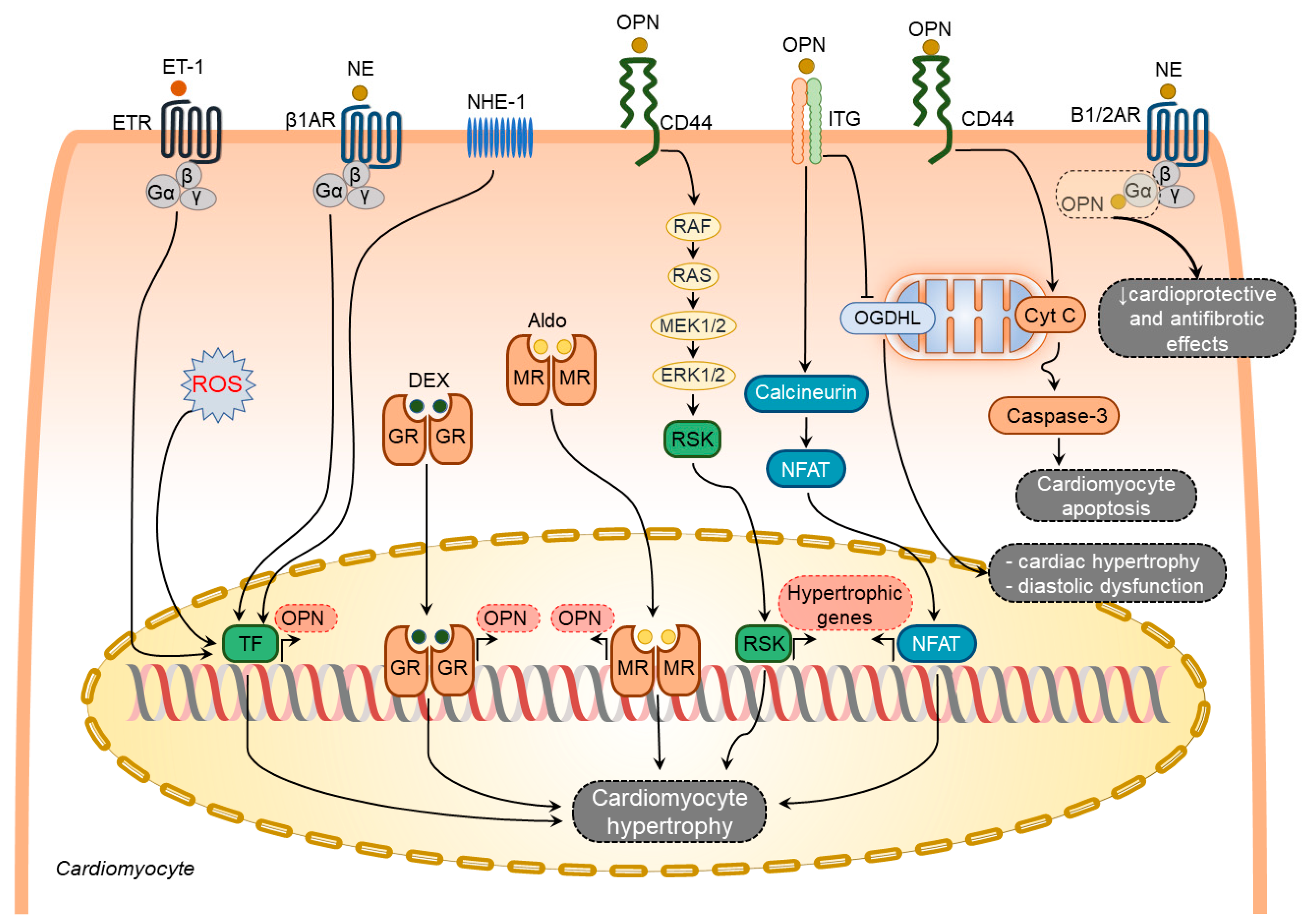
2.1.1. Osteopontin in Cardiomyocytes

Several factors have been shown to induce osteopontin expression in cardiomyocytes including aldosterone [49], dexamethasone [40], endothelin-1 [34], norepinephrine (NE) [34], and ROS [50] (Figure 1). In contrast, other known stimulators of osteopontin expression such as Ang-II [41][51], interleukin-1β (IL-1β), and interferon-γ (IFN-γ) [40] failed to induce osteopontin expression in cardiomyocytes.
Figure 1. Osteopontin signaling in cardiomyocytes. Various factors such as endothelin-1 (ET-1), norepinephrine (NE), sodium-hydrogen exchanger isoform-1 (NHE-1), reactive oxygen species (ROS), dexamethasone (DEX), and aldosterone (Aldo) upregulate osteopontin (OPN) in cardiomyocytes. In turn, osteopontin regulates several processes in cardiomyocytes via modulating certain signaling pathways. These pathways include induction of hypertrophic genes expression via integrin- and CD44-mediated signaling pathways, cardiomyocyte hypertrophy and diastolic dysfunction via blocking mitochondrial protein oxoglutarate dehydrogenase-like (OGDHL), and cardiomyocyte apoptosis via mitochondrial apoptosis signaling pathway, attenuation of the beneficial effects of adrenergic signaling by directly binding and blocking G-α subunit of the adrenergic receptors. The overall effect of osteopontin signaling in cardiomyocytes results in cardiomyocyte hypertrophy, cardiomyocyte apoptosis, and cardiomyocyte mitochondrial dysfunction.
Endothelin-1 and NE-mediated osteopontin upregulation in rat cardiomyocytes coincide with the induction of atrial natriuretic peptide expression [34], suggesting an active involvement of osteopontin in hypertrophic responses of the cardiomyocytes. Cardiomyocyte hypertrophy, LV dilatation, and dysfunction caused by Na+/H+ exchanger isoform 1 (NHE1) overexpression in mice were associated with osteopontin upregulation and osteopontin-CD44 mediated activation of p90-ribosomal S6 kinase signaling pathways [52]. All these pathological changes were reversed in double transgenic mice expressing active NHE1 and osteopontin knockout [52]. Hypertrophic effects of osteopontin on cardiomyocytes can also be mediated through the calcineurin/NFAT pathway [53][54]. In addition, osteopontin can diminish the cardioprotective and antifibrotic effects of β1- and β2-adrenoceptors signaling pathways by direct binding to their Gαs subunits [49].
Several studies have demonstrated an association of increased osteopontin expression with enhanced cardiomyocyte apoptosis in different HF models [29][43][46]. Cardiomyocyte-specific osteopontin overexpression in mice was associated with decreased LV function and increased cardiomyocyte apoptosis [55]. In support of these findings, treatment of adult rat cardiomyocytes with purified osteopontin or adenoviral-mediated expression of osteopontin induced cell apoptosis via CD44-mediated activation of the mitochondrial death pathway and endoplasmic reticulum stress [55].
Col4a3-deficiency in mice recapitulates multiple features of HF with preserved ejection fraction and is associated with cardiac hypertrophy, diastolic dysfunction, myocardial fibrosis, and mitochondrial dysfunction [56]. Targeting osteopontin significantly ameliorated the HF phenotype in these mice [56]. This entry revealed that osteopontin induced mitochondrial dysfunction in cardiomyocytes by downregulating myocardial 2-oxoglutarate dehydrogenase-like, a vital protein in normal mitochondrial function [56].
When taken together, osteopontin serves as an upstream and downstream of diverse signaling pathways involved in cardiomyocyte hypertrophy, apoptosis, and mitochondrial dysfunction (Figure 1).

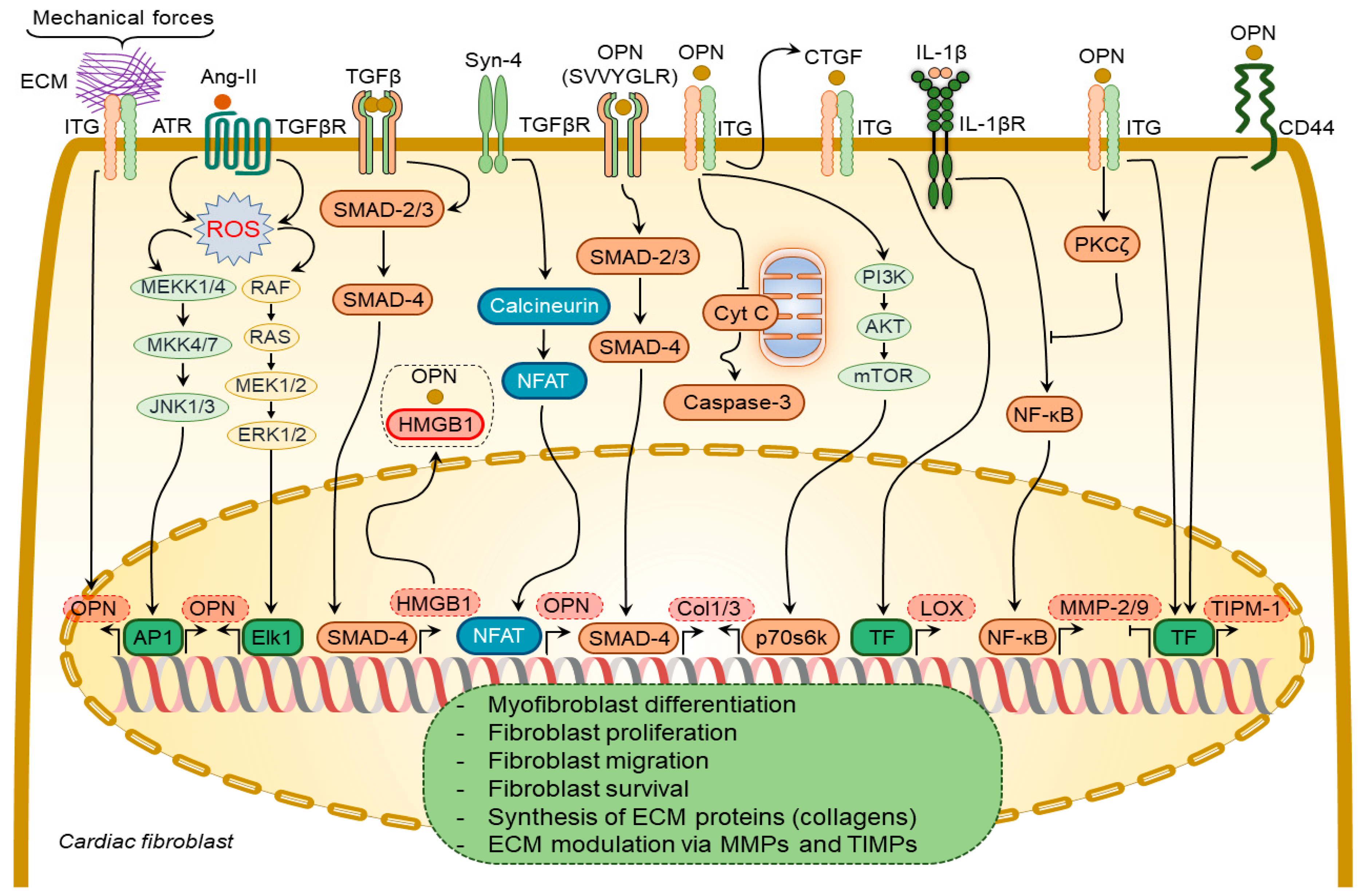
2.1.2. Osteopontin in Cardiac Fibroblasts

In DCM patients, increased levels of myocardial osteopontin strongly correlated with collagen expression [57], suggesting osteopontin involvement in cardiac fibrogenesis. Plasma from aged osteopontin-null mice failed to cause age-specific activation of cardiac fibroblasts [58]. Similarly, cardiac fibroblasts isolated from osteopontin-null mice displayed decreased proliferation and adhesion to ECM [35][59], less spreading, less resistance to detachment by shear stress, and a reduction in collagen gel contraction, which could be partially restored by ectopic osteopontin expression [59].
Several growth factors and cytokines have been shown to regulate osteopontin expression in cardiac fibroblasts, including Ang-II [41][51] and syndecan-4 [60] (Figure 2). Ang-II enhances osteopontin expression in cardiac fibroblasts via NADPH-ROS-mediated activation of ERK1/2 and JNKs pathways [41]. Syndecan-4 increases osteopontin expression via calcineurin/NFAT signaling pathways [60]. Mechanical stretch can also induce osteopontin expression in cardiac fibroblasts [61].
Figure 2. Osteopontin signaling in cardiac fibroblasts. A number of factors induce osteopontin (OPN) expression in cardiac fibroblasts including angiotensin-II (Ang-II), syndecan-4 (Syn-4), and mechanical forces. In turn, osteopontin regulates several processes in cardiac fibroblasts via modulating certain signaling pathways. These processes include binding of osteopontin to high mobility group box 1 (HMGB1) to form focal adhesions in cardiac fibroblasts; TGFβR activation via its SVVYGLR motif and subsequent induction of collagen-1 and -3 (Col1/3) expression; enhancement of Col1/3 expression via integrin receptor-mediated signaling pathways; inhibition of cardiac fibroblast apoptosis via blocking the release of cytochrome C (Cyt C) from mitochondria; inhibition of the activity and expression of MMP-2/9 induced by interleukin-1β (IL-1β) signaling pathway; induction of tissue inhibitor of metalloproteinases 1 (TIMP-1) expression via integrin- and CD44-mediated signaling pathways. The overall effect of osteopontin signaling in cardiac fibroblasts results in myofibroblast differentiation, proliferation, migration, and survival, synthesis of extracellular matrix (ECM) proteins, and modulation of ECM via regulating expression of MMPs and TIMPs.
Transforming growth factor-β (TGF-β) is a multifunctional cytokine mediating myofibroblast transformation and collagen synthesis. TGF-β1 failed to induce myofibroblast differentiation of osteopontin-depleted fibroblasts [59], suggesting a pivotal role of osteopontin in myofibroblast differentiation. Osteopontin operates along with high mobility group box 1 intracellularly to form focal adhesions in the myofibroblasts in response to TGF-β1 stimulation [59].
Osteopontin causes collagen-I synthesis and secretion in fibroblasts through a focal adhesion kinase (FAK)/protein kinase B (Akt)-dependent pathway [62]. The thrombin-cleaved N-terminal osteopontin fragment can induce collagen-I and -III expression in cardiac fibroblasts via activation of the TGFβR signaling pathway [61]. Ang-II-induced cardiac fibroblast proliferation and contraction are mediated by osteopontin RGD domain-β3-integrin receptor signaling pathway [63]. A combination of osteopontin and Ang-II promoted the contraction of three-dimensional collagen gels and cardiac fibroblast growth via β3 integrin-mediated signaling pathways [63].
Osteopontin inhibited the expression and activity of MMP-2 and MMP-9 in cardiac fibroblasts induced by IL-1β stimulation [64]. These effects were mediated by β3 integrins-induced activation of the PKC-ζ signaling pathway [64]. Osteopontin upregulated tissue inhibitor matrix metalloproteinase 1 (TIMP-1) and downregulated MMP-1 expression in fibroblasts via αvβ3 and CD44-receptor signaling [65]. Remarkably, MMP-9-cleaved osteopontin fragments containing the RGD motif induced more pronounced cardiac fibroblast migration than the full-length osteopontin [66].
Osteopontin is a strong regulator of lysyl oxidase expression and activity in cardiac fibroblasts, which is responsible for the formation of cross-linked, insoluble collagen and increased ECM accumulation and myocardial stiffness [60][67]. It induces lysyl oxidase upregulation in cardiac fibroblasts [67] via increasing connective tissue growth factor expression [59].

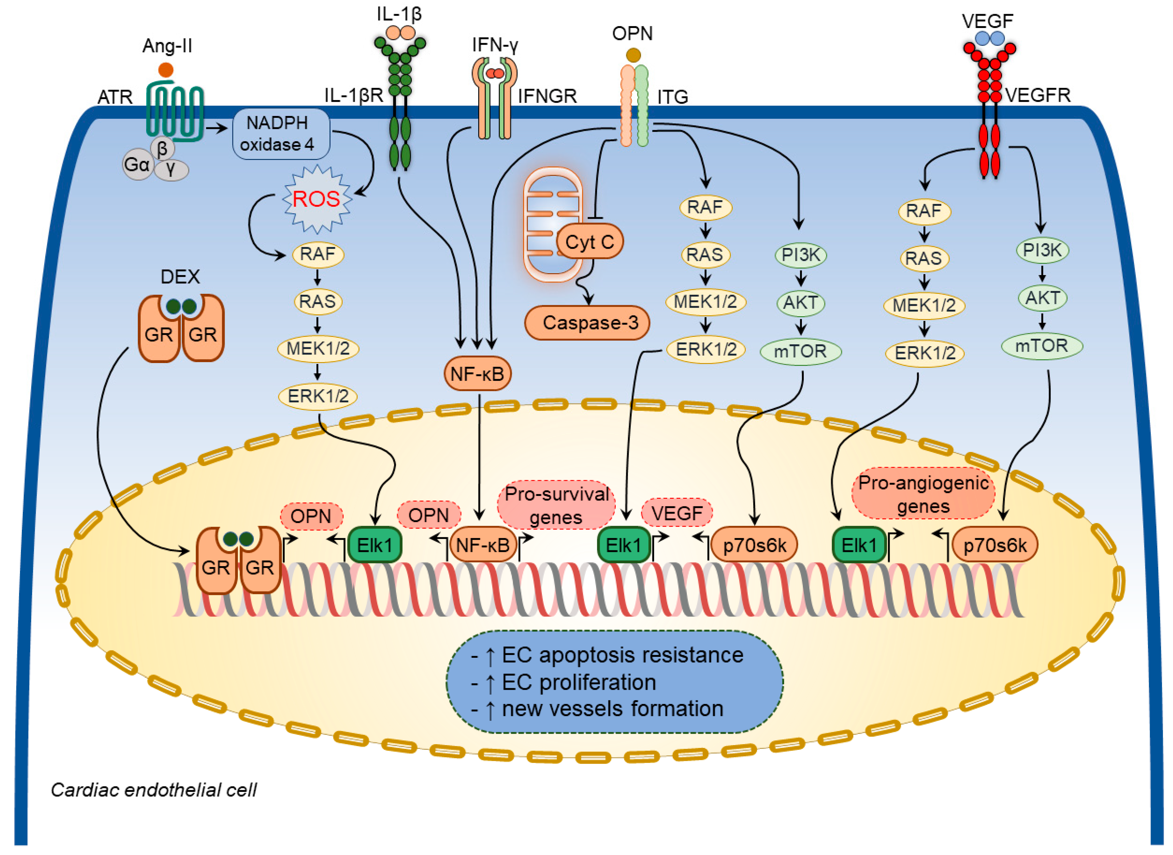
2.1.3. Osteopontin in Cardiac Endothelial Cells

Various growth factors and inflammatory mediators have been shown to regulate osteopontin expression in cardiac endothelial cells [40][41][51] (Figure 3). Ang-II increased osteopontin expression in cardiac endothelial cells [41][51] through NADPH-ROS-mediated activation of the Erk1/2 signaling pathway [51]. A combination of IL-1β and IFN-γ also augmented osteopontin expression in cardiac endothelial cells [40]. Moreover, dexamethasone significantly increased osteopontin expression in in vitro experiments [68].
Figure 3. Osteopontin signaling in cardiac endothelial cells. A number of factors induce osteopontin (OPN) expression in cardiac endothelial cells including angiotensin-II (Ang-II), interleukin-1β (IL-1β), interferon-γ (IFN-γ), and dexamethasone (DEX). In turn, osteopontin regulates several cellular processes in cardiac endothelial cells via modulating certain signaling pathways. These pathways include osteopontin-induced vascular endothelial growth factor (VEGF) expression and subsequent VEGF-mediated expression of pro-angiogenic factors, inhibition of cardiac endothelial cell apoptosis via blocking mitochondria-mediated apoptosis pathway. Osteopontin signaling in cardiac endothelial cells results in increased apoptosis resistance, increased cell proliferation, and new vessels formation.
Osteopontin exerted cytoprotective effects on endothelial cells and promoted angiogenesis by enhancing vascular endothelial growth factor (VEGF) expression through the phosphatidylinositol 3-kinase (PI3K)/protein kinase B (Akt)- and ERK-mediated pathways [69]. In addition, osteopontin facilitated the survival of stressed endothelial cells [70]. Endothelial cells plated on osteopontin-coated surfaces become apoptosis-resistant to serum deprivation via integrin-mediated activation of the nuclear factor kappa B (NF-κB) pathway [71]. Similarly, soluble osteopontin inhibited apoptosis of endothelial cells deprived of essential growth factors [70]. Osteopontin also promoted endothelial regeneration of the injured vessels through activation of RGD-αvβ3 signaling pathways [72]. In line with these findings, osteopontin-null mice showed impaired myocardial angiogenic response post-MI, which resulted in adverse LV remodeling, suggesting that osteopontin plays a crucial role in maintaining and restoring myocardial capillarization following MI [73]. Thus, osteopontin regulates coronary microvascular homeostasis by exerting cytoprotective and pro-survival effects on endothelial cells (Figure 3).

2.2. Implications of Osteopontin in Specific Pathological Processes

2.2.1. Osteopontin in Cardiac Hypertrophy

Cardiac hypertrophy is an adaptive response to hemodynamic load characterized by the thickening of the heart walls and enlargement of the individual cardiomyocytes, which has a compensatory role in maintaining cardiac performance and attenuating ventricular wall stress and chamber dilatation. At the cellular level, cardiomyocyte hypertrophy is characterized by an increase in cell dimensions mainly due to augmented contractile protein synthesis [74]. However, sustained hemodynamic stress in pathological conditions such as hypertension and valvular heart disease drives adverse alterations in the myocardium characterized by excessive cardiomyocyte apoptosis, myocardial inflammation, and fibrosis, eventually culminating in ventricular dilation and dysfunction.
Robust upregulation of myocardial osteopontin expression upon mechanical stress was clearly shown in several rodent models [34][44][61]. Pressure overload induced an acute and strong elevation of osteopontin expression in the myocardium in rodents following TAC [34][44][61] and in rats with renovascular hypertension [34]. The increase in myocardial osteopontin expression was reported to be proportional to the degree of the afterload [75]. In line with these findings, there was a strong correlation between expression levels of osteopontin and atrial natriuretic peptide in the LV myocardium in renovascular and TAC-induced LV remodeling models [34]. In the latter models, cardiomyocytes were the main cell type in the heart producing osteopontin [34]. In contrast, in spontaneously hypertensive rats with cardiac hypertrophy, osteopontin expression was identified primarily in non-myocytes in the interstitial and perivascular space (possibly infiltrating macrophages and fibroblasts) [76].
The exploitation of osteopontin-null mice in several animal models of cardiac hypertrophy revealed that osteopontin is a key mediator in the mechanical stress-induced myocardial hypertrophic response [35][44][52][56]. In mice subjected to TAC, a lack of osteopontin was associated with attenuated cardiac hypertrophic response [44]. Knocking out osteopontin in transgenic mice with NHE1 overexpression significantly reduced cardiac hypertrophy, attenuated collagen deposition, and improved cardiac function [52]. Similarly, in another genetic mouse model of HF with preserved ejection fraction induced by Col4a3 deficiency, osteopontin deletion was associated with improved parameters of LV diastolic function and cardiac hypertrophy reduction [56]. Controversial data were obtained in the model of Ang-II infusion [29][35]. Thus, in one study, osteopontin deficiency attenuated cardiac hypertrophy induced by Ang-II infusion [35], whereas, in another study, osteopontin deletion did not prevent cardiac hypertrophy, despite attenuated myocardial fibrosis [29].

2.2.2. Osteopontin in Cardiac Inflammation

Heart diseases are associated with inflammation, which contributes to cardiac dysfunction and myocardial fibrosis [10][77][78]. Numerous cytokines and chemokines are actively involved in the onset and progression of myocardial fibrosis [10][78]. Osteopontin has been shown to exert cytokine- and chemokine-like functions [79].
Among various inflammatory cells, macrophages represent the dominant inflammatory cell population in the remodeled myocardium [80]. Macrophage infiltration of the myocardium has been shown in various models, including spontaneously hypertensive rats and murine TAC [80][81]. Complete abolition of CD45+ monocytes recruitment to the myocardium in response to Ang-II infusion in osteopontin-null mice [82] suggested the involvement of osteopontin in this process.
Osteopontin expressed in circulating leucocytes may also be actively involved in inflammatory cell recruitment to the myocardium and systemic inflammation in patients with various cardiac diseases [83]. Circulating CD4+ T lymphocytes expressing osteopontin and circulating osteopontin levels both correlate with the New York Heart Association Functional Class (NYHA FC) in HF patients and are associated with plaque instability in coronary artery disease patients [83][84].
Inflammatory cells have been identified as one of the major contributors to fibrogenesis in different tissues and organs under various disease conditions [85]. During the acute inflammatory phase of wound healing processes upon tissue injury in different tissues and organs, osteopontin expression is enhanced in infiltrating leukocytes, while during the chronic inflammatory phase, its upregulation is observed in resident macrophages [86]. Increased osteopontin expression by resident and recruited inflammatory cells increases collagen deposition and accumulation in the tissues [83][84][87].
Osteopontin expression by immune/inflammatory cells is associated with cardiac hypertrophic and fibrotic responses in the settings of a number of heart diseases [38][39][45][82][88]. Studies exploiting mice with a genetically modified osteopontin expression provided important insights into its role in myocardial inflammation in diverse cardiac conditions [45][55][87][89][90][91][92]. Mice overexpressing osteopontin in cardiomyocytes spontaneously develop severe cardiomyopathy, which is characterized by enhanced recruitment of inflammatory cells to the myocardium and excessive collagen accumulation, subsequently leading to chronic myocarditis and eventually premature death [55][87]. In line with these reports, deletion of osteopontin in desmin deficient mice, which spontaneously develop DCM with increased myocardial inflammation and fibrosis, was associated with an attenuation of myocardial inflammation and improvement in LV systolic function [45].
Cardiomyocyte-specific integrin-linked kinase (ILK) deficient mice spontaneously develop lethal cardiomyopathy characterized by excessive inflammatory cell accumulation, myocardial fibrosis, and cardiomyocyte apoptosis [90]. Augmented osteopontin expression in cardiomyocytes revealed by comprehensive profiling and mitigation of HF severity in these mice by application of anti-osteopontin antibodies implicated osteopontin as a major contributor to this phenotype [90]. Likewise, in other murine models, including those of Chagas heart disease and viral myocarditis, osteopontin-null mice displayed improved cardiac remodeling and attenuated myocardial inflammation [91] [92].
When taken together, osteopontin is highly upregulated in acute and chronic inflammatory phases of the myocardial injury and can contribute to adverse myocardial remodeling and cardiac dysfunction by promoting the recruitment of inflammatory cells to the myocardium.

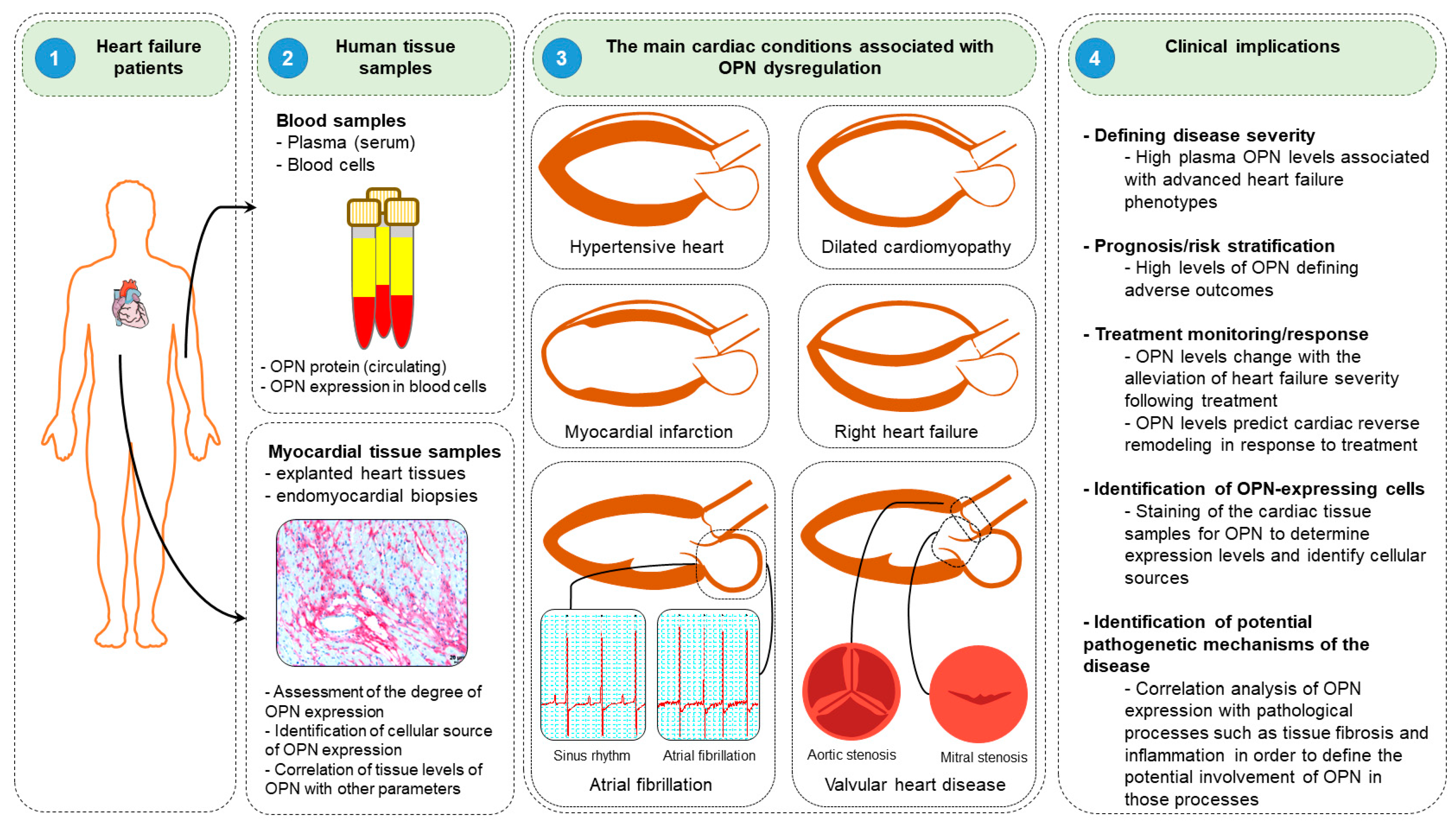
2.3. Clinical Implication of Osteopontin

Clinical studies have suggested that osteopontin might serve as a potent diagnostic and prognostic biomarker in diverse HF conditions. In this section, researchers discuss the roles and clinical implications of osteopontin in various HF diseases, including DCM, hypertensive HF, MI, and right HF (Figure 4).
Figure 4. Clinical implications of osteopontin. (1) Blood samples are collected from the peripheral blood, while myocardial tissues are obtained from explanted hearts or endomyocardial biopsies during cardiac catheterization or cardiac surgery. (2) Plasma/serum and blood cells are isolated from blood samples to measure osteopontin (OPN) levels. Similarly, osteopontin expression levels are evaluated and cellular sources of osteopontin are identified in myocardial tissues. (3) Assessment of osteopontin levels might be useful in several cardiac conditions, including hypertensive heart, dilated cardiomyopathy, myocardial infarction, and right heart failure, atrial fibrillation and valvular heart disease. (4) Assessment of osteopontin levels might be useful in determining diseases severity, prognostic/risk stratification, monitoring of treatment response, identification of cellular sources of osteopontin in various cardiac conditions and help to understand the mechanisms of the disease pathogenesis.

This entry is adapted from the peer-reviewed paper 10.3390/cimb44080245

References

  1. Lim, S.S.; Vos, T.; Flaxman, A.D.; Danaei, G.; Shibuya, K.; Adair-Rohani, H.; Amann, M.; Anderson, H.R.; Andrews, K.G.; Aryee, M.; et al. A comparative risk assessment of burden of disease and injury attributable to 67 risk factors and risk factor clusters in 21 regions, 1990–2010: A systematic analysis for the Global Burden of Disease Study 2010. Lancet 2012, 380, 2224–2260.
  2. Vos, T.; Abajobir, A.A.; Abate, K.H.; Abbafati, C.; Abbas, K.M.; Abd-Allah, F.; Abdulkader, R.S.; Abdulle, A.M.; Abebo, T.A.; Abera, S.F. Global, regional, and national incidence, prevalence, and years lived with disability for 328 diseases and injuries for 195 countries, 1990–2016: A systematic analysis for the Global Burden of Disease Study 2016. Lancet 2017, 390, 1211–1259.
  3. Yancy, C.W.; Jessup, M.; Bozkurt, B.; Butler, J.; Casey, D.E.; Drazner, M.H.; Fonarow, G.C.; Geraci, S.A.; Horwich, T.; Januzzi, J.L. 2013 ACCF/AHA guideline for the management of heart failure: A report of the American College of Cardiology Foundation/American Heart Association Task Force on Practice Guidelines. J. Am. Coll. Cardiol. 2013, 62, e147–e239.
  4. Corris, P.A.; Seeger, W. Call It by the Correct Name—Pulmonary Hypertension Not Pulmonary Arterial Hypertension: Growing Recognition of the Global Health Impact for a Well-Recognized Condition and the Role of the Pulmonary Vascular Research Institute; American Physiological Society Bethesda: Rockville, MD, USA, 2020.
  5. Friedberg, M.K.; Redington, A.N. Right versus left ventricular failure: Differences, similarities, and interactions. Circulation 2014, 129, 1033–1044.
  6. Nakamura, M.; Sadoshima, J. Mechanisms of physiological and pathological cardiac hypertrophy. Nat. Rev. Cardiol. 2018, 15, 387–407.
  7. Vonk-Noordegraaf, A.; Haddad, F.; Chin, K.M.; Forfia, P.R.; Kawut, S.M.; Lumens, J.; Naeije, R.; Newman, J.; Oudiz, R.J.; Provencher, S.; et al. Right heart adaptation to pulmonary arterial hypertension: Physiology and pathobiology. J. Am. Coll. Cardiol. 2013, 62 (Suppl. S25), D22–D33.
  8. Egemnazarov, B.; Crnkovic, S.; Nagy, B.M.; Olschewski, H.; Kwapiszewska, G. Right ventricular fibrosis and dysfunction: Actual concepts and common misconceptions. Matrix Biol. J. Int. Soc. Matrix Biol. 2018, 68–69, 507–521.
  9. Travers, J.G.; Kamal, F.A.; Robbins, J.; Yutzey, K.E.; Blaxall, B.C. Cardiac Fibrosis: The Fibroblast Awakens. Circ. Res. 2016, 118, 1021–1040.
  10. Sydykov, A.; Mamazhakypov, A.; Petrovic, A.; Kosanovic, D.; Sarybaev, A.S.; Weissmann, N.; Ghofrani, H.A.; Schermuly, R.T. Inflammatory Mediators Drive Adverse Right Ventricular Remodeling and Dysfunction and Serve as Potential Biomarkers. Front. Physiol. 2018, 9, 609.
  11. Carrillo-Salinas, F.J.; Ngwenyama, N.; Anastasiou, M.; Kaur, K.; Alcaide, P. Heart Inflammation: Immune Cell Roles and Roads to the Heart. Am. J. Pathol. 2019, 189, 1482–1494.
  12. Frump, A.L.; Bonnet, S.; de Jesus Perez, V.A.; Lahm, T. Emerging role of angiogenesis in adaptive and maladaptive right ventricular remodeling in pulmonary hypertension. Am. J. Physiol. Lung Cell. Mol. Physiol. 2018, 314, L443–L460.
  13. Oka, T.; Akazawa, H.; Naito, A.T.; Komuro, I. Angiogenesis and cardiac hypertrophy: Maintenance of cardiac function and causative roles in heart failure. Circ. Res. 2014, 114, 565–571.
  14. Viswanathan, G.; Mamazhakypov, A.; Schermuly, R.T.; Rajagopal, S. The Role of G Protein-Coupled Receptors in the Right Ventricle in Pulmonary Hypertension. Front. Cardiovasc. Med. 2018, 5, 179.
  15. Hartupee, J.; Mann, D.L. Neurohormonal activation in heart failure with reduced ejection fraction. Nat. Rev. Cardiol. 2017, 14, 30–38.
  16. Ryan, J.J.; Archer, S.L. Emerging concepts in the molecular basis of pulmonary arterial hypertension: Part I: Metabolic plasticity and mitochondrial dynamics in the pulmonary circulation and right ventricle in pulmonary arterial hypertension. Circulation 2015, 131, 1691–1702.
  17. Lopaschuk, G.D.; Karwi, Q.G.; Tian, R.; Wende, A.R.; Abel, E.D. Cardiac Energy Metabolism in Heart Failure. Circ. Res. 2021, 128, 1487–1513.
  18. Piao, L.; Marsboom, G.; Archer, S.L. Mitochondrial metabolic adaptation in right ventricular hypertrophy and failure. J. Mol. Med. 2010, 88, 1011–1020.
  19. Sabbah, H.N. Targeting the Mitochondria in Heart Failure: A Translational Perspective. JACC Basic Transl. Sci. 2020, 5, 88–106.
  20. Shults, N.V.; Melnyk, O.; Suzuki, D.I.; Suzuki, Y.J. Redox Biology of Right-Sided Heart Failure. Antioxidants 2018, 7, 106.
  21. van der Pol, A.; van Gilst, W.H.; Voors, A.A.; van der Meer, P. Treating oxidative stress in heart failure: Past, present and future. Eur. J. Heart Fail. 2019, 21, 425–435.
  22. Esfandiary, A.; Kutsche, H.S.; Schreckenberg, R.; Weber, M.; Pak, O.; Kojonazarov, B.; Sydykov, A.; Hirschhauser, C.; Wolf, A.; Haag, D.; et al. Protection against pressure overload-induced right heart failure by uncoupling protein 2 silencing. Cardiovasc. Res. 2019, 115, 1217–1227.
  23. López, B.; Ravassa, S.; Moreno, M.U.; José, G.S.; Beaumont, J.; González, A.; Díez, J. Diffuse myocardial fibrosis: Mechanisms, diagnosis and therapeutic approaches. Nat. Rev. Cardiol. 2021, 18, 479–498.
  24. Frangogiannis, N.G. The extracellular matrix in myocardial injury, repair, and remodeling. J. Clin. Investig. 2017, 127, 1600–1612.
  25. Frangogiannis, N.G. The Extracellular Matrix in Ischemic and Nonischemic Heart Failure. Circ. Res. 2019, 125, 117–146.
  26. Frangogiannis, N.G. Matricellular proteins in cardiac adaptation and disease. Physiol. Rev. 2012, 92, 635–688.
  27. Singh, M.; Dalal, S.; Singh, K. Osteopontin: At the cross-roads of myocyte survival and myocardial function. Life Sci. 2014, 118, 1–6.
  28. Zahradka, P. Novel role for osteopontin in cardiac fibrosis. Circ. Res. 2008, 102, 270–272.
  29. Matsui, Y.; Jia, N.; Okamoto, H.; Kon, S.; Onozuka, H.; Akino, M.; Liu, L.; Morimoto, J.; Rittling, S.R.; Denhardt, D.; et al. Role of osteopontin in cardiac fibrosis and remodeling in angiotensin II-induced cardiac hypertrophy. Hypertension 2004, 43, 1195–1201.
  30. Anwar, A.; Li, M.; Frid, M.G.; Kumar, B.; Gerasimovskaya, E.V.; Riddle, S.R.; McKeon, B.A.; Thukaram, R.; Meyrick, B.O.; Fini, M.A.; et al. Osteopontin is an endogenous modulator of the constitutively activated phenotype of pulmonary adventitial fibroblasts in hypoxic pulmonary hypertension. Am. J. Physiol. Lung Cell. Mol. Physiol. 2012, 303, L1–L11.
  31. Singh, M.; Foster, C.R.; Dalal, S.; Singh, K. Role of osteopontin in heart failure associated with aging. Heart Fail. Rev. 2010, 15, 487–494.
  32. Giachelli, C.M.; Schwartz, S.M.; Liaw, L. Molecular and cellular biology of osteopontin Potential role in cardiovascular disease. Trends Cardiovasc. Med. 1995, 5, 88–95.
  33. Thayer, J.M.; Giachelli, C.M.; Mirkes, P.E.; Schwartz, S.M. Expression of osteopontin in the head process late in gastrulation in the rat. J. Exp. Zool. 1995, 272, 240–244.
  34. Graf, K.; Do, Y.S.; Ashizawa, N.; Meehan, W.P.; Giachelli, C.M.; Marboe, C.C.; Fleck, E.; Hsueh, W.A. Myocardial osteopontin expression is associated with left ventricular hypertrophy. Circulation 1997, 96, 3063–3071.
  35. Collins, A.R.; Schnee, J.; Wang, W.; Kim, S.; Fishbein, M.C.; Bruemmer, D.; Law, R.E.; Nicholas, S.; Ross, R.S.; Hsueh, W.A. Osteopontin modulates angiotensin II-induced fibrosis in the intact murine heart. J. Am. Coll. Cardiol. 2004, 43, 1698–1705.
  36. Singh, M.; Foster, C.R.; Dalal, S.; Singh, K. Osteopontin: Role in extracellular matrix deposition and myocardial remodeling post-MI. J. Mol. Cell. Cardiol. 2010, 48, 538–543.
  37. Denhardt, D.T.; Guo, X. Osteopontin: A protein with diverse functions. FASEB J. 1993, 7, 1475–1482.
  38. Williams, E.B.; Halpert, I.; Wickline, S.; Davison, G.; Parks, W.C.; Rottman, J.N. Osteopontin expression is increased in the heritable cardiomyopathy of Syrian hamsters. Circulation 1995, 92, 705–709.
  39. Murry, C.E.; Giachelli, C.M.; Schwartz, S.M.; Vracko, R. Macrophages express osteopontin during repair of myocardial necrosis. Am. J. Pathol. 1994, 145, 1450–1462.
  40. Singh, K.; Balligand, J.L.; Fischer, T.A.; Smith, T.W.; Kelly, R.A. Glucocorticoids increase osteopontin expression in cardiac myocytes and microvascular endothelial cells. Role in regulation of inducible nitric oxide synthase. J. Biol. Chem. 1995, 270, 28471–28478.
  41. Xie, Z.; Singh, M.; Singh, K. ERK1/2 and JNKs, but not p38 kinase, are involved in reactive oxygen species-mediated induction of osteopontin gene expression by angiotensin II and interleukin-1beta in adult rat cardiac fibroblasts. J. Cell Physiol. 2004, 198, 399–407.
  42. Lund, S.A.; Giachelli, C.M.; Scatena, M. The role of osteopontin in inflammatory processes. J. Cell Commun. Signal. 2009, 3, 311–322.
  43. Sam, F.; Xie, Z.; Ooi, H.; Kerstetter, D.L.; Colucci, W.S.; Singh, M.; Singh, K. Mice lacking osteopontin exhibit increased left ventricular dilation and reduced fibrosis after aldosterone infusion. Am. J. Hypertens. 2004, 17, 188–193.
  44. Xie, Z.; Singh, M.; Singh, K. Osteopontin modulates myocardial hypertrophy in response to chronic pressure overload in mice. Hypertension 2004, 44, 826–831.
  45. Psarras, S.; Mavroidis, M.; Sanoudou, D.; Davos, C.H.; Xanthou, G.; Varela, A.E.; Panoutsakopoulou, V.; Capetanaki, Y. Regulation of adverse remodelling by osteopontin in a genetic heart failure model. Eur. Heart J. 2012, 33, 1954–1963.
  46. Subramanian, V.; Krishnamurthy, P.; Singh, K.; Singh, M. Lack of osteopontin improves cardiac function in streptozotocin-induced diabetic mice. Am. J. Physiol. Heart Circ. Physiol. 2007, 292, H673–H683.
  47. Trueblood, N.A.; Xie, Z.; Communal, C.; Sam, F.; Ngoy, S.; Liaw, L.; Jenkins, A.W.; Wang, J.; Sawyer, D.B.; Bing, O.H.; et al. Exaggerated left ventricular dilation and reduced collagen deposition after myocardial infarction in mice lacking osteopontin. Circ. Res. 2001, 88, 1080–1087.
  48. Duerr, G.D.; Mesenholl, B.; Heinemann, J.C.; Zoerlein, M.; Huebener, P.; Schneider, P.; Feisst, A.; Ghanem, A.; Tiemann, K.; Dewald, D.; et al. Cardioprotective effects of osteopontin-1 during development of murine ischemic cardiomyopathy. BioMed Res. Int. 2014, 2014, 124063.
  49. Pollard, C.M.; Desimine, V.L.; Wertz, S.L.; Perez, A.; Parker, B.M.; Maning, J.; McCrink, K.A.; Shehadeh, L.A.; Lymperopoulos, A. Deletion of Osteopontin Enhances beta(2)-Adrenergic Receptor-Dependent Anti-Fibrotic Signaling in Cardiomyocytes. Int. J. Mol. Sci. 2019, 20, 1396.
  50. Jiang, P.; Zhang, D.; Qiu, H.; Yi, X.; Zhang, Y.; Cao, Y.; Zhao, B.; Xia, Z.; Wang, C. Tiron ameliorates high glucose-induced cardiac myocyte apoptosis by PKCdelta-dependent inhibition of osteopontin. Clin. Exp. Pharmacol. Physiol. 2017, 44, 760–770.
  51. Xie, Z.; Pimental, D.R.; Lohan, S.; Vasertriger, A.; Pligavko, C.; Colucci, W.S.; Singh, K. Regulation of angiotensin II-stimulated osteopontin expression in cardiac microvascular endothelial cells: Role of p42/44 mitogen-activated protein kinase and reactive oxygen species. J. Cell Physiol. 2001, 188, 132–138.
  52. Abdulrahman, N.; Jaspard-Vinassa, B.; Fliegel, L.; Jabeen, A.; Riaz, S.; Gadeau, A.P.; Mraiche, F. Na(+)/H(+) exchanger isoform 1-induced osteopontin expression facilitates cardiac hypertrophy through p90 ribosomal S6 kinase. Physiol. Genom. 2018, 50, 332–342.
  53. Mohamed, I.A.; Gadeau, A.P.; Fliegel, L.; Lopaschuk, G.; Mlih, M.; Abdulrahman, N.; Fillmore, N.; Mraiche, F. Na+/H+ exchanger isoform 1-induced osteopontin expression facilitates cardiomyocyte hypertrophy. PLoS ONE 2015, 10, e0123318.
  54. Mlih, M.; Abdulrahman, N.; Gadeau, A.P.; Mohamed, I.A.; Jaballah, M.; Mraiche, F. Na(+)/H (+) exchanger isoform 1 induced osteopontin expression in cardiomyocytes involves NFAT3/Gata4. Mol. Cell. Biochem. 2015, 404, 211–220.
  55. Dalal, S.; Zha, Q.; Daniels, C.R.; Steagall, R.J.; Joyner, W.L.; Gadeau, A.P.; Singh, M.; Singh, K. Osteopontin stimulates apoptosis in adult cardiac myocytes via the involvement of CD44 receptors, mitochondrial death pathway, and endoplasmic reticulum stress. Am. J. Physiol. Heart Circ. Physiol. 2014, 306, H1182–H1191.
  56. Yousefi, K.; Irion, C.I.; Takeuchi, L.M.; Ding, W.; Lambert, G.; Eisenberg, T.; Sukkar, S.; Granzier, H.L.; Methawasin, M.; Lee, D.I.; et al. Osteopontin Promotes Left Ventricular Diastolic Dysfunction Through a Mitochondrial Pathway. J. Am. Coll. Cardiol. 2019, 73, 2705–2718.
  57. Cabiati, M.; Svezia, B.; Matteucci, M.; Botta, L.; Pucci, A.; Rinaldi, M.; Caselli, C.; Lionetti, V.; Del Ry, S. Myocardial Expression Analysis of Osteopontin and Its Splice Variants in Patients Affected by End-Stage Idiopathic or Ischemic Dilated Cardiomyopathy. PLoS ONE 2016, 11, e0160110.
  58. Sawaki, D.; Czibik, G.; Pini, M.; Ternacle, J.; Suffee, N.; Mercedes, R.; Marcelin, G.; Surenaud, M.; Marcos, E.; Gual, P.; et al. Visceral Adipose Tissue Drives Cardiac Aging Through Modulation of Fibroblast Senescence by Osteopontin Production. Circulation 2018, 138, 809–822.
  59. Lenga, Y.; Koh, A.; Perera, A.S.; McCulloch, C.A.; Sodek, J.; Zohar, R. Osteopontin expression is required for myofibroblast differentiation. Circ. Res. 2008, 102, 319–327.
  60. Herum, K.M.; Lunde, I.G.; Skrbic, B.; Louch, W.E.; Hasic, A.; Boye, S.; Unger, A.; Brorson, S.H.; Sjaastad, I.; Tonnessen, T.; et al. Syndecan-4 is a key determinant of collagen cross-linking and passive myocardial stiffness in the pressure-overloaded heart. Cardiovasc. Res. 2015, 106, 217–226.
  61. Herum, K.M.; Romaine, A.; Wang, A.; Melleby, A.O.; Strand, M.E.; Pacheco, J.; Braathen, B.; Duner, P.; Tonnessen, T.; Lunde, I.G.; et al. Syndecan-4 Protects the Heart From the Profibrotic Effects of Thrombin-Cleaved Osteopontin. J. Am. Heart Assoc. 2020, 9, e013518.
  62. Zhao, H.; Wang, W.; Zhang, J.; Liang, T.; Fan, G.P.; Wang, Z.W.; Zhang, P.D.; Wang, X.; Zhang, J. Inhibition of osteopontin reduce the cardiac myofibrosis in dilated cardiomyopathy via focal adhesion kinase mediated signaling pathway. Am. J. Transl. Res. 2016, 8, 3645–3655.
  63. Ashizawa, N.; Graf, K.; Do, Y.S.; Nunohiro, T.; Giachelli, C.M.; Meehan, W.P.; Tuan, T.L.; Hsueh, W.A. Osteopontin is produced by rat cardiac fibroblasts and mediates A(II)-induced DNA synthesis and collagen gel contraction. J. Clin. Investig. 1996, 98, 2218–2227.
  64. Xie, Z.; Singh, M.; Siwik, D.A.; Joyner, W.L.; Singh, K. Osteopontin inhibits interleukin-1beta-stimulated increases in matrix metalloproteinase activity in adult rat cardiac fibroblasts: Role of protein kinase C-zeta. J. Biol. Chem. 2003, 278, 48546–48552.
  65. Pardo, A.; Gibson, K.; Cisneros, J.; Richards, T.J.; Yang, Y.; Becerril, C.; Yousem, S.; Herrera, I.; Ruiz, V.; Selman, M.; et al. Up-regulation and profibrotic role of osteopontin in human idiopathic pulmonary fibrosis. PLoS Med. 2005, 2, e251.
  66. Lindsey, M.L.; Zouein, F.A.; Tian, Y.; Padmanabhan Iyer, R.; de Castro Bras, L.E. Osteopontin is proteolytically processed by matrix metalloproteinase 9. Can. J. Physiol. Pharmacol. 2015, 93, 879–886.
  67. Lopez, B.; Gonzalez, A.; Lindner, D.; Westermann, D.; Ravassa, S.; Beaumont, J.; Gallego, I.; Zudaire, A.; Brugnolaro, C.; Querejeta, R.; et al. Osteopontin-mediated myocardial fibrosis in heart failure: A role for lysyl oxidase? Cardiovasc. Res. 2013, 99, 111–120.
  68. Kim, J.S.; Bashir, M.M.; Werth, V.P. Gottron’s papules exhibit dermal accumulation of CD44 variant 7 (CD44v7) and its binding partner osteopontin: A unique molecular signature. J. Investig. Dermatol. 2012, 132, 1825–1832.
  69. Dai, J.; Peng, L.; Fan, K.; Wang, H.; Wei, R.; Ji, G.; Cai, J.; Lu, B.; Li, B.; Zhang, D.; et al. Osteopontin induces angiogenesis through activation of PI3K/AKT and ERK1/2 in endothelial cells. Oncogene 2009, 28, 3412–3422.
  70. Khan, S.A.; Lopez-Chua, C.A.; Zhang, J.; Fisher, L.W.; Sorensen, E.S.; Denhardt, D.T. Soluble osteopontin inhibits apoptosis of adherent endothelial cells deprived of growth factors. J. Cell. Biochem. 2002, 85, 728–736.
  71. Scatena, M.; Almeida, M.; Chaisson, M.L.; Fausto, N.; Nicosia, R.F.; Giachelli, C.M. NF-kappaB mediates alphavbeta3 integrin-induced endothelial cell survival. J. Cell Biol. 1998, 141, 1083–1093.
  72. Liaw, L.; Lindner, V.; Schwartz, S.M.; Chambers, A.F.; Giachelli, C.M. Osteopontin and beta 3 integrin are coordinately expressed in regenerating endothelium in vivo and stimulate Arg-Gly-Asp-dependent endothelial migration in vitro. Circ. Res. 1995, 77, 665–672.
  73. Zhao, X.; Johnson, J.N.; Singh, K.; Singh, M. Impairment of myocardial angiogenic response in the absence of osteopontin. Microcirculation 2007, 14, 233–240.
  74. Frey, N.; Katus, H.A.; Olson, E.N.; Hill, J.A. Hypertrophy of the heart: A new therapeutic target? Circulation 2004, 109, 1580–1589.
  75. Ndisang, J.F.; Chibbar, R.; Lane, N. Heme oxygenase suppresses markers of heart failure and ameliorates cardiomyopathy in L-NAME-induced hypertension. Eur. J. Pharmacol. 2014, 734, 23–34.
  76. Singh, K.; Sirokman, G.; Communal, C.; Robinson, K.G.; Conrad, C.H.; Brooks, W.W.; Bing, O.H.; Colucci, W.S. Myocardial osteopontin expression coincides with the development of heart failure. Hypertension 1999, 33, 663–670.
  77. Prabhu, S.D.; Frangogiannis, N.G. The Biological Basis for Cardiac Repair After Myocardial Infarction: From Inflammation to Fibrosis. Circ. Res. 2016, 119, 91–112.
  78. Adamo, L.; Rocha-Resende, C.; Prabhu, S.D.; Mann, D.L. Reappraising the role of inflammation in heart failure. Nat. Rev. Cardiol. 2020, 17, 269–285.
  79. Icer, M.A.; Gezmen-Karadag, M. The multiple functions and mechanisms of osteopontin. Clin. Biochem. 2018, 59, 17–24.
  80. Weisheit, C.; Zhang, Y.; Faron, A.; Köpke, O.; Weisheit, G.; Steinsträsser, A.; Frede, S.; Meyer, R.; Boehm, O.; Hoeft, A.; et al. Ly6C(low) and not Ly6C(high) macrophages accumulate first in the heart in a model of murine pressure-overload. PLoS ONE 2014, 9, e112710.
  81. Yang, G.H.; Zhou, X.; Ji, W.J.; Zeng, S.; Dong, Y.; Tian, L.; Bi, Y.; Guo, Z.Z.; Gao, F.; Chen, H.; et al. Overexpression of VEGF-C attenuates chronic high salt intake-induced left ventricular maladaptive remodeling in spontaneously hypertensive rats. Am. J. Physiol. Heart Circ. Physiol. 2014, 306, H598–H609.
  82. Lorenzen, J.M.; Schauerte, C.; Hubner, A.; Kolling, M.; Martino, F.; Scherf, K.; Batkai, S.; Zimmer, K.; Foinquinos, A.; Kaucsar, T.; et al. Osteopontin is indispensible for AP1-mediated angiotensin II-related miR-21 transcription during cardiac fibrosis. Eur. Heart J. 2015, 36, 2184–2196.
  83. Soejima, H.; Irie, A.; Fukunaga, T.; Oe, Y.; Kojima, S.; Kaikita, K.; Kawano, H.; Sugiyama, S.; Yoshimura, M.; Kishikawa, H.; et al. Osteopontin expression of circulating T cells and plasma osteopontin levels are increased in relation to severity of heart failure. Circ. J. Off. J. Jpn. Circ. Soc. 2007, 71, 1879–1884.
  84. Soejima, H.; Irie, A.; Fukunaga, T.; Sugamura, K.; Kojima, S.; Sakamoto, T.; Yoshimura, M.; Kishikawa, H.; Nishimura, Y.; Ogawa, H. Elevated plasma osteopontin levels were associated with osteopontin expression of CD4+ T cells in patients with unstable angina. Circ. J. Off. J. Jpn. Circ. Soc. 2006, 70, 851–856.
  85. Van Linthout, S.; Miteva, K.; Tschöpe, C. Crosstalk between fibroblasts and inflammatory cells. Cardiovasc. Res. 2014, 102, 258–269.
  86. Mazzali, M.; Kipari, T.; Ophascharoensuk, V.; Wesson, J.; Johnson, R.; Hughes, J. Osteopontin—A molecule for all seasons. Qjm 2002, 95, 3–13.
  87. Renault, M.A.; Robbesyn, F.; Reant, P.; Douin, V.; Daret, D.; Allieres, C.; Belloc, I.; Couffinhal, T.; Arnal, J.F.; Klingel, K.; et al. Osteopontin expression in cardiomyocytes induces dilated cardiomyopathy. Circ. Heart Fail. 2010, 3, 431–439.
  88. Satoh, M.; Nakamura, M.; Akatsu, T.; Shimoda, Y.; Segawa, I.; Hiramori, K. Myocardial osteopontin expression is associated with collagen fibrillogenesis in human dilated cardiomyopathy. Eur. J. Heart Fail. 2005, 7, 755–762.
  89. Papathanasiou, S.; Rickelt, S.; Soriano, M.E.; Schips, T.G.; Maier, H.J.; Davos, C.H.; Varela, A.; Kaklamanis, L.; Mann, D.L.; Capetanaki, Y. Tumor necrosis factor-alpha confers cardioprotection through ectopic expression of keratins K8 and K18. Nat. Med. 2015, 21, 1076–1084.
  90. Dai, J.; Matsui, T.; Abel, E.D.; Dedhar, S.; Gerszten, R.E.; Seidman, C.E.; Seidman, J.G.; Rosenzweig, A. Deep sequence analysis of gene expression identifies osteopontin as a downstream effector of integrin-linked kinase (ILK) in cardiac-specific ILK knockout mice. Circ. Heart Fail. 2014, 7, 184–193.
  91. Caballero, E.P.; Santamaria, M.H.; Corral, R.S. Endogenous osteopontin induces myocardial CCL5 and MMP-2 activation that contributes to inflammation and cardiac remodeling in a mouse model of chronic Chagas heart disease. Biochim. Biophys. Acta Mol. Basis Dis. 2018, 1864, 11–23.
  92. Szalay, G.; Sauter, M.; Haberland, M.; Zuegel, U.; Steinmeyer, A.; Kandolf, R.; Klingel, K. Osteopontin: A fibrosis-related marker molecule in cardiac remodeling of enterovirus myocarditis in the susceptible host. Circ. Res. 2009, 104, 851–859.
More
This entry is offline, you can click here to edit this entry!
Video Production Service