Erythropoietin in Optic Neuropathies: History
Please note this is an old version of this entry, which may differ significantly from the current revision.
Subjects: Ophthalmology
Contributor: , , , , ,

Erythropoietin (EPO) is known as a hormone for erythropoiesis in response to anemia and hypoxia. EPO could interact with its heterodimer receptor (EPOR/βcR) to exert its anti-apoptosis, anti-inflammation and anti-oxidation effects in preventing retinal ganglion cells death through different intracellular signaling pathways.

  • erythropoietin
  • neuroprotection
  • retinal ganglion cell
  • optic neuropathy
  • optic nerve protection

1. Introduction

Erythropoietin (EPO) is a hormone that can stimulate erythropoiesis [1]. Its expression is regulated by hypoxia-inducible factors (HIF), a transduction factor sensitive to anemia and hypoxia [2]. EPO is produced by interstitial cells in the adult kidney [3]. It is secreted into the plasma and stimulates hematopoietic stem cell differentiation into red blood cells; however, the effect of EPO is not limited to erythroid tissues. EPO and EPO receptors (EPOR) have autocrine and paracrine functions in extra-hematopoietic tissues such as the endothelium, the heart, and the central nervous system, including the retina [4,5,6,7]. The role of EPO in paracrine signaling in the retina, which occurs inside the blood-retinal barrier, suggests its physiological roles other than erythropoiesis. An abundance of EPO and EPOR has been demonstrated in the retina of humans [8]. EPO, the product of ganglion cells and retinal pigment epithelium cells is capable of targeting EPOR on photoreceptor cells, bipolar cells and amacrine cells [9].

2. EPOR: Different Isoforms with Pleiotropic Functions

The structure of EPOR consists of a cytoplasmic domain with 235 amino acids, a single transmembrane domain with 23 amino acids, and an extracellular domain with 225 amino acids [17]. There are two subdomains, D1 and D2, in the extracellular domain, both of which are necessary for EPO binding [17]. Different isoforms of EPOR have been identified and characterized to have pleiotropic functions:

2.1. The Homodimer Isoform: EPOR2

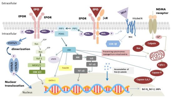
The homodimer isoform is present in erythroblasts [17]. During hematopoiesis, EPO binds to its receptor and results in homodimerization of EPOR (Figure 1) [18]. Following binding, Janus kinase-2 (JAK-2) activates several secondary signal molecules [17,19], such as STAT5 [20], MAPK, and PI3-K/Akt [21]. The activation of these molecules contributes to the differentiation and maturation of erythroid progenitor cells [22].
Figure 1. Binding of EPO to EPOR induces JAK-2 phosphorylation, dimerization, and subsequently activate STAT5/3, Ras/Raf/MEK/ERK, PI3-K/Akt, and NF-κB pathways. JAK-2 phosphorylates STAT5 or STAT3, leading to the dimerization of STAT5 (STAT3). STAT5 (STAT3) and the last signaling molecule in the MAPK pathway translocate into the nucleus and upregulate the expression of antiapoptotic Bcl-2 and Bcl-xL. Activation of PI3-k/Akt pathway increases endothelial nitric oxide synthase (eNOS) protein expression and NO production, which could increase blood flow and attenuate regional injury. PI3-k/Akt pathway also phosphorylates transcription factor GATA-1 and Foxo3 A, which enhance the expression of antiapoptosis proteins. Activation of the IKK complex by Akt phosphorylates IκB, resulting in its ubiquitination, and degradation, and in the releases of bound NF-κB. Free NF-κB translocates into the nucleus and exerts its antiapoptosis activity through the expression of inhibitors of apoptotic proteins (IAPs). Furthermore, binding of EPO to EPOR/βcR activates Wnt signaling, which inhibits GSK-3β phosphorylation and allow β-catenin to stabilize and accumulate in the cytoplasm in a non-phosphorylated form. Free β-catenin translocates into the nucleus and trigger transcription of Wnt-target gene responsible for cell antiapoptosis and the development of nervous system. Activation of NMDA receptors allows the influx of Ca2+, which induces excitotoxicity via initiation of the μ-calpain/Bax/cytochrome c/caspase-9 pathway. The caspases result in DNA fragmentation and lead to cell apoptosis. Activation of PI3-K/Akt pathway could also inhibit caspase activity by preventing cytochrome c leakage from mitochondria, thus inhibiting DNA degradation.

2.2. Heterodimer Isoform: EPOR/βcR

In addition to the homodimerization, there are other types of EPO receptors, which involve affinities 8–16 times weaker [23,24]. A study confirmed the receptor was a heterodimer consisting of EPOR and the β common receptor (βcR), a subunit of granulocyte- macrophage colony stimulating factor, interleukin 5, and interleukin 3 [25]. Since the EpoR/βcR heterodimer has the properties against tissue injury and inflammation, some authors named it as the “tissue-protective receptor” or “innate repair receptor” [26]. Recent data found the presence of this heterodimer complex in the RGCs, inner nuclear layer and photoreceptors [27].

2.3. Extracellular Soluble Isoform: sEPOR

The extracellular soluble isoform of EPOR (sEPOR), which lacks the transmembrane and cytoplasmic domains, is found in human plasma [30]. During hypoxia, in contrast to the expression of the full-length form increased through HIF transduction, the expression of the sEPOR is downregulated. In many studies, sEPOR is viewed as an endogenous antagonist of EPO, which blocks the neuroprotective effects of EPO. This form interacts with EPO without further activation of any downstream pathways. Moreover, its binding with EPO restricts the interaction of EPO with other receptor isoforms, resulting in a lower availability and bioactivity of EPO [10,31].

3. Effects of EPO

3.1. Angiogenic Effects

The transcription factor, HIF, has a vital role in hematopoiesis. In normoxic conditions, prolyl hydroxylase domain proteins (PHDs) hydroxylate all HIF-α subunit. After binding to von Hippel–Lindau tumor suppressor protein (VHL), hydroxylated HIF-α is then ubiquitinated. The ubiquitination of hydroxylated HIFs results in its degradation by the proteasome. In hypoxic conditions, the action of PHDs is inhibited. The stabilized HIF-α thus binds to HIF-β and translocates into the nucleus to regulate erythropoiesis via regulation of the expression of the EPO gene, vascular endothelial growth factor (VEGF) [32], as well as genes coding for proteins involved in iron metabolism [33], which are important for tissue oxygenation. After translation of EPO, the EPO is secreted into the circulation to reach hematopoietic cells. EPO binds with EPOR on erythroid cells, which triggers homodimerization of EPOR and activates the EPOR-associated JAK-2 by autophosphorylation (Figure 1) [17]. The active kinase JAK-2 results in the phosphorylation of tyrosine residues on the cytoplasmic portion of the EPOR [17]. The phospho-tyrosine residues recruit various proteins, which subsequently activate a series of pathways, including JAK-2/STAT5 (STAT3), PI3-K/Akt, and MAPK pathway [20,21]. JAK-2 phosphorylates STAT5 or STAT3, once it binds to the cytoplasmic portion of EPOR. STAT5 (STAT3) homodimerizes and translocates into the nucleus as a gene transcription factor. Activating the JAK-2/STAT5 (STAT3) pathway also leads to the upregulation of the antiapoptotic B-cell lymphoma-extra-large (Bcl-XL) protein, therefore protecting proerythroblasts from apoptosis [34]. After the activation of Ras by adaptor proteins, initiation of RAF/mitogen-activated protein kinase (MEK)/extracellular signal-regulated kinase (ERK) pathway would occur. RAF-1 protein kinase phosphorylates MEK, which subsequently phosphorylates MAPK/ERK1/2 [35]. The last molecules in the cascades translocate into the nucleus and activate various gene transcription factors for erythropoiesis regulation. PI3-K/Akt pathway is one of the main activating signaling pathways. PI3-K could lead to the phosphorylation of Akt, which could activate other proteins involved in erythropoiesis regulation. Akt phosphorylates the transcription factor GATA binding protein-1 (GATA-1), which is an important transcription factor for the anti-apoptotic Bcl-XL expression and erythroid-specific genes. Phosphorylation of GATA-1 could enhance GATA-1 activity in erythroid cell [36]. The forkhead box O3A (Foxo3A), another Akt targeted transcription factor, has proapoptotic functions; in contrast, phosphorylation of Foxo3A inhibits its transcriptional activity [37]. Figure 1 illustrates the intracellular signaling pathway of EPO. Dysfunction in these signaling pathway leads to abnormal erythropoiesis by disrupting cells proliferation and apoptosis.

3.2. Antiapoptotic Effects

Binding of EPO to EPOR results in JAK2 phosphorylation and initiates STAT5 (STAT3), MAPK, PI3-K/Akt and nuclear factor kappa-light-chain-enhancer (NF-κB) downstream pathways, which execute the antiapoptotic effect of EPO (Figure 1) [38]. The last molecules in the STAT5 (STAT3) and MAPK pathways could translocate into the nucleus and activate the apoptotic regulators of Bcl-2 family, antiapoptotic Bcl-2 and Bcl-XL, to inhibit apoptosis [39]. Activation of PI3-K/Akt pathway also prevents cell apoptosis. Cell death signaling can be initiated by caspases or mitochondrial membrane depolarization. When the mitochondria membrane is depolarized, cytochrome c would be released into the cytoplasm and form the apoptosome complex with apoptotic protease activating factor-1 (Apaf-1) [40]. Pro-caspase-9 is activated by the apoptosome, which initiates downstream caspase activation. The activated caspases would cause DNA fragmentation and lead to cell apoptosis [40]. Activation of PI3-K/Akt pathway could inhibit caspase activity by preventing cytochrome c leakage from the mitochondria [41]. IκB kinase (IKK), another Akt target, is also associated with cell survival. In resting cells, NF-κB is held by the IκB. Activation of the IKK complex phosphorylates IκB, resulting in its ubiquitination and degradation, and in the releases of bound NF-κB. NF-κB exerts its protective effects through the increase in inhibitors of apoptotic protein (IAPs) [42], blocking of caspase activity [42], suppression of TNF-α related apoptosis [42], direct enhancing activation of Bcl-XL, and removal of cellular reactive oxygen species (ROS) [43].

3.3. Anti-Inflammatory Effects

In inflammatory conditions, EPO was detected at the borders of the injury sites. Hence, the potential anti-inflammatory effect of EPO has also been investigated. EPO was found to decrease pro-inflammatory cytokine production, including intercellular adhesion molecule-1 (ICAM-1) [46], interleukin-6 (IL-6) [47], and TNF-α [48,49]. EPO also increased the production of the anti-inflammatory cytokine IL-10 [48]. Additionally, EPO could increase endothelial nitric oxide synthase (eNOS) protein expression (Figure 1) [50], which increase nitric oxide production. Nitric oxide could increase blood flow, and attenuate regional injury [51]. As regards the innate immune system, EPO could facilitate phagocytosis in macrophages [52], mediate dendritic cell maturation and immunomodulation [53], and reduce inflammation caused by mast cells [54].

3.4. Antioxidant Effects

EPO has the ability to attenuate oxidative stress, allowing it to be categorized as a cytoprotective agent [59,60]. EPO could induce heme oxygenase-1 expression via PI3K/Akt pathway [61], which could provide a cytoprotective effect in astrocytes [62]. EPO also increases the level of glutathione peroxidase, a potent antioxidant protein, which can decrease the toxic activity of ROS [63].

4. Current Strategy of EPO for Optic Nerve Protection and Repair

Encouraging results of EPO from basic research support the possibility of integrating its therapeutic effects in glaucomatous optic neuropathy, optic neuritis, non-arteritic anterior ischemic optic neuropathy (NAION), and traumatic optic neuropathy (TON).
Glaucomatous optic neuropathy, a neurodegenerative disease, is characterized by progressive loss of RGCs. Elevated intraocular pressure (IOP) is considered the most important risk factor of glaucomatous optic neuropathy. However, some patients experienced continued RGCs loss despite good intraocular pressure control, suggesting the presence of other complicated mechanisms stimulating RGC death. Multifactorial mechanisms have been postulated for glaucomatous optic neuropathy, including vascular insufficiency, inflammation [66,67], excitotoxicity [68] and neurotrophic factor withdrawal [69]. Due to the complex pathogenesis of glaucoma, EPO was developed to prevent the IOP-independent RGCs loss. Several studies have reported that the EPO level in the aqueous humor increased in patients with glaucoma [70]. The cause of the elevated aqueous EPO in glaucomatous eyes might be related to the ischemia, hypoxia, or elevated ROS caused by glaucomatous damage [71]. The increase in EPO is identified as a compensatory response due to the presence of glutamate, nitric oxide and the free radicals after the glaucomatous damage [72]. EPO is found to have neuroprotective effects regardless of the EPO administration methods. However, the discussion of EPO in the treatment of glaucoma is limited to animal studies. In humans, there are only a few observational studies investigating the correlation between EPO and glaucoma, especially neovascular glaucoma [79,80,81]. To date, human studies using EPO for the treatment of glaucoma are still lacking. Future studies could focus the application of EPO in patients with primary open-angle glaucoma to see if EPO exhibits the same neuroprotective effects in animal experiments.
Optic neuritis is another high occurring disease among the world population. For optic neuritis, methylprednisolone is the standard treatment in clinical practices. Although steroid treatment could accelerate visual acuity recovery, recent study demonstrates that steroids could not influence the visual outcome or atrophy of the optic nerve [82]. An animal study even demonstrated that methylprednisolone could increase RGCs degeneration by inhibiting the neurotrophin pathway [83]. Since EPO has shown multiple neurotrophin-like properties in various neuronal disorders, the efficacy of EPO is evaluated as an add-on therapy to methylprednisolone in autoimmune optic neuritis by investigators. In an experimental autoimmune encephalomyelitis (EAE) rat model, intraperitoneal injection of EPO (5000 U/kg) significantly increased the survivability and functionality of RGCs in rats afflicted with myelin oligodendrocyte glycoprotein (MOG)-induced optic neuritis [84]. In the model of MOG-EAE, Sättler et al. concluded that the PI3-K/Akt pathway plays an important role in RGCs survivability under systemic treatment with EPO [84]. Establishment of potentially relevant intracellular conduction pathways might make the application of EPO more feasible in MOG-EAE. Human studies have been performed, but the results were not conclusive.
Non-arteritic anterior ischemic optic neuropathy is thought to result from vascular insufficiency. Patients with hypertension or obstructive sleep apnea have higher risk of developing NAION since the disease could result in hypoperfusion of the optic nerve. The hypoperfusion causes ischemia and swelling of the axons, thus increasing the pressure on the nervous tissues confined within the tight borders of the posterior scleral outlet. The axon swelling results in further ischemia and neuron swelling. The vicious cycle leads to severe ganglion cells damage. Due to the evidence showing neuroprotection effect of EPO, investigators also determined the efficacy of EPO in NAION. Modarres et al. conducted a prospective interventional case series by intravitreally injecting EPO (2000 IU/0.2 mL) into thirty-one patients within 1 month of the NAION onset [89].
For traumatic optic neuropathy, indirect TON is the more common type. The shearing force could lead to small vessel and neuron axon injury around the optic nerve by inducing ischemia, inflammation, and oxidative stress, all of which result in ganglion cell death. Currently, the common treatments are observation, corticosteroids and optic canal decompression. However, none of these managements are proven to be effective. Since EPO has shown to be neuroprotective, EPO might play a role in treating indirect TON. Intravenous EPO was first commenced in patients with indirect TON by Kashkouli et al. in 2011 [92]. Indirect TON patients with intravenous EPO (10,000 IU in 3 days) were compared to indirect TON patients without treatment. They found that the EPO-treated group has higher BCVA than that in the observation group. They advocated intravenous EPO may be a new effective and safe treatment in patients with indirect TON [92].

5. Advances in EPO Derivatives

Epoetin alfa (Epogen), a type of ESA medicine, has been the standard of care for patients with kidney disease and cancer-related anemia. Epoetin alfa-epbx (RetacritTM) shares the same amino acid sequence and similar carbohydrate composition as epoetin alfa (EpogenTM). In 2018, the protein was approved by the FDA, making it the first biosimilar EPO molecules approved in the USA [97]. Darbepoetin alfa (DA, Aranesp), an alternative agent of Epoetin alfa and a hyperglycosylated EPO analog, is a novel ESA with two additional N-glycosylation sites accompanied by 22 sialic acid moieties. In the attempt to extend the molecule’s half-life by three-fold longer than EPO in vivo, glycoengineering was conducted to increase the structure’s resistance to degradation. Darbepoetin alfa was approved for treating anemia resulting from renal diseases and cancer chemotherapy. The treatment protocol only requires a once-per-week visit and is accompanied by lower clinical costs [98,99]. C.E.R.A. (continuous erythropoietin receptor activator), a third-generation ESA, is an EPO (~34 kDa) integrated with methoxypolyethylene glycol (PEG, 30 kDa). Compared with other EPO derivatives, C.E.R.A. has a unique pharmacological profile with the longest half-life and slowest clearance rate. These unique pharmacological properties exist because of methoxypolyethylene glycol (PEG) integration into EPO. Notably, EPO pegylation (the process of connecting a hydrophilic polymer to EPO) significantly prolongs the duration of EPO action, and enhances proteolytic resistance in cell-free plasma [100].

Asialerythropoietin (asialoEPO) was evaluated to be a safe drug for clinical treatments. However, asialoEPO’s half-life (t1/2~1.14 min) is much shorter than that of EPO (t1/2~5.6 h). The short half-life gives asialoEPO insufficient persistence time to stimulate hematopoiesis. Based on the above concept, researchers found that chemical modification of the EPO binding sites could abolish erythropoiesis function but retain the tissue-protective effect. Carbamylated EPO (cEpo), a chemically modified derivative of EPO’s lysine residues, was found to act through the heterodimeric EPOR/βcR rather than classical EPOR2 primarily because of the modified structure of cEpo. The study has confirmed that cEpo possesses neuron anti-apoptotic effects similar to EPO but instead does not induce neovascularization [101].

6. Advances in EPO Delivery

6.1. Protein-Based Ocular Delivery

Many EPO studies involve frequent injections of ophthalmic proteins via invasive procedures, which might result in a variety of adverse effects and increase the probability of irreversible damage to the patient’s eye. Topical sustain released formulations are non-invasive drugs, that effectively reaches the posterior segment of the eye. According to Silva et al., mucoadhesive polymers such as chitosan and hyaluronic acid can improve the ocular bioavailability of drugs with the support of nanoparticulate delivery systems [106]. The formulation was found to be non-cytotoxic toward ARPE-19 and HaCa T cell lines. CS/HA6-rhEPO may be a promising topical formulation after enhancing its bioavailability through different ocular barriers. For the intraocular route of administration, De Julius et al. developed two polymer microparticles, poly (propylene sulfide) (PPS) and poly (lactic-co-glycolic acid) (PLGA), to prolong His-tagged rhEPO-R76E (42kDa) release [107]. The rhEPO-R76E was loaded into the polymeric microparticles to prolong in vivo release for at least 28 days to resolve the issue involving short half-life of the rhEPO-R76E (t1/2~13 min). PPS-based microparticles platform is especially promising because it is degradable by ROS. The delivery system provides extended neuroprotection and inherent antioxidant benefits, which reinforces its ability in ocular delivery of EPO.

6.2. Gene-Based Ocular Delivery

Under the gene delivery approaches, EPO has significant therapeutic potential in neurodegenerative diseases due to its neuroprotective effects. However, recombinant EPO is limited in clinical treatment of glaucoma patients due to its short half-life. As regards this issue, Bond et al. constructed a viral gene delivery system for EPO-R76E [108]. Treatment with recombinant adeno-associated virus (rAAV) provides sustainable, long-term delivery of EPO-R76E without a critical rise in hematocrit [108,109]. AAV-mediated long-term EPO expression is achievable in animal models with the primary functions of promoting red blood cells proliferation and neuroprotection.
Another challenge in applying gene therapy in humans is the improvement of drug selectivity. For systemically secreted hormone, such as EPO, it is vital to use an inducible genetic delivery system to avoid excess expression and side effects. However, precise expression control is highly desirable when maintaining steady-state red blood cell counts within a narrow therapeutic window. Hines-Beard et al. packaged EPOR76E into a recombinant adeno-associated viral vector under the control of the tetracycline inducible promoter [110]. In the retina, tetracycline-controlled expression of green fluoresce protein (GFP) in retinal pigmented epithelium and photoreceptor cells becomes apparent in rats following subretinal injections of rAAV-2/2 vector. The outer nuclear layer in the eyes was approximately 8 μm thicker in mice that received doxycycline water as compared to the control groups.

6.3. Surface Receptor-Targeted Ocular Delivery

Through surface receptor-targeted delivery results, most suggest that the tissue-protective effect of EPO and injury response are mediated by the EPOR/βcR heterodimer and not by the EPOR homodimer [112]. In addition to some well-known derivatives of EPO with better affinity for the EPOR/βcR heterodimer, such as asialoEPO, cEpo and HBSP (see above), traptamers of transmembrane domain (TMD) proteins of EPOR/βcR are also an option. He et al. constructed ELI-3 traptamer that specifically targets the TMD of human EPOR and triggers cooperative JAK/STAT signaling for proliferation and tissue protection [113].

6.4. Cell-Based Ocular Delivery

Mesenchymal stem cell (MSC) therapy is potential in treating optic neuropathies. MSCs have demonstrated to possess neuroprotective effects in numerous neurodegenerative diseases while maintaining retinal morphology [114,115]. They could also regulate inflammatory responses [116] and increase the secretion of neurotrophic factors [117]. Furthermore, they could transdifferentiate into retinal progenitor cells [118]. MSCs could be the vector of EPO because these cells could cross the brain-retinal barrier and localize into the inflamed sites [121]. The phenomenon emphasizes a mutualistic relationship between MSCs and EPO. Hence, some investigators attempt to evaluate the efficacy of EPO-expressing MSC in treating retinal degenerative diseases. Ding et al. transduced MSCs with lentiviral particles encoding EPO. They found that co-treatment with EPO and MSCs instead of only with MSC could attenuate human retinal neuron apoptosis by restoring the mitochondrial membrane potential and protect human retinal neurons from glutamate neurotoxicity [122]. The ability for EPO to synchronize with MSCs may become beneficial in producing medical protocols involving the treatment of patients with glaucomatous optic neuropathy. Given their endogenous long-term therapeutic effects, MSCs-based therapy could be the future direction in optic nerve repair.
This entry is adapted from the peer-reviewed paper 10.3390/ijms23137143

This entry is offline, you can click here to edit this entry!
ScholarVision Creations