Electrochemical Biosensing of SARS-CoV-2 Virus for COVID-19 Management: History
Please note this is an old version of this entry, which may differ significantly from the current revision.
Contributor:

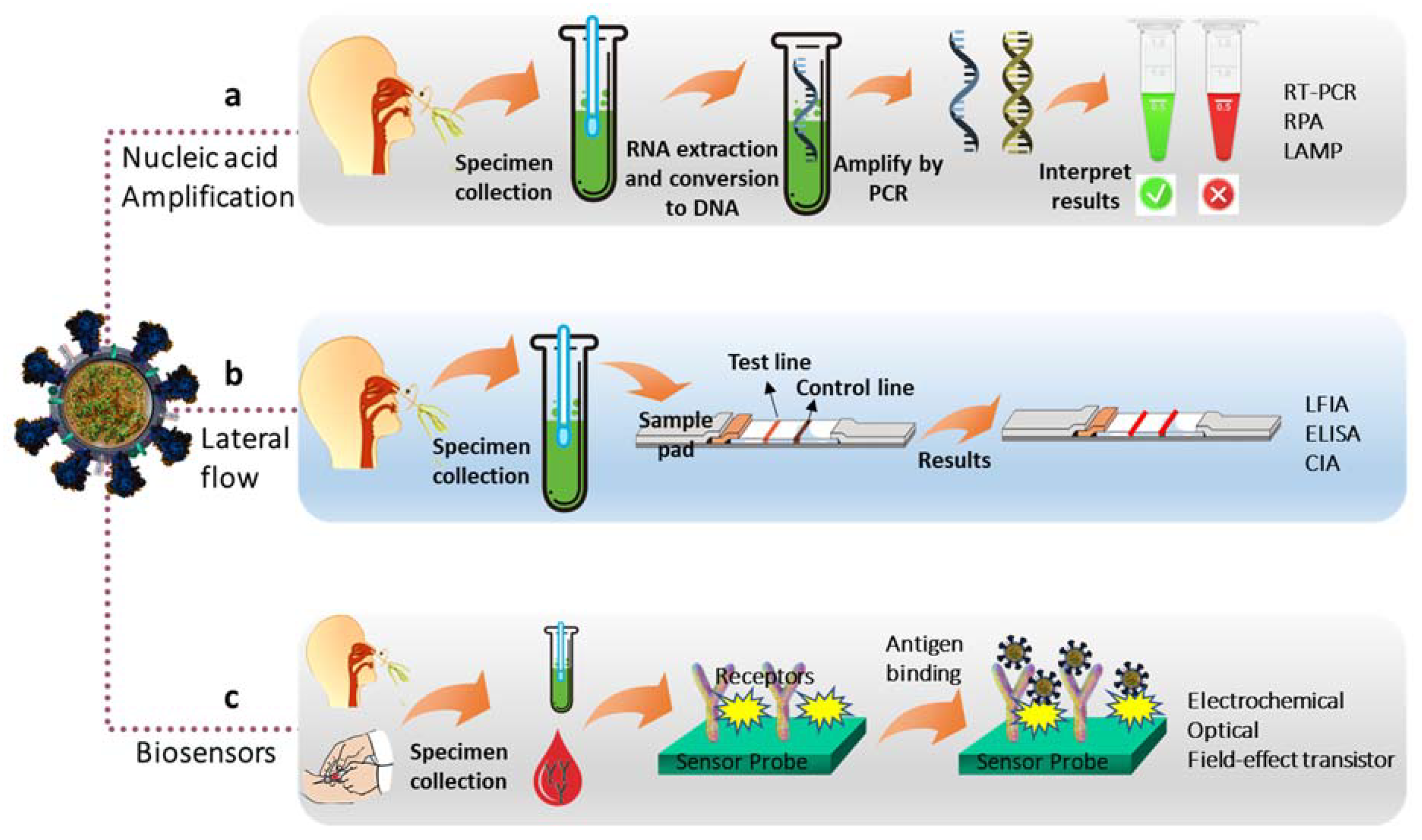
Rapid and early diagnosis of lethal coronavirus disease-19 (COVID-19) caused by the severe acute respiratory syndrome coronavirus 2 (SARS-CoV-2) is an important issue considering global human health, economy, education, and other activities. The advancement of understanding of the chemistry/biochemistry and the structure of the SARS-CoV-2 virus has led to the development of low-cost, efficient, and reliable methods for COVID-19 diagnosis over “gold standard” real-time reverse transcription-polymerase chain reaction (RT-PCR) due to its several limitations. This led to the development of electrochemical sensors/biosensors for rapid, fast, and low-cost detection of the SARS-CoV-2 virus from the patient’s biological fluids by detecting the components of the virus, including structural proteins (antigens), nucleic acid, and antibodies created after COVID-19 infection. 

  • SARS-CoV-2
  • electrochemical transduction
  • immunosensors
  • aptasensors
  • bioreceptors
  • serological test

1. Introduction

Since the first case in December 2019 in Wuhan, China, the global outbreak of severe acute respiratory syndrome coronavirus 2 (SARS-CoV-2) has resulted in a life-threatening respiratory infectious coronavirus disease-19 (COVID-19) that has significantly affects the global human health, socio-economy, education, national financial policies, and other activities [1][2]. World Health Organization (WHO) declared COVID-19 as a pandemic on 12 March 2020, due to the rapid human-to-human transmission of the virus with the primary symptoms of fever, coughing, short breathing, etc. [3]. The human-to-human rapid transmission of this virus can occur through the droplet, contact, airborne, fomite, fecal-oral, and bloodborne transmissions.
A person affected with SARS-CoV-2 virus or a COVID-19 patient can remain asymptomatic without showing any signs or mild symptoms [1][2][3]. These asymptomatic COVID-19 patients are the major spreaders of the SARS-CoV-2 virus. Therefore, within a short time, the SARS-CoV-2 virus spread all the six continents of the world with the total number of cases as of 29 May 2022 was over 531 million and total deaths of over 6.31 million [4]. Even though about 65% of the world population has been vaccinated with at least one dose of WHO-approved vaccines [5], the number of virus-infected people and the associated deaths are still increasing. This is mainly due to the mutation of the SARS-CoV-2 virus over time by genetic variation in the population of circulating viral strains that limit the efficacy of COVID-19 vaccines [6].
Thus, the best solution to control this lethal disease is still isolation of the infected patients through the earlier detection of COVID-19. At present, real-time reverse transcription-polymerase chain reaction (RT-PCR) is the ‘’gold standard’’ method for diagnosing COVID-19 disease that is based on the molecular testing of single-strand ribonucleic acid (ssRNA) from the SARS-CoV-2 virus (Figure 1a). Nevertheless, RT-PCR testing is time-consuming, costly, and requires a specialized laboratory setup with expensive instrumentations and trained personnel [7][8]. Furthermore, the highly contagious nature SARS-CoV-2 virus could enable its faster human-to-human transmission during the sample collection and analyses.
Figure 1. Schematic of the diagnosis methods of COVID-19. (a) Molecular testing based on nucleic acid amplification assays, (b) lateral flow immunoassay, and (c) biosensors.
To overcome these drawbacks, several other molecular testing based on isothermal nucleic acid (NA) amplification assays, such as loop-mediated isothermal amplification (LAMP), recombinase polymerase amplification (RPA), deoxyribonucleic acid (DNA) nano-scaffold-based hybrid chain reaction, and NA sequence-based amplification, have already been reported (Figure 1a) [9][10][11][12]. These methods also exhibit some certain limitations along with their advantages of being low cost, rapid analyses, and highly sensitive and specific. For example, LAMP and RPA methods require the design of a complex primer, and the LAMP method is unable to perform multiplex amplification [10].
In comparison to the above diagnosis methods, lateral flow immunoassay (LFIA) platforms with optical detection (colorimetry/fluorescence), enzyme-linked immunosorbent assay (ELISA), chemiluminescent immunoassay (CIA), and electrochemistry-based serological test (detection of antibody and antigen) have received much attention for diagnosis COVID-19 (Figure 1b) [13][14][15][16][17]. In particular, electrochemistry-based SARS-CoV-2 virus detection systems have received great potential over ‘gold standard’ RT-PCR, NA amplification assays, and optical methods.
This is due to the superior advantages of electrochemical biosensors and immunosensors, including a short reading time, require a small volume of samples, miniaturization ability, point-of-care (POC) and point-of-need (PON) testing, and high sensitivity and specificity [16][17][18][19][20].
Furthermore, electrochemical biosensors and/or immuno-sensors are capable of label-free and label-based diagnosis of SARS-CoV-2 virus by exploiting redox indicators and labels [21][22]. Accordingly, a larger number of electrochemical biosensors and immuno-sensors have been developed for diagnosing COVID-19 by detecting antibodies, antigens (structural proteins), and nucleic acids of SARS-CoV-2 along with the development of novel electrode modifiers and fabrication methods (Figure 1c) [9][16][17][23][24].

2. Designing Electrochemical SARS-CoV-2 Virus Biosensors

2.1. Antibody Biosensors

The antibody test is principally based on the detection of antibodies developed in individuals due to exposure to the SARS-CoV-2 virus [25]. The electrochemical antibody-based biosensors probe is based on the utilization of suitable nanomaterials (e.g., self-assembled monolayer, functionalized graphene, and conducting polymer) modified electrodes for the immobilization of SARS-CoV-2 structural proteins [26][27][28]. Subsequently, the SARS-CoV-2 structural proteins are used to anchor the antibodies (anti-IgG or anti-IgM), created in the biological body fluids of COVID-19 infected patients (Figure 2) [27].
Figure 2. Schematic of the electrochemical strategies of COVID-19 diagnosis.
For a label-free detection, the anchored SARS-CoV-2 antibodies induce a variation in current, potential, and resistance of the redox indicator (e.g., [Fe(CN)6]3−/4−, [Ru(NH3)6]3+, etc.) and enabling the label-free diagnosis of COVID-19 [21][22]. In contrast, for label-based detection of SARS-CoV-2 antibodies, the representatives’ antibodies can be functionalized with labels (e.g., quantum dots, redox-active molecules, and low-dimensional carbon materials) prior to the attachment with the biosensor probe [29]. Upon anchoring the labeled antibodies with a biosensor probe, the labels generate current responses suitable for the detection/determination of the SARS-CoV-2 virus with high specificity and selectivity.
Label-free sensors are preferable to label-based sensors due to their less complicated designs, short preparation time, and low cost [30]. However, lack of sensitivity, cross-reactivity, and interference are major disadvantages of label-free sensors for their practical application [29]. Even though anybody-based label-free and label-based diagnostic systems for SARS-CoV-2 and other viruses are rapid and highly specific, their clinical efficacy for SARS-CoV-2 infection testing is restricted, as it may take several days to weeks to develop a detectable antibody response in COVID-19 patients after starting to show the symptoms [25].

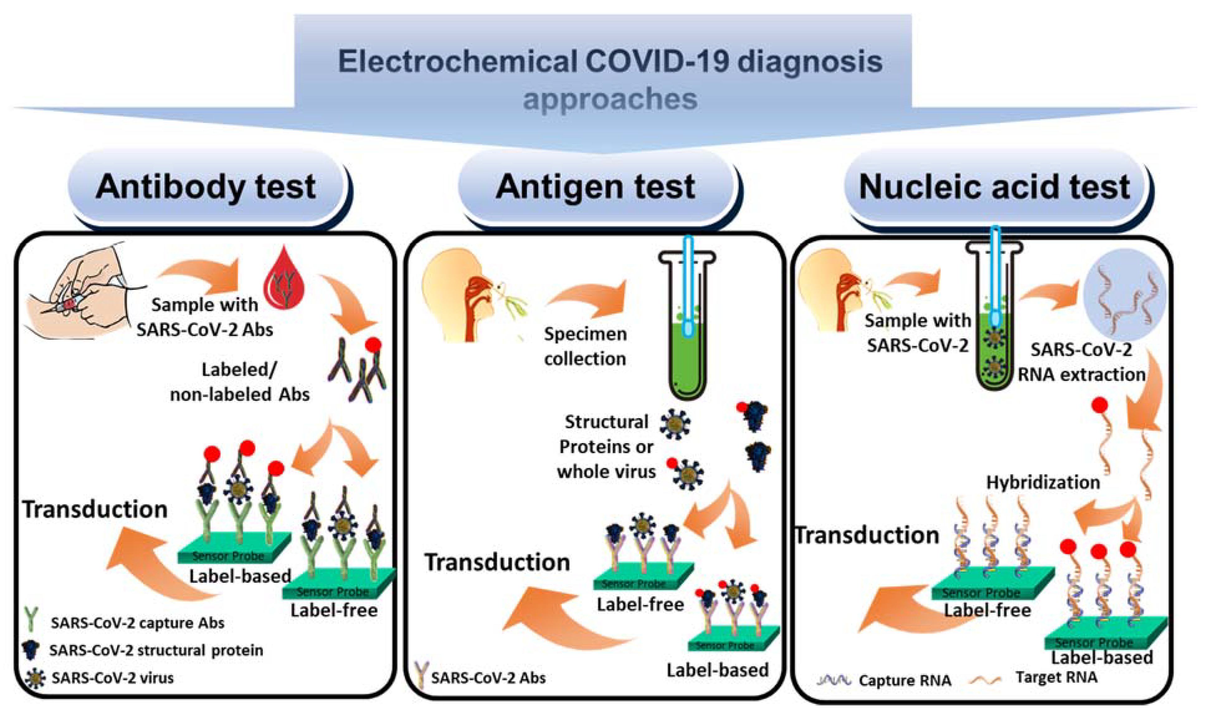
2.2. Antigen Biosensors

The antigen test for the diagnosis of SARS-CoV-2 infection is highly sensitive and accurate and capable of rapid detection of the virus in clinical samples [23]. Therefore, antigen tests have received emergency authorization from the food and drug administration (FDA) to diagnose SARS-CoV-2 [31]. For a simple label-free electrochemical detection of the SARS-CoV-2 virus, the protein receptor antibodies (anti- SARS-CoV-2 SP, anti- SARS-CoV-2 NP, anti- SARS-CoV-2 EP, and anti- SARS-CoV-2 MP) can be immobilized onto nanomaterial-modified electrodes. Subsequently, the whole SARS-CoV-2 virus or the corresponding structural proteins can bind with the sensors probe (Figure 2) [23][30].
This induces discrimination of the current, potential, and resistance signals of the redox indicators, enable the label-free diagnosis of COVID-19 [21][22]. Aaptamers, molecularly imprinted polymer (MIP), and angiotensin-converting enzyme 2 (ACE2) can also be used as receptors for the construction of antigen biosensors. In contrast, for label-based detection of the SARS-CoV-2 virus and the corresponding structural proteins, prior labeling of them is required before anchoring to the sensor probe. Upon anchoring the labeled-virus or labeled-structural proteins attached to the biosensor probe can generate a current response suitable for the detection/determination of the virus with high specificity and selectivity [29][30].

3. Electrochemical Biosensors for the Detection of SARS-CoV-2 Virus

3.1. Electrochemical Antibody-Based Detection of SARS-CoV-2 Virus

Antibodies, such as anti-IgG, anti-IgM, and immunoglobin A (anti-IgA), can be developed from the COVID-19 patients’ body fluids that can be used to detect SARS-CoV-2. The recent immuno-chromatographic study suggests that both IgG and IgM antibodies exhibit 11.1%, 92.9%, and 96.8% sensitivity of SARS-CoV-2 detection at the early stage (several days to weeks after the COVID-19 infection), intermediate stage (1–2 weeks after the onset), and late-stage (more than 2 weeks), respectively [32][33].
Anti-IgA is another major antibody in the respiratory tract that is produced by B-lymphocytes and expressed after 2 weeks of COVID-19 infection [32][33]. Thus, the detection of antibodies in human blood serum samples by developing highly sensitive electrochemical biosensors can be an effective tool for diagnosing COVID-19 infection at the early stage of infection, in which SP and NP can serve as antigens for the specific binding of antibodies.
The IgG antibody is a lighter and smaller (~150 kDa) antibody compared to the IgM antibody (~900 kDa) with two antigen-binding sites [34][35]. The anti-IgG can be detected only after a week of infection without altering its concentration for a long period after infection and after several weeks, anti-IgG reactivity reaches >98% [36]. Thus, the detection of anti-IgG has attracted considerable attention over IgM and IgA antibodies. Some selected examples for the detection of anti-IgG are outlined below.
Electrode materials play a crucial role in improving the sensitivity of electrochemical detection. This is because electrode materials with high surface area and functionality could increase the amount of immobilized target-specific antigens or antibodies [37][38] and induce to enhance the detection sensitivity with a wide dynamic range and low limit of detection (LOD). The high electrical conductivity is also crucial for obtaining high sensitivity, which can be achieved by providing an efficient electron transport channel for the redox reaction of a redox probe or target analyte.
Among the various nanomaterials for the modification of electrode surfaces for biosensor development, graphene, and its related materials, including graphene oxide (GO) and reduced graphene oxide (rGO), have attracted significant interest [39][40]. This is mainly due to their high chemical functionality and surface area, solution processability, and excellent electron transporting capability [39]. Accordingly, Yakoh et al. reported a label-free electrochemical immunosensor based on a GO-modified paper electrode that can successfully detect IgG antibodies [41].
The IgG antibody can specifically bind with the SARS-CoV-2 SP antigen-modified GO/paper immunosensor probe that induces the high specificity and sensitivity of COVID-19 diagnosis in clinical serum samples. Upon forming the anti-IgG/SP antigen complex, the redox activity of [Fe(CN)6]3−/4− is decreased with increasing the concentration of IgG antibodies. This can be ascribed to the insulating nature of antibodies, which induce a decrease in the redox peak current by increasing the charge transfer resistance (Rct) as the electrode/electrolyte interface.
Concurrently, the immunosensor exhibits a wide linear range for the detection of anti-IgG with the LOD of 1 ng/mL. In another report, Ali et al. prepared an ultrasensitive and label-free 3D biosensor based on a micropillar array of Au nanoparticles (AuNPs) coated with rGO sheets [42]. Subsequently, the SARS-CoV-2 NP was immobilized onto the AuNPs/rGO-modified micropillar array via (3-dimethylaminopropyl)-N′-ethylcarbodiimide (EDC)-N-hydroxysulfosuccinimide (NHS) coupling chemistry. Finally, the immunosensor array was integrated into a microfluidic channel to complete the electrochemical cell.
The as-prepared immunosensor could selectively bind IgG antibodies, which induces to increase in the Rct for [Fe(CN)6]3−/4− redox couple with increasing the concentration of IgG antibodies in the detection range of 100 fM to 1 nM and the LOD of 13 fM. The same research group utilized a similar rGO-modified Au micropillar array for the immobilization of SARS-CoV-2 SP antigen via EDC-NHS coupling [43].
This label-free electrochemical microfluidic immunosensors probe could selectively bind the IgG antibodies which induces to increase in the Rct values for the redox reaction of [Fe(CN)6]3−/4− with increasing the concentration of anti-IgG. The corresponding LOD of the immunosensor was 2.8 × 10⁻¹⁵ M for the detection of anti-IgG. A similar sensing electrode based on GO and Au nanostar-modified carbon-based screen-printed electrode (SPE) was developed by Hashemi et al. for the label-free detection of IgG antibodies specific for SARS-CoV-2 SP antigen with a low LOD and high sensitivity [44].
Other than GO and rGO, metal-oxide, conducting polymers, and molecular functionalization of electrodes can be effective substrates for the development of COVID-19 antibody sensors [45][46][47][48]. Li et al. prepared a label-free COVID-19 IgG antibody sensing platform based on microfluidic paper-based analytical devices (μPADs) [45]. The carbon-coated μPAD was modified with ZnO nanowires (NWs) using the hydrothermal method. Then, the glutaraldehyde and (3-aminopropyl)-trimethoxysilane-modified ZnO NWs electrode surface was functionalized with SARS-CoV-2 SP for the selective binding of anti-IgG, specific for COVID-19 infection. The as-prepared electrochemical impedance spectroscopy (EIS) based label-free immunosensor platform could detect IgG antibodies in human serum samples up to 1 μg/mL.
To date, electrochemical label-based detection of COVID-19 antibody is less developed compared to the label-free approaches. This is possibly due to the complicated labeling steps, long preparation time, and high cost even though label-based detection exhibited high specificity and selectivity. Ameku et al. reported a label-based method for the detection of SARS-CoV-2 specific anti-IgG based on a polyglutaraldehyde SPE [46].

3.2. Electrochemical Antigen-Based Detection of SARS-CoV-2 Virus

The detection of the SARS-CoV-2 virus protein antigens, including SP, NP, and whole virus, providing the possibility of early diagnosis of COVID-19. Accordingly, various strategies have recently been developed, and many of them are now commercially available for the early diagnosis of COVID-19 [49]. This section of the entry discusses the recently developed antigen-based electrochemical sensors/biosensors for COVID-19 diagnosis by dividing into three subsections, namely the detection of SP, detection of NP, and detection of whole virus or virus particles.

3.2.1. Detection of Spike Proteins

The SP is the key trans-membrane protein of the SARS-CoV-2 virus with diverse amino acid sequences providing the precise diagnosis of COVID-19 infection [50][51]. Therefore, intensive efforts have been made for the development of electrochemical SP sensors by developing novel chemistry, electrode materials, and capture probe (e.g., antibody, aptamer, and angiotensin-converting enzyme).
Antibody capture probe-based electrochemical biosensors or immunosensors display high specificity and reliability for the detection of target biomolecules [52]. Accordingly, anti-SARS-CoV-2 capture probe-based immunosensors have received potential interest for screening SARS-CoV-2 protein antigens. Adeel et al. developed an ultrasensitive and label-free electrochemical SP sensing platform based on a functionalized self-supported graphitic carbon foil electrode [53].
The electrode was prepared by mild acidic treatment with partial oxidation and exfoliation of graphitic carbon foil electrode. The subsequent ethylenediamine functionalization of the electrode provided a suitable platform for the covalent attachment of the anti-SARS-CoV-2 SP capture probe. Upon the binding of specific SARS-CoV-2 SP onto the sensor probe, the electrochemical redox activity of the [Fe(CN)6]3−/4− redox couple was varied with the concentration of SP.
This enabled the detection of SP within the concentration range from 0.2–100 ng/mL with a LOD (27 pg/mL) in diluted blood plasma. A similar antibody-capture probe-based field-effect transistor (FET) was developed by Seo et al. to detect SARS-CoV-2 SP in COVID-19 infected patients’ samples [54]. This graphene-based FET device was coated with anti-SARS-CoV-2 SP against the SARS-CoV-2 SP using the 1-pyrenebutyric acid N-hydroxysuccinimide ester.
The effectiveness of the FET device for binding SARS-CoV-2 SP and SARS-CoV-2 was tested using a cultivated virus, viral antigen, and nasopharyngeal swab samples. The biosensor could detect SARS-CoV-2 SP at a concentration of 100 fg/mL clinical transport medium. Similar antibody capture probe-based SARS-CoV-2 SP detection platform was developed by Malla et al. [55]. This label-based and POC biosensor is prepared using an SPE electrode modified with antibody-peroxidase-loaded magnetic beads (MBs) that could detect SARS-CoV-2 SP in the range from 3.12–200 ng/mL with the LOD of 0.20, 0.31, and 0.54 ng/mL in human saliva, urine, and serum, respectively.
Even if antibody capture probes exhibit high specificity, several limitations of antibodies, such as low stability, high cost, short shelf-life, and immunogenicity prompted to use of alternative capture probes for the development of biosensors and other biological applications [38][52]. The aptamer is one of the suitable alternatives to antibodies that can overcome the intrinsic limitations of immunosensors. Aptamers are peptide or NA (DNA or RNA) molecules with high stability, specificity, long-shelf-life, low immunogenicity, and easy synthesis and functionalization [39].
Thus, aptamers have attracted wide interest in developing electrochemical biosensors to detect various target biomolecules [38][39][52]. Idili et al. prepared a label-based electrochemical aptasensor for the detection of SARS-CoV-2 SP that is based on an Au electrode modified with methylene blue derivative (MB2) labeled aptamer [56]. The binding event of SARS-CoV-2 SP with the aptamer induces the variation of aptamer conformation, which in turn varies the position of the redox label MB2. This generated a quantitative electrochemical signal related to the variation of the concertation of SARS-CoV-2 SP.
The aptasensor could detect the picomolar level of SP with high specificity. In another report, Curti et al. reported another label-based aptasensor based on single-walled carbon nanotube screen-printed electrodes (SWCNT-SPEs) functionalized with a redox-tagged DNA aptamer [57]. The selective binding of SARS-CoV-2 SP folded the DNA aptamer which reduces the efficiency of the electron transfer between the AttoMB2 redox label and the electrode surface. Thus, the redox signal of the redox tag is suppressed by increasing the concertation of SARS-CoV-2 SP and forms the basis of SP quantification. The aptasensor exhibited high selectivity and specificity with the LOD of 7 nM.
ACE2 is another potential and selective receptor for binding SARS-CoV-2 SP with nanomolar range affinity [58]. Furthermore, the active site of the ACE2 is located away from the SARS-CoV-2 binding site, thus, resulting in an effect on the electrochemical signal upon binding with SARS-CoV-2. Inspired by this advantage of ACE2, Vezza et al. developed an accurate and rapid diagnostic device for the detection of SARS-CoV-2 SP that is based on a printed circuit board (PCB) electrode [59].
For the fabrication of the sensing platform, the PCB electrode was modified with 1H,1H,2H,2H-perfluorodecanethiol (PFDT) followed by the ACE2 functionalization via physisorption. The selective binding of SARS-CoV-2 SP onto the sensor probe induces to decrease in the redox activity of [Fe(CN)6]3−/4− and increases the Rct in EIS measurement by increasing the concentration of SP. This enabled the detection of SARS-CoV-2 SP with the LOD of 1.68 ng/mL. Similarly, another SP sensing platform was developed that is based on MBs and AuNPs conjugated to ACE2 for the capturing and detection of SARS-CoV-2 SP [60].
This magneto-assay modified SPE exhibited 93.7% specificity for SARS-CoV-2 SP with the LOD of 0.35 ag/mL. In another report, Lima et al. prepared a low-cost advanced diagnostic device based on AuNP-modified graphite leads. The cysteamine modification of AuNPs enabled the covalent immobilization of ACE2. The subsequent binding of SARS-CoV-2 SP to the ACE2 receptor induces a decrease in the electron transfer kinetics of [Fe(CN)6]3−/4− redox probe and decreases the oxidation current by increasing the concentration of SP. This label-free approach is capable of detecting SP in clinical saliva and nasopharyngeal/oropharyngeal with excellent sensitivity, specificity, and accuracy. This research further demonstrated that ACE2 is an effective, specific, and selective receptor for anchoring SARS-CoV-2 SP.
In the quest of replacing the labile and expensive biological receptor, recently, synthetic receptor or plastic antibodies, such as molecularly imprinted polymer (MIP) have received potential interest for biosensors, bioanalyses, drug delivery, and disease diagnosis [61]. In particular, MIP has attracted significant interest as a receptor for biosensors development, mainly due to its antibody-like ability to bind and discriminate between molecules, excellent chemical and thermal stability, and low cost [62]. Taking the advantages of MIPs, Ayankojo et al. developed an electrochemical biosensor for the detection of SARS-CoV-2 SP that is based on disposable Au-based thin-film electrodes (Au-TFME) chip modified with MIP film [63].

This entry is adapted from the peer-reviewed paper 10.3390/chemosensors10070287

References

  1. Hu, B.; Guo, H.; Zhou, P.; Shi, Z.-L. Characteristics of SARS-CoV-2 and COVID-19. Nat. Rev. Microbiol. 2021, 19, 141–154.
  2. Feng, W.; Zong, W.; Wang, F.; Ju, S. Severe acute respiratory syndrome coronavirus 2 (SARS-CoV-2): A review. Mol. Cancer 2020, 19, 100.
  3. Cucinotta, D.; Vanelli, M. WHO Declares COVID-19 a Pandemic. Acta Biomed. 2020, 91, 157–160.
  4. COVID-19 Coronavirus Pandemic. Available online: https://www.worldometers.info/coronavirus/ (accessed on 29 May 2022).
  5. Available online: https://www.axios.com/2022/04/13/world-covid-cases-surpass-500-million (accessed on 29 May 2022).
  6. Harvey, W.T.; Carabelli, A.M.; Jackson, B.; Gupta, R.K.; Thomson, E.C.; Harrison, E.M.; Ludden, C.; Reeve, R.; Rambaut, A.; COVID-19 Genomics UK (COG-UK) Consortium; et al. SARS-CoV-2 variants, spike mutations and immune escape. Nat. Rev. Microbiol. 2021, 19, 409–424.
  7. Taleghani, N.; Taghipour, F. Diagnosis of COVID-19 for controlling the pandemic: A review of the state-of-the-art. Biosens. Bioelectron. 2021, 174, 112830.
  8. Yüce, M.; Filiztekin, E.; Özkaya, K.G. COVID-19 diagnosis-a review of current methods. Biosens. Bioelectron. 2021, 172, 112752.
  9. Kaushik, A.K.; Dhau, J.S.; Gohel, H.; Mishra, Y.K.; Kateb, B.; Kim, N.-Y.; Goswami, D.Y. Electrochemical SARS-CoV-2 Sensing at Point-of-Care and Artificial Intelligence for Intelligent COVID-19 Management. ACS Appl. Bio. Mater. 2020, 3, 7306–7325.
  10. Kashir, J.; Yaquinuddin, A. Loop mediated isothermal amplification (LAMP) assays as a rapid diagnostic for COVID-19. Med. Hypotheses 2020, 141, 109786.
  11. Sun, Y.; Yu, L.; Liu, C.; Ye, S.; Chen, W.; Li, D.; Huang, W. One-tube SARS-CoV-2 detection platform based on RT-RPA and CRISPR/Cas12a. J. Transl. Med. 2021, 19, 74.
  12. Jiao, J.; Duan, C.; Xue, L.; Liu, Y.; Sun, W.; Xiang, Y. DNA nanoscaffold-based SARS-CoV-2 detection for COVID-19 diagnosis. Biosens. Bioelectron. 2020, 167, 112479.
  13. Serebrennikova, K.V.; Byzova, N.A.; Zherdev, A.V.; Khlebtsov, N.G.; Khlebtsov, B.N.; Biketov, S.F.; Dzantiev, B.B. Lateral Flow Immunoassay of SARS-CoV-2 Antigen with SERS-Based Registration: Development and Comparison with Traditional Immunoassays. Biosensors 2021, 11, 510.
  14. Alharbi, S.A.; Almutairi, A.Z.; Jan, A.A.; Alkhalify, A.M. Enzyme-Linked Immunosorbent Assay for the Detection of Severe Acute Respiratory Syndrome Coronavirus 2 (SARS-CoV-2) IgM/IgA and IgG Antibodies Among Healthcare Workers. Cureus 2020, 12, e10285.
  15. Infantino, M.; Grossi, V.; Lari, B.; Bambi, R.; Perri, A.; Manneschi, M.; Terenzi, G.; Liotti, I.; Ciotta, G.; Taddei, C.; et al. Diagnostic accuracy of an automated chemiluminescent immunoassay for anti-SARS-CoV-2 IgM and IgG antibodies: An Italian experience. J. Med. Virol. 2020, 92, 1–5.
  16. Drobysh, M.; Ramanaviciene, A.; Viter, R.; Chen, C.-F.; Samukaite-Bubniene, U.; Ratautaite, V.; Ramanavicius, A. Biosensors for the Determination of SARS-CoV-2 Virus and Diagnosis of COVID-19 Infection. Int. J. Mol. Sci. 2022, 23, 666.
  17. Jiang, C.; Mu, X.; Du, B.; Tong, Z. A review of electrochemical biosensor application in the detection of the SARS-CoV-2. Micro Nano Lett. 2022, 17, 49–58.
  18. Adeel, M.; Asif, K.; Rahman, M.M.; Daniele, S.; Canzonieri, V.; Rizzolio, F. Glucose Detection Devices and Methods Based on Metal–Organic Frameworks and Related Materials. Adv. Funct. Mater. 2021, 31, 2106023.
  19. Rahman, M.M.; Lopa, N.S.; Lee, J.-J. Advances in electrochemical aptasensing for cardiac biomarkers. Bull. Korean Chem. Soc. 2022, 43, 51–68.
  20. Adeel, M.; Rahman, M.M.; Caligiuri, I.; Canzonieri, V.; Rizzolio, F.; Daniele, S. Recent advances of electrochemical and optical enzyme-free glucose sensors operating at physiological conditions. Biosens. Bioelectron. 2020, 165, 112331.
  21. Lopa, N.S.; Rahman, M.M.; Ahmed, F.; Ryu, T.; Sutradhar, S.C.; Lei, J.; Kim, J.; Kim, D.H.; Lee, Y.H.; Kim, W. Simple, low-cost, sensitive and label-free aptasensor for the detection of cardiac troponin I based on a gold nanoparticles modified titanium foil. Biosens. Bioelectron. 2019, 126, 381–388.
  22. Rahman, M.M.; Lopa, N.S.; Kim, Y.J.; Choi, D.-K.; Lee, J.-J. Label-Free DNA Hybridization Detection by Poly(Thionine)-Gold Nanocomposite on Indium Tin Oxide Electrode. J. Electrochem. Soc. 2016, 163, B153–B157.
  23. Kumar, N.; Shetti, N.P.; Jagannath, S.; Aminabhavi, T.M. Electrochemical sensors for the detection of SARS-CoV-2 virus. Chem. Eng. J. 2022, 430, 132966.
  24. Madhurantakam, S.; Muthukumar, S.; Prasad, S. Emerging Electrochemical Biosensing Trends for Rapid Diagnosis of COVID-19 Biomarkers as Point-of-Care Platforms: A Critical Review. ACS Omega 2022, 7, 12467–12473.
  25. Kontou, P.I.; Braliou, G.G.; Dimou, N.L.; Nikolopoulos, G.; Bagos, P.G. Antibody Tests in Detecting SARS-CoV-2 Infection: A Meta-Analysis. Diagnostics 2020, 10, 319.
  26. Kim, Y.J.; Rahman, M.M.; Lee, J.-J. Ultrasensitive and label-free detection of annexin A3 based on quartz crystal microbalance. Sens. Actuators B Chem. 2013, 177, 172–177.
  27. Ruiz de Eguilaz, M.; Cumba, L.R.; Forster, R.J. Electrochemical detection of viruses and antibodies: A mini review. Electrochem. Commun. 2020, 16, 106762.
  28. Ozer, T.; Geiss, B.J.; Henry, C.S. Review-Chemical and Biological Sensors for Viral Detection. J. Electrochem. Soc. 2020, 167, 037523.
  29. Koyappayil, A.; Lee, M.-H. Ultrasensitive Materials for Electrochemical Biosensor Labels. Sensors 2021, 21, 89.
  30. Ehsan Shabani, E.; Dowlatshahi, S.; Abdekhodaie, M.J. Laboratory detection methods for the human coronaviruses. Eur. J. Clin. Microbiol. Infect. Dis. 2021, 40, 225–246.
  31. Coronavirus Disease 2019 (COVID-19) Emergency Use Authorizations for Medical Devices FDA. 2020. Available online: https://www.fda.gov/medical-devices/emergency-use-authorizations-medical-devices/coronavirus-disease-2019-covid-19-emergency-use-authorizations-medical-devices (accessed on 29 May 2022).
  32. Xu, L.; Li, D.; Ramadan, S.; Li, Y.; Klein, N. Facile biosensors for rapid detection of COVID-19. Biosens. Bioelectron. 2020, 170, 112673.
  33. Pan, Y.; Li, X.; Yang, G.; Fan, J.; Tang, Y.; Zhao, J.; Long, X.; Guo, S.; Zhao, Z.; Liu, Y.; et al. Serological immunochromatographic approach in diagnosis with SARSCoV-2 infected COVID-19 patients. J. Infect. 2020, 81, e28–e32.
  34. Burrell, C.J.; Howard, C.R.; Murphy, F.A. Chapter 6-Adaptive Immune Responses to Infection; In Fenner and White’s Medical Virology, 5th ed.; Burrell, C.J., Howard, C.R., Murphy, F.A., Eds.; Academic Press: London, UK, 2017; pp. 65–76.
  35. Casali, P. IgM. In Encyclopedia of Immunology, 2nd ed.; Delves, P.J., Ed.; Elsevier: Oxford, UK, 1998; pp. 1212–1217.
  36. Neumann, F.; Rose, R.; Römpke, J.; Grobe, O.; Lorentz, T.; Fickenscher, H.; Krumbholz, A. Development of SARS-CoV-2 Specific IgG and Virus-Neutralizing Antibodies after Infection with Variants of Concern or Vaccination. Vaccines 2021, 9, 700.
  37. Ge, C.-Y.; Rahman, M.M.; Zhang, W.; Lopa, N.S.; Jin, L.; Yoon, S.; Jang, H.; Xu, G.-R.; Kim, W. An Electrochemical Immunosensor Based on a Self-Assembled Monolayer Modified Electrode for Label-Free Detection of α-Synuclein. Sensors 2020, 20, 617.
  38. Adeel, M.; Rahman, M.M.; Lee, J.-J. Label-free aptasensor for the detection of cardiac biomarker myoglobin based on gold nanoparticles decorated boron nitride nanosheets. Biosens. Bioelectron. 2019, 126, 143–150.
  39. Rahman, M.M.; Liu, D.; Lopa, N.S.; Baek, J.-B.; Nam, C.-H.; Lee, J.-J. Effect of the carboxyl functional group at the edges of graphene on the signal sensitivity of dopamine detection. J. Electroanal. Chem. 2021, 898, 115628.
  40. Rahman, M.M.; Lopa, N.S.; Ju, M.J.; Lee, J.-J. Highly sensitive and simultaneous detection of dopamine and uric acid at graphene nanoplatelet-modified fluorine-doped tin oxide electrode in the presence of ascorbic acid. J. Electroanal. Chem. 2017, 792, 54–60.
  41. Yakoh, A.; Pimpitak, U.; Rengpipat, S.; Hirankarn, N.; Chailapakul, N.; Chaiyo, S. Paper-Based Electrochemical Biosensor for Diagnosing COVID-19: Detection of SARS-CoV-2 Antibodies and Antigen. Biosens. Bioelectron. 2021, 176, 112912.
  42. Ali, M.A.; Hu, C.; Zhang, F.; Jahan, S.; Yuan, B.; Saleh, M.S.; Gao, S.-J.; Panat, R. N protein-based ultrasensitive SARS-CoV-2 antibody detection in seconds via 3D nanoprinted, microarchitected array electrodes. J. Med. Virol. 2022, 94, 2067–2078.
  43. Ali, M.A.; Hu, C.; Jahan, S.; Yuan, B.; Saleh, M.S.; Ju, E.; Gao, S.-J.; Panat, R. Sensing of COVID-19 Antibodies in Seconds via Aerosol Jet Nanoprinted Reduced-Graphene-Oxide-Coated 3D Electrodes. Adv. Mater. 2021, 33, 2006647.
  44. Hashemi, S.A.; Bahrani, S.; Mousavi, S.M.; Omidifar, N.; Behbahan, N.G.G.; Arjmand, M.; Ramakrishna, S.; Lankarani, K.B.; Moghadami, M.; Shokripour, M.; et al. Ultra-precise label-free nanosensor based on integrated graphene with Au nanostars toward direct detection of IgG antibodies of SARS-CoV-2 in blood. J. Electroanal. Chem. 2021, 894, 115341.
  45. Li, X.; Qin, Z.; Fu, H.; Li, T.; Peng, R.; Li, Z.; Rini, J.M.; Liu, X. Enhancing the performance of paper-based electrochemical impedance spectroscopy nanobiosensors: An experimental approach. Biosens. Bioelectron. 2021, 177, 112672.
  46. Ameku, W.A.; Provance, D.W.; Morel, C.M.; De-Simone, S.G. Rapid Detection of Anti-SARS-CoV-2 Antibodies with a Screen-Printed Electrode Modified with a Spike Glycoprotein Epitope. Biosensors 2022, 12, 272.
  47. Tran, V.V.; Tran, N.H.T.; Hwang, H.S.; Chang, M. Development strategies of conducting polymer-based electrochemical biosensors for virus biomarkers: Potential for rapid COVID-19 detection. Biosens. Bioelectron. 2021, 182, 113192.
  48. Rashed, M.Z.; Kopechek, J.A.; Priddy, M.C.; Hamorsky, K.T.; Palmer, K.E.; Mittal, N.; Valdez, J.; Flynn, J.; Williams, S.J. Rapid detection of SARS-CoV-2 antibodies using electrochemical impedance-based detector. Biosens. Bioelectron. 2021, 171, 112709.
  49. Find. Test Directory, FIND. (n.d.). Available online: https://www.finddx.org/test-directory/ (accessed on 25 May 2022).
  50. Abdelhamid, H.N.; Badr, G. Nanobiotechnology as a platform for the diagnosis of COVID-19: A review. Nanotechnol. Environ. Eng. 2021, 6, 19.
  51. Liu, G.; Rusling, J.F. COVID-19 antibody tests and their limitations. ACS Sens. 2021, 6, 593–612.
  52. Rahman, M.M.; Kim, Y.J.; Lee, J.-J. Sensitivity Control of Label-free DNA Hybridization Detection Based on Poly(thionine)-Modified Glassy Carbon and Gold Electrodes. Bull. Korean Chem. Soc. 2017, 38, 27–32.
  53. Adeel, M.; Asif, K.; Canzonieri, V.; Barai, H.S.; Rahman, M.M.; Daniele, S.; Rizzolio, F. Controlled, partially exfoliated, self-supported functionalized flexible graphitic carbon foil for ultrasensitive detection of SARS-CoV-2 spike protein. Sens. Actuators B Chem. 2022, 359, 131591.
  54. Seo, G.; Lee, G.; Kim, M.J.; Baek, S.-H.; Choi, M.; Ku, K.B.; Lee, C.-S.; Jun, S.; Park, D.; Kim, H.G.; et al. Rapid Detection of COVID-19 Causative Virus (SARS-CoV-2) in Human Nasopharyngeal Swab Specimens Using Field-Effect Transistor- Based Biosensor. ACS Nano 2020, 14, 5135–5142.
  55. Malla, P.; Liao, H.-P.; Liu, C.-H.; Wu, W.-C.; Sreearunothai, P. Voltammetric biosensor for coronavirus spike protein using magnetic bead and screen-printed electrode for point-of-care diagnostics. Microchim. Acta 2022, 189, 168.
  56. Idili, A.; Parolo, C.; Alvarez-Diduk, R.; MerkoçI, A. Rapid and Efficient Detection of the SARS-CoV-2 Spike Protein Using an Electrochemical Aptamer-Based Sensor. ACS Sens. 2021, 6, 3093–3101.
  57. Curti, F.; Fortunati, S.; Knoll, W.; Giannetto, M.; Corradini, R.; Bertucci, A.; Careri, M. A Folding-Based Electrochemical Aptasensor for the Single-Step Detection of the SARS-CoV-2 Spike Protein. ACS Appl. Mater. Interfaces 2022, 14, 19204–19211.
  58. Lan, J.; Ge, J.; Yu, J.; Shan, S.; Zhou, H.; Fan, S.; Zhang, Q.; Shi, X.; Wang, Q.; Zhang, L.; et al. Structure of the SARS-CoV-2 spike receptor-binding domain bound to the ACE2 receptor. Nature 2020, 581, 215–220.
  59. Vezza, V.J.; Butterworth, A.; Lasserre, P.; Blair, E.O.; MacDonald, A.; Hannah, S.; Rinaldi, C.; Hoskisson, P.A.; Ward, A.C.; Longmuir, A.; et al. An electrochemical SARS-CoV-2 biosensor inspired by glucose test strip manufacturing processes. Chem. Commun. 2021, 57, 3704–3707.
  60. Nascimento, E.D.; Fonseca, W.T.; de Oliveira, T.R.; de Correia, C.R.S.T.B.; Faça, V.M.; de Morais, B.P.; Silvestrini, V.C.; Pott-Junior, H.; Teixeira, F.R.; Faria, R.C. COVID-19 diagnosis by SARS-CoV-2 Spike protein detection in saliva using an ultrasensitive magneto-assay based on disposable electrochemical sensor. Sens. Actuators B Chem. 2022, 353, 131128.
  61. Pan, J.; Chen, W.; Ma, Y.; Pan, G. Molecularly imprinted polymers as receptor mimics for selective cell recognition. Chem. Soc. Rev. 2018, 47, 5574–5587.
  62. Yano, K.; Karube, I. Molecularly imprinted polymers for biosensor applications. Trends Anal. Chem. 1999, 18, 199–204.
  63. Ayankojo, A.G.; Boroznjak, R.; Reut, J.; Opik, A.; Syritski, V. Molecularly imprinted polymer based electrochemical sensor for quantitative detection of SARS-CoV-2 spike protein. Sens. Actuators B Chem. 2022, 353, 131160.
More
This entry is offline, you can click here to edit this entry!