Cytoskeleton as a Potential Therapeutic Target against Glioblastoma: History
Please note this is an old version of this entry, which may differ significantly from the current revision.
Contributor: , , , , , , ,

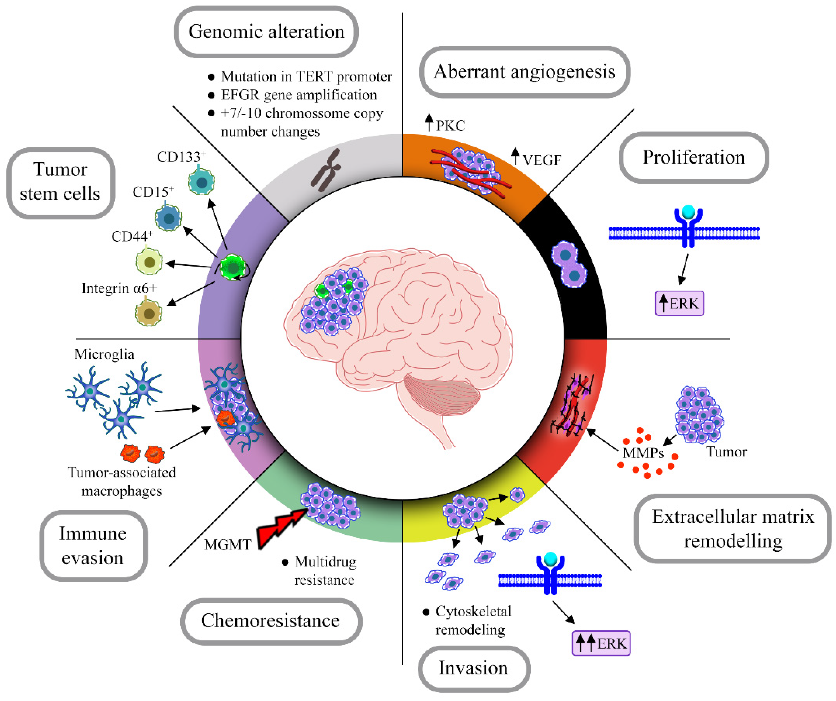
Glioblastomas are considered the most common and aggressive primary brain tumor in adults, with an average of 15 months’ survival rate. The treatment is surgery resection, followed by chemotherapy with temozolomide, and/or radiotherapy. Glioblastoma must have wild-type IDH gene and some characteristics, such as TERT promoter mutation, EGFR gene amplification, microvascular proliferation, among others. Glioblastomas have great heterogeneity at cellular and molecular levels, presenting distinct phenotypes and diversified molecular signatures in each tumor mass, making it difficult to define a specific therapeutic target. It is believed that the main responsibility for the emerge of these distinct patterns lies in subcellular populations of tumor stem cells, capable of tumor initiation and asymmetric division. 

  • glioblastoma
  • brain tumor
  • cancer stem cells
  • molecular oncology
  • chemoresistance

1. Introduction

1.1. Overview of Glioblastoma Classification, Mutations and Treatment

More than 120 types of central nervous system tumors have been described. In spite of the high variability, these tumors do not always represent a diagnosis of malignancy as only 32% of them are malignant. Among brain tumors, there are those originating from glial cells, known as gliomas. Glioblastoma (GBM), a high-grade subtype of glioma, presents astrocytic differentiation and stands out as the most common and aggressive primary brain tumor in adults, representing more than 50% of all gliomas. According to the World Health Organization (WHO), there is no prevention protocol for GBMs, and most patients only become aware of the tumor with the onset of the first symptoms, such as cognitive impairments, neurological deficits and headaches [1]. These symptoms often emerge when the tumor is already properly installed and cell proliferation has escaped the control steps. Despite treatment, only 5% of patients survive for more than 5 years [2][3].
Historically, histopathological analysis considers GBMs as astrocytomas due to their high morphological similarity to astrocytes. In hematoxylin–eosin (H&E) staining, it is possible to detect polygonal or spindle-shaped cells with irregular borders and an acidophilic fibrillar cytoplasm, nuclear atypia, microvascular proliferation with newly developed vessels, necrotic areas and high mitotic activity. These characteristics constitute their main histopathological features in which pathologists rely for diagnosis [4][5][6][7].
However, new classifications have emerged, especially after technical advances in molecular profiling. The 2021 WHO classification takes histological and molecular characteristics in consideration and divides the diffuse adult gliomas into three subtypes: (1) GBM IDH-wild type, (2) astrocytoma IDH-mutant and (3) oligodendroglioma IDH-mutant 1p19q co-deleted. This new classification differs from the previous 2007 WHO classification because it takes into account analysis of mutations in Isocitrate dehydrogenase 1 (IDH) gene, in Telomerase reverse transcriptase (TERT) promoter and co-deletion of chromosomal regions 1p and 19q [8]. Thus, the 2021 WHO classification states that GBMs must have wild-type IDH gene and at least one of the following characteristics: microvascular proliferation, necrosis, TERT promoter mutation, EGFR gene amplification or combined copy number alteration of chromosomes 7 and 10 [9].
The discovery of IDH mutations configurated a major breakthrough in the glioma field. The genetic status of IDH is currently one of the most important aspects that have to be taken into consideration to evaluate the prognosis and treatment of different subclasses of gliomas [9][10]. In the 2021 WHO classification, GBM is no longer divided into IDH-mutant and IDH-wild type. IDH-mutant tumors are now classified as astrocytomas or oligodendrogliomas depending on other markers and, in general, the presence of IDH-mutant genes confers a better prognosis and an increase in life expectancy for the patient. The importance of IDH and the consequences of having this mutation will be further discussed in a separate topic because it is still an important line of investigation for the discovery of treatment strategies against GBM.
Another frequent mutation in GBM is the one that occurs in the TERT promoter. This gene encodes the subcatalytic unit of telomerase, an enzyme involved in the “reconstruction” of telomeric subunits [11][12]. This is the most common mutation in GBMs and it results in increased expression of telomerase, present in more than 69% of all GBMs. The physiological meaning of this mutation is extremely important due to its impact on cell proliferation. Since telomere attrition is one of the key signals to senescence, the ability to stably elongate these structures ensures cell immortality and tumor maintenance [12][13][14].
The most common chromosomal abnormalities found in GBMs are gain of chromosome 7p (trisomy) and loss of chromosome 10q (monosomy). One of the oncogenes on 7p is Epidermal Growth Factor Receptor (EGFR), which is amplified in about one-third of GBMs, and in 10q there is the inactivation of tumor-suppressor genes in the telomeric region of 10q, especially Phosphatase and Tensin Homolog (PTEN) [15]. The large number of brain tumor subtypes and the multiplicity of mutations do not always reflect in a variety of treatments, which are multimodal, consisting of maximum surgical resection, radiotherapy and chemotherapy. For GBMs, this same treatment is maintained in most patients, using the chemotherapeutic drug temozolomide (TMZ) [16].
TMZ is an alkylating agent, applied in the clinic since 2005 as the gold-standard treatment for most brain tumors, including GBMs. Once inside the cell, the TMZ molecule undergoes conversion to monomethyl-triacene-imidazol-carboxamide (MTIC), which acts on DNA and is based on the alkylation capacity of the O6 and N7 positions of guanine, methylation and, consequently, induction of cell cycle arrest during the G2/M phase, before completing cell replication [17][18].
GBM has some characteristics that increase its malignancy leading to a poor prognosis, such as chromosomal aberrations, chemoresistance, high proliferation and intrinsic and extrinsic heterogeneity, caused mainly by subpopulations of tumor stem cells, in addition to being highly infiltrative and modulating the environment around it, recruiting immune cells and modifying the extracellular matrix (Figure 1).
Figure 1. Hallmarks of GBM. GBM has some striking features that contribute to its aggressive phenotype and that could be therapeutic targets. Infiltrating cells, genomic alterations, aberrant angiogenesis, immune evasion and modulation of the extratumoral environment are some of the characteristics present in the vast majority of these tumors.
Several compounds are being tested to treat GBM, mostly natural compounds, which have been successful in in vitro approaches. Conjugated treatments with TMZ and new compounds have also been explored, with promising results, since TMZ was approved for clinical use in 2005 and there is no other chemotherapeutic agent for GBM therapy [19].
The gold-standard treatment for GBM only provides an average of 15 months’ survival for patients. Several other therapies, including those acting on GBM cytoskeletal proteins, GBM stem cells and IDH mutations, together with other strategies such as those to overcome the blood–brain barrier or to use oncolytic virus have been widely tested.

1.2. GBM Stem Cell Population: Implications in Tumor Resistance and Recurrence

GBMs are tumors that show great heterogeneity at cellular and molecular levels. They present a very complex and diversified molecular signature among different patients and also an intrinsic cellular heterogeneity of each tumor mass, formed by cell niches with distinct phenotypic characteristics. It is believed that the main responsibity for the emergence of these niches belongs to subpopulations of tumor stem cells (TSC), also called tumor initiating cells [20]. They have been classified as a tumor cell subpopulation capable of tumor initiation and asymmetric division. Like normal stem cells, TSCs have the capacity of self-renewal, to originate several phenotypically different cell types and to establish contact with the microenvironment, in addition to having specific stem cell markers. GBM initiating cells are also capable of forming oncospheres from a single CD133+ cell and generate a tumor when at least 100 cells are transplanted into the brain of immunodeficient mice, which does not occur with CD133- cells [21][22][23]. It has been shown that these cells are resistant to several chemotherapeutic drugs such as TMZ, carboplatin, paclitaxel (Taxol) and etoposide (VP16), as well as radiotherapy [24][25]. The wide variety found among GBMs from different patients in relation to the percentage of tumor stem cells is remarkable. Some articles found that samples of the primary GBM tumor mass obtained from three different patients had between 10 and 69.7% of CD133+ cells. These cells express 32 to 56 times more of the MGMT protein responsible for DNA repair and escape from TMZ treatment [25]. Furthermore, anti-apoptotic genes, including FLIP, BCL-2 and BCL-XL, were also found at higher levels in CD133+ cells than in CD133- cells from the same tumor mass [25].
Due to the high cellular heterogeneity of GBM, 40% of primary GBM samples do not have CD133+ cells, however, they have cells within the tumor mass with characteristics of stem cells. Thus, Son and colleagues screened neural stem cell markers in 24 GBM samples and identified CD15 as a marker for another GBM stem cell lineage that is CD133- [26].
The microenvironment of GBM stem cell populations plays a crucial role in tumor maintenance. A subpopulation of GBM stem cells, preferentially located in perivascular regions, expresses high levels of CD44 and ID1 [27]. Another highly expressed marker found in such subpopulations close to perivascular regions is the α6 integrin. This marker may co-localize with CD133, however, even when cells are negative for CD133, those cells still have a high capacity to generate tumors. Knockdown of α6 integrin with shRNA inhibits cell growth, induces apoptosis in culture and inhibits tumor formation in vivo [28].
Assuming that neural stem cells lose their self-renewal capacity when differentiated into neuronal-like or glial-like phenotypes, one of the strategies to limit tumor growth is to induce tumor stem cells to a postmitotic mature cell fate. Bone morphogenetic protein (BMP) is a member of the TGF-β superfamily capable of inducing differentiation of GBM stem cells into astrocytes [29][30]; however, astrocytes are able to re-enter in the cell cycle, again inducing tumorigenesis. Achaete-scute homolog 1 (ASCL1) is a growth factor that participates in the neural differentiation process during embryonic development. A subpopulation of GBM tumor mass cells expressing high levels of ASCL1 has been found [31]. Patients with high levels of ASCL1 had longer survival than patients with low expression of ASCL1. ASCL1 is able to induce GBM stem cell differentiation, decrease proliferation rate, increase neurospheres formation in vitro, and tumor mass growth in vivo is inhibited when it is overexpressed [31].
Given the high rates of tumor recurrence and low patient survival with conventional treatment, new therapeutic approaches targeting TSCs are being proposed and developed. The aim of new therapeutic approaches is to combine classic with new drugs that target TSCs [32]. Thus, its recurrence with a more malignant phenotype in patients is avoided, increasing their survival.

2. IDH Mutation and Resistance Outcome

Parsons and colleagues were the first to identify IDH genetic alteration in GBM specifically [33]. A study of genome-wide exon sequencing was performed in 22 GBM samples from different patients, and one of the biggest surprises was the discovery of this novel mutation, which resulted in the replacement of an arginine 132 by one histidine in amino acid 132 of IDH1 (R132H IDH1 mutation). R132H IDH1 was present in 12% of the GBM samples, suggesting an important role in glioma transformation process [33].
The IDH1 gene is located at chromosome 2q33 and encodes IDH1 protein which resides in the cytoplasm [34]. IDH1 catalyzes the oxidative carboxylation of isocitrate to α-ketoglutarate (α-KG), which is noteworthy involved in the activity of tricarboxylic acid cycle (also known as Krebs cycle or citric acid cycle) [35]. Further works showed that the R132H IDH1 mutation was also a frequent feature of acute myeloid leukemia (AML), encountered in 16% of the studied samples [36].
A mutant IDH2 gene (IDH2 R172K or R172M) has additionally been identified in glioma subsets, although less frequently than IDH1 mutations [37]. IDH2 is analogous to IDH1. Like IDH1, IDH2 catalyzes the formation of α-KG from isocitrate but is localized in the mitochondria. A spectrum of mutations is observed at IDH1 and IDH2 in cancers. For instance, common IDH1 mutations include R132H and R132C, and common IDH2 mutations include R172K and R172M [38].
Almost all reported cases of gliomas bearing IDH mutations have been heterozygous, and inactivating alterations such as deletions or nonsense mutations were not observed for this gene in any cancer. The evidence of a possible genetic selection led to the conclusion that the IDH mutations confer the enzyme with an oncogenic gain of function [39].
Zhao and colleagues reported a dominant-negative role for mutant IDH1 R132H, the function of the wild-type enzyme being inhibited in vitro in presence of the mutant enzyme [39]. They additionally showed that IDH1/2 wild-type isoforms prevented the accumulation and overexpression of the hypoxia-inducible factor-1α (HIF-1α), a transcription factor associated with aggressive types of cancer [39]. These results led the authors to suggest a tumor suppression function for IDH1/2. In parallel studies, Yan and colleagues showed that IDH1/2 mutations reduced the enzyme’s ability to generate α-KG [37]. Dang and colleagues provided the missing piece of the puzzle, showing that IDH1/2 mutants gain the neomorphic enzymatic activity to reduce α-KG to R(–)-2-hydroxyglutarate (2HG) [40]. Consistent with this finding, measurement of 2HG in IDH1/2 mutated gliomas or leukemia samples revealed 2HG levels higher than in neoplasms bearing wild-type IDH1/2 [40][41]. The fact that tumors with IDH1/2 mutations overexpressed HIF-1α when compared to wild-type IDH1/2 tumors suggested that 2HG could act as an oncometabolite, promoting malignant transformation.
The IDH1/2 mutations are a dividing point to categorize gliomas. Virtually all tumors presenting wild-type copies are classified as GBMs. On the other hand, gliomas harboring IDH mutations can be subdivided into two subclasses: those presenting chromosome 1p/19q co-deletion, referred historically as oligodendrogliomas; and those without 1p/19q co-deletion, representing astrocytomas [42]. The presence or absence of the co-deletion determines the clinical evolution whereas around 94% of IDH-mutant non-1p/19q co-deleted gliomas present TP53 mutations and 86% present inactivating ATRX mutations [42]. These genetic alterations, highly frequent in such tumors, strongly suggest that IDH mutation is likely to be at the origin of such malignancies and could give rise to tumors of both lineages astrocytic and oligodendrocytic. Clinically, IDH mutation is a favorable prognostic factor when compared to wild-type gliomas, as patients respond better to chemotherapy as explained next [37].
The wild-type IDH1/2 catalyzes the conversion of isocitrate to α-KG and, at the same time, the reduction of NADP+ to NADPH with the production of CO2. They can promote the formation of hydrogen bonds with the β-carboxyl of isocitrate. Mutations replacing the R132 of IDH1, as well as the R140 and R172 of IDH2, confer to the enzymes the ability to convert α-KG into 2HG concomitantly with the oxidation of NADPH to NAD+. 2HG and α-KG share a very similar chemical structure, but differ only in the presence of a hydroxyl group in 2HG at the same carbon that establishes a bound with an oxygen atom in the α-KG molecule [40].
The discovery of these intriguing data involving the neomorphic activity of IDH1/2 raised the question of the mechanisms by which enhanced 2HG levels could lead to the accumulation of HIF-1α. Several studies from different laboratories have shown that 2HG impairs the catalytic function of α-KG-dependent enzymes. It acts as a competitor of α-KG, occupying its binding site in these enzymes, a competition authorized by the structural similarity of 2HG to α-KG [43][44]. Interestingly, prolyl hydroxylases (PDH) are among the enzymes susceptible to have their activity modified by 2HG. Those enzymes are responsible for targeting HIF-1α to degradation [45], an activity consistent with the high levels of HIF-1α observed in IDH mutated tumors [39]. PHD inhibition by 2HG remains however debated. Koivunen and colleagues have indeed provided convincing data showing decreased HIF-1α levels in cells treated with 2HG [46]. PHDs are not the only enzymes that can potentially be affected by the overproduction of 2HG. 2HG accumulation due to IDH mutated isoforms has additionally been shown to inhibit the catalytic activity of the ten-eleven translocation (TET) enzyme family. TET family components are key mediators of DNA demethylation and upon its inhibition, 2HG accumulation produces a hypermethylated profile in such cells. [47][48][49]. This finding raises 2HG to a whole new level, because it implies a role for this metabolite in gene expression control, and brings forward a role for 2HG and α-KG-dependent enzymes in epigenetic control of cancer cell behavior.
Similar to 2HG, other metabolites have been shown as potent modulators of epigenetic-related enzymes, especially those that depend on α-KG. Recently, the metabolite gamma-hydroxybutyrate (GHB) was demonstrated as an inhibitor of TET2 enzymes in GBM stem-like cells (GSC) [50]. The authors have shown that more differentiated glioma cells decrease the expression of succinic semi-aldehyde dehydrogenase (SSADH), an enzyme involved in GABA metabolism [51]. Upon this downregulation, GHB is overproduced and acts as a competitor to α-KG resulting in a hypermethylation profile and a decrease in proliferation and tumorigenic properties of GSC [50].
Elucidating the function of 2HG in cancer cells is further complicated by the occurrence of enantiomers. High levels of the right-handed enantiomer of 2HG, D-2HG, have been reported to cause oxidative stress in rat brains [52], an event that could potentially promote oncogenesis. In the human clinics, high levels of 2HG have been linked to a rare metabolic disorder known as D-2-hydroxyglutaric aciduria [53][54], resulting in most cases from a mutation in D-2-hydroxyglutarate dehydrogenase. Although these patients showed significantly higher levels of D-2HG compared to leukemia or glioma patients harboring IDH1/2 mutations, they do not have a predisposition to developing gliomas, leukemia or other malignancies [55]. Moreover, patients bearing gliomas with wild-type isoforms of IDH1 present a poorer prognosis when compared to patients with IDH1 mutated tumors [33]. Even with the demonstration that an accumulation of D-2HG, in the case of the D-2-hydroxyglutaric aciduria, does not predispose individuals to develop tumors and those patients’ bearing gliomas with IDH1 mutations have better survival expectancies than those bearing the wild-type ones, the overproduction of 2HG in gliomas, driven by the IDH mutations, has been used as an argument to explain the genesis of glioma.
As a byproduct derived from the TCA cycle, 2HG is among the metabolites that have been shown to be important in the progression of gliomas. Notably, they change glioma physiology by interfering in the functions of αKG-dependent enzymes, impairing epigenetic modifications. The discovery of the influence of 2HG and other metabolites in tumor behavior, together with data acquired in genetics, brought back the cancer metabolism as one of the major effectors in tumorigenesis, rendering important the study of the reactions involved in the cellular reprogramming of energy metabolism pathways.
The discovery of mutations in IDH1 and 2 revolutionized the treatment of malignancies bearing such genetic alterations. They have been observed in multiple types of cancer such as gliomas, AML, cholangiosarcomas, myelodysplastic syndromes and chondrosarcoma. The FDA-approved inhibitory drugs used in trials in the last few years are Ivosidenib [56] for IDH1 mutations and Enasidenib in the case of IDH2 mutations [57]. Despite the relative success of these small drugs in the treatment of AML, they have failed to treat gliomas and other types of IDH mutated cancers. New drugs are in development, such as Vorasidenib, a dual inhibitor of mutant IDH1 and 2. Results of a phase I clinical trial have shown a favorable safety profile for treating gliomas and tumor shrinkage in patients with non-enhancing gliomas [58]. These data are promising and perhaps IDH inhibitors can become a pathway to treat such disease. Beyond small molecule inhibitors, recently, it was reported the development of a new vaccine against gliomas carrying IDH1 mutation. Platten and colleagues have demonstrated that a vaccine against the IDH R132H isoform was able to induce immune response in 93% of IDH mutant glioma cases. Moreover, they reported high three-year progression-free and death-free rates [59].
Accumulation of evidence regarding the metabolic and physiologic consequences of IDH mutations has enabled scientists to advance in new therapeutic avenues, but it is still necessary to understand how they are acquired and what are the selective advantages that such mutation brings to cells.

3. The Cytoskeleton as a Potential Therapeutic Target against Glioblastoma

Another broad group of therapeutic targets against GBM is cytoskeletal proteins. The cytoskeleton is composed by three main components: microfilaments, intermediate filaments, and microtubules, together with their accessory proteins. Its major functions are to sustain the shape of cells and also to allow cells to resist against mechanical deformations. The cytoskeletal proteins span throughout the cytoplasm, cell nucleus and plasma membrane, connecting and integrating intracellular structures with the extracellular environment. As it is involved in several cellular processes, such as transport of molecules, cell migration, differentiation and proliferation, abnormalities in its structure and function are often the cause of many diseases [60].
The highly infiltrative and recurrence capacities of GBM are strongly associated with aberrant expression of several cytoskeletal proteins [61][62]. Thus, agents that act in the cytoskeleton have been used against GBM due to their capacity to interfere with cell proliferation [63].

3.1. Microtubules as Therapeutic Targets against Glioblastoma

Microtubules are essential cytoskeletal elements in mitosis and cell division, therefore, are one of the best and most important targets in several cancer therapies. Drugs that target microtubules are mainly divided into microtubule destabilizing and stabilizing drugs [64].
Among microtubule stabilizing drugs, Paclitaxel (also known as Taxol) and its different derivatives has been tested for GBM therapy. The soluble form of the drug was extensively tested against GBM, but it turned out to be unable to properly penetrate the blood–brain barrier [65]. Thus, improvements have been developed such as other forms associated with copolymers made of PLGA (poly (d,l lactide-co-glycolide)) and PEG (polyethylene glycol) [66], conjugated with other molecules, like poly-L-glutamic acid [67][68] or the peptide Angiopep-2 [69]; however, the clinical trials using these Taxol variations have been terminated or have yet to yield conclusive results.
Other taxane analogue molecules such as Ortataxelis [70], Cabazitaxel [71] and TPI-287 [72] have been tested. While the first two drugs did not show significant effects on patients’ survival, the third drug was well-tolerated and three patients out of seven had partial response. Epotilones is another class of tubulin stabilizing drugs that have also been used against GBM. Patupiloneis [73], Sagopilone [74] and Ixabepilone [75] have been tested in phase I/II clinical trials but there were mild or no significant effects on patients’ survival.
Like microtubule stabilizing drugs, several microtubule destabilizing drugs are being explored in GBM treatment. Vincristine has been used in cancer treatment together with procarbazine and lomustine forming what is called the PCV treatment. However, in a large clinical trial, 447 patients with recurrent glioma were treated with PCV but no significant effects in patients’ survival were observed [76]. Other drugs, such as Verubulin [77], Batabulin [78] and 2-methoxyestradiol [79], also had little or no efficacy in patients’ survival. On the other hand, Lexibulin (CYT997) [80], Lisavanbulin (BAL101553) [81] and Mebendazole [82] have shown promising results in vitro and in mouse models but no results from clinical trials have been released yet.

3.2. Intermediate Filaments as Therapeutic Targets against Glioblastoma

Apart from microtubules, another therapeutic target against GBM could be the intermediate filaments. Indeed, effects on vimentin have been studied. Withaferin-A, a steroidal lactone compound isolated from Withania somnifera, is an inhibitor of a variety of proteins, but its most studied target is vimentin [63]. In vitro studies showed that Withaferin-A interferes with the migratory capacity of U251 and U87 GBM cell lines [83]; however, other basic studies are still needed before starting clinical trials.
Pritumumab, an anti-vimentin antibody, shows specificity for GBM but does not show specificity for normal adult cells (neurons, astrocytes or other cells) [84]. This is due to the fact that Pritumumab binds to cell surface vimentin, only expressed in glioma cells. This antibody has been tested in clinical trials to treat patients diagnosed with GBM, astrocytoma or neuroblastoma [84]. In general, Pritumumab showed a 5% increase in patients’ survival. Thus, administration of higher doses of Pritumumab has the potential to improve the life expectancy in GBM patients, although side-effects may appear and are to be determined using other clinical trials.

3.3. Other Alternative Cytoskeletal Therapeutic Targets against Glioblastoma

A new mode of GBM therapy is the application of low-intensity (1–3 V/cm) and alternating electric field of the order of 100–300 kHz applied via cutaneous arrays to provide optimal tumor site coverage [85].
Although the tumor treating fields (TTF) therapy was demonstrated to inhibit cancer proliferation by interfering with the mitotic spindle, it is now clear that TTF act in several other biological processes, including DNA repair, permeability of cell membranes, and intracellular molecules known as dipoles. Specifically, for GBM, TTF is delivered at an optimal frequency of 200 kHz and intensity of 1–2 V/cm [86]. Indeed, a clinical trial using this therapy against GBM was the first, after the introduction of temozolomide chemotherapy [19], to show an increase in patients’ survival [87][88] without any strong systemic adverse events [89].
The main cellular and molecular mechanism through which TTF therapy acts is during mitosis, particularly in the mitotic spindle. When TTFs are applied, tubulin proteins tend to align with the electric field, thus interfering with the microtubule polymerization/depolymerization, resulting in spindle malformation and cell cycle arrest. Moreover, failure of the spindle assembly checkpoint [90] leads to incorrect chromosome segregation and possibly cell death [90]. TTF has also been described as capable of interfering with the septin protein complex, known to participate in the cleavage furrow [91][92], again leading to anomalous chromosomal segregation and possibly structural changes to the membrane of cells [92].
The use of TTF therapy still has some disadvantages such as the difficulty to treat more diffuse tumors located at different sites into the brain and also the high costs of treatment per month per patient [93]. Despite these issues, TTF has become “the fourth cancer treatment modality” against GBM [94].
A new mechanism of intercellular communication known as tunneling nanotubes (TNTs), has been described to play a pivotal role in cell–cell interaction [95][96]. TNTs are thin, open-ended, actin-based membrane nanotubes allowing the exchange of various molecules or even organelles, as bridges between cells [97][98].
TNTs were first identified by Rustom et al. in pheochromocytoma PC12 cells [95]. Since then, a variety of in vitro studies have reported the presence of TNTs in several cell types, including GBM [99][100][101][102]. Moreover, reports have shown that communication between GBM and other central nervous system cells such as astrocytes [102][103] and microglia [100] can occur through TNTs. The transfer of mitochondria appears to modulate GBM drug-resistant state [104]. Indeed, it was recently described the capacity of TNTs to transfer mitochondria between GBM stem cells in organoids [105]. It was also demonstrated that TNTs allow GBM cells to adapt to temozolomide and ionizing radiation treatments through transmission of the MGMT protein from the MGMT mRNA-positive cells to other cells expressing low MGMT levels [106].
Thus, TNTs seem to be very important for GBM progression and treatment resistance. In the next years, a better comprehension of TNT functions may allow the development of new therapies against glioblastoma.

This entry is adapted from the peer-reviewed paper 10.3390/cancers14133203

References

  1. Alexander, B.M.; Cloughesy, T.F. Adult glioblastoma. J. Clin. Oncol. 2017, 35, 2402–2409.
  2. Ohgaki, H.; Kleihues, P. Genetic Pathways to Primary and Secondary Glioblastoma. Am. J. Pathol. 2007, 170, 1445–1453.
  3. Ostrom, Q.; Gittleman, H.; Liao, P.; Rouse, C.; Chen, Y.; Dowling, J.; Wolinsky, Y.; Kruchko, C.; Barnholtz-Sloan, J. CBTRUS Statistical Report: Primary Brain and Central Nervous System Tumors Diagnosed in the United States in 2007–2011. Neuro-Oncology 2014, 16, iv1–iv63.
  4. Kleihues, P.; Louis, D.N.; Scheithauer, B.W.; Rorke, L.B.; Reifenberger, G.; Burger, P.C.; Cavenee, W.K. The WHO Classification of Tumors of the Nervous System. J. Neuropathol. Exp. Neurol. 2002, 61, 215–225.
  5. Brat, D.J.; Van Meir, E.G. Vaso-occlusive and prothrombotic mechanisms associated with tumor hypoxia, necrosis, and accelerated growth in glioblastoma. Lab. Investig. 2004, 84, 397–405.
  6. Schultz, S.; Pinsky, G.S.; Wu, N.C.; Chamberlain, M.C.; Rodrigo, A.S.; Martin, S.E. Fine needle aspiration diagnosis of extracranial glioblastoma multiforme: Case report and review of the literature. CytoJournal 2005, 2, 19.
  7. Linkous, A.G.; Yazlovitskaya, E.M. Angiogenesis in glioblastoma multiforme: Navigating the maze. Anti-Cancer Agents Med. Chem. 2011, 11, 712–718.
  8. Molinaro, A.M.; Taylor, J.W.; Wiencke, J.K.; Wrensch, M.R. Genetic and molecular epidemiology of adult diffuse glioma. Nat. Rev. Neurol. 2019, 15, 405–417.
  9. Louis, D.N.; Perry, A.; Wesseling, P.; Brat, D.J.; Cree, I.A.; Figarella-Branger, D.; Hawkins, C.; Ng, H.K.; Pfister, S.M.; Reifenberger, G.; et al. The 2021 WHO Classification of Tumors of the Central Nervous System: A summary. Neuro-Oncology 2021, 23, 1231–1251.
  10. Louis, D.N.; Perry, A.; Reifenberger, G.; Von Deimling, A.; Figarella-Branger, D.; Cavenee, W.K.; Ohgaki, H.; Wiestler, O.D.; Kleihues, P.; Ellison, D.W. The 2016 World Health Organization Classification of Tumors of the Central Nervous System: A summary. Acta Neuropathol. 2016, 131, 803–820.
  11. Weinrich, S.L.; Pruzan, R.; Ma, L.; Ouellette, M.; Tesmer, V.M.; Holt, S.E.; Bodnar, A.G.; Lichtsteiner, S.; Kim, N.W.; Trager, J.B.; et al. Reconstitution of human telomerase with the template RNA component hTR and the catalytic protein subunit hTRT. Nat. Genet. 1997, 17, 498–502.
  12. Bell, R.J.; Rube, H.T.; Xavier-Magalhães, A.; Costa, B.M.; Mancini, A.; Song, J.S.; Costello, J.F. Understanding TERT Promoter Mutations: A Common Path to Immortality. Mol. Cancer Res. 2016, 14, 315–323.
  13. Killela, P.J.; Reitman, Z.J.; Jiao, Y.; Bettegowda, C.; Agrawal, N.; Diaz, L.A., Jr.; Friedman, A.H.; Friedman, H.; Gallia, G.L.; Giovanella, B.C.; et al. TERT promoter mutations occur frequently in gliomas and a subset of tumors derived from cells with low rates of self-renewal. Proc. Natl. Acad. Sci. USA 2013, 110, 6021–6026.
  14. Yuan, Y.; Qi, C.; Maling, G.; Xiang, W.; Yanhui, L.; Ruofei, L.; Yunhe, M.; Jiewen, L.; Qing, M. TERT mutation in glioma: Frequency, prognosis and risk. J. Clin. Neurosci. 2016, 26, 57–62.
  15. Inda, M.D.M.; Fan, X.; Muñoz, J.; Perot, C.; Fauvet, D.; Danglot, G.; Palacio, A.; Madero, P.; Zazpe, I.; Portillo, E.; et al. Chromosomal abnormalities in human glioblastomas: Gain in chromosome 7p correlating with loss in chromosome 10q. Mol. Carcinog. 2002, 36, 6–14.
  16. Stupp, R.; Hegi, M.E.; Mason, W.P.; van den Bent, M.J.; Taphoorn, M.J.B.; Janzer, R.C.; Ludwin, S.K.; Allgeier, A.; Fisher, B.; Belanger, K.; et al. Effects of radiotherapy with concomitant and adjuvant temozolomide versus radiotherapy alone on survival in glioblastoma in a randomised phase III study: 5-year analysis of the EORTC-NCIC trial. Lancet Oncol. 2009, 10, 459–466.
  17. Wesolowski, J.; Rajdev, P.; Mukherji, S. Temozolomide (Temodar). Am. J. Neuroradiol. 2010, 31, 1383–1384.
  18. Lee, S.Y. Temozolomide resistance in glioblastoma multiforme. Genes Dis. 2016, 3, 198–210.
  19. Cohen, M.H.; Johnson, J.R.; Pazdur, R. Food and Drug Administration Drug Approval Summary: Temozolomide Plus Radiation Therapy for the Treatment of Newly Diagnosed Glioblastoma Multiforme. Clin. Cancer Res. 2005, 11, 6767–6771.
  20. Abou-Antoun, T.; Hale, J.S.; Lathia, J.D.; Dombrowski, S.M. Brain Cancer Stem Cells in Adults and Children: Cell Biology and Therapeutic Implications. Neurotherapeutics 2017, 14, 372–384.
  21. Singh, S.K.; Clarke, I.D.; Terasaki, M.; Bonn, V.E.; Hawkins, C.; Squire, J.; Dirks, P.B. Identification of a cancer stem cell in human brain tumors. Cancer Res. 2003, 63, 5821–5828.
  22. Yuan, X.; Curtin, J.; Xiong, Y.; Liu, G.; Waschsmann-Hogiu, S.; Farkas, D.L.; Black, K.L.; Yu, J.S. Isolation of cancer stem cells from adult glioblastoma multiforme. Oncogene 2004, 23, 9392–9400.
  23. Singh, S.K.; Hawkins, C.; Clarke, I.D.; Squire, J.A.; Bayani, J.; Hide, T.; Henkelman, R.M.; Cusimano, M.D.; Dirks, P.B. Identification of human brain tumour initiating cells. Nature 2004, 432, 396–401.
  24. Bao, S.; Wu, Q.; McLendon, R.E.; Hao, Y.; Shi, Q.; Hjelmeland, A.B.; Dewhirst, M.W.; Bigner, D.D.; Rich, J.N. Glioma stem cells promote radioresistance by preferential activation of the DNA damage response. Nature 2006, 444, 756–760.
  25. Liu, G.; Yuan, X.; Zeng, Z.; Tunici, P.; Ng, H.; Abdulkadir, I.R.; Lu, L.; Irvin, D.; Black, K.L.; Yu, J.S. Analysis of gene expression and chemoresistance of CD133+ cancer stem cells in glioblastoma. Mol. Cancer 2006, 5, 67.
  26. Son, M.J.; Woolard, K.; Nam, D.-H.; Lee, J.; Fine, H.A. SSEA-1 Is an Enrichment Marker for Tumor-Initiating Cells in Human Glioblastoma. Cell Stem Cell 2009, 4, 440–452.
  27. Anido, J.; Sáez-Borderías, A.; Gonzàlez-Juncà, A.; Rodón, L.; Folch, G.; Carmona, M.A.; Prieto-Sánchez, R.M.; Barba, I.; Martinez-Saez, E.; Prudkin, L.; et al. TGF-β Receptor Inhibitors Target the CD44high/Id1high Glioma-Initiating Cell Population in Human Glioblastoma. Cancer Cell 2010, 18, 655–668.
  28. Lathia, J.D.; Gallagher, J.; Heddleston, J.M.; Wang, J.; Eyler, C.E.; MacSwords, J.; Wu, Q.; Vasanji, A.; McLendon, R.E.; Hjelmeland, A.B.; et al. Integrin Alpha 6 Regulates Glioblastoma Stem Cells. Cell Stem Cell 2010, 6, 421–432.
  29. Lee, J.; Son, M.J.; Woolard, K.; Donin, N.M.; Li, A.; Cheng, C.H.; Kotliarova, S.; Kotliarov, Y.; Walling, J.; Ahn, S.; et al. Epigenetic-Mediated Dysfunction of the Bone Morphogenetic Protein Pathway Inhibits Differentiation of Glioblastoma-Initiating Cells. Cancer Cell 2008, 13, 69–80.
  30. Piccirillo, S.G.M.; Reynolds, B.A.; Zanetti, N.; Lamorte, G.; Binda, E.; Broggi, G.; Brem, H.; Olivi, A.; DiMeco, F.; Vescovi, A.L. Bone morphogenetic proteins inhibit the tumorigenic potential of human brain tumour-initiating cells. Nature 2006, 444, 761–765.
  31. Park, N.I.; Guilhamon, P.; Desai, K.; McAdam, R.F.; Langille, E.; O’Connor, M.; Lan, X.; Whetstone, H.; Coutinho, F.J.; Vanner, R.J.; et al. ASCL1 Reorganizes Chromatin to Direct Neuronal Fate and Suppress Tumorigenicity of Glioblastoma Stem Cells. Cell Stem Cell 2017, 21, 411.
  32. Sundar, S.J.; Hsieh, J.K.; Manjila, S.; Lathia, J.D.; Sloan, A. The role of cancer stem cells in glioblastoma. Neurosurg. Focus 2014, 37, E6.
  33. Parsons, D.W.; Jones, S.; Zhang, X.; Lin, J.C.-H.; Leary, R.J.; Angenendt, P.; Mankoo, P.; Carter, H.; Siu, I.-M.; Gallia, G.L.; et al. An Integrated Genomic Analysis of Human Glioblastoma Multiforme. Science 2008, 321, 1807–1812.
  34. Narahara, K.; Kimura, S.; Kikkawa, K.; Takahashi, Y.; Wakita, Y.; Kasai, R.; Nagai, S.; Nishibayashi, Y.; Kimoto, H. Probable assignment of soluble isocitrate dehydrogenase (IDH1) to 2q33.3. Qual. Life Res. 1985, 71, 37–40.
  35. Geisbrecht, B.V.; Gould, S.J. The Human PICD Gene Encodes a Cytoplasmic and Peroxisomal NADP+-dependent Isocitrate Dehydrogenase. J. Biol. Chem. 1999, 274, 30527–30533.
  36. Mardis, E.R.; Ding, L.; Dooling, D.J.; Larson, D.E.; McLellan, M.D.; Chen, K.; Koboldt, D.C.; Fulton, R.S.; Delehaunty, K.D.; McGrath, S.D.; et al. Recurring Mutations Found by Sequencing an Acute Myeloid Leukemia Genome. N. Engl. J. Med. 2009, 361, 1058–1066.
  37. Yan, H.; Parsons, D.W.; Jin, G.; McLendon, R.; Rasheed, B.A.; Yuan, W.; Kos, I.; Batinic-Haberle, I.; Jones, S.; Riggins, G.J.; et al. IDH1 and IDH2 Mutations in Gliomas. N. Engl. J. Med. 2009, 360, 765–773.
  38. Reitman, Z.J.; Parsons, D.W.; Yan, H. IDH1 and IDH2: Not Your Typical Oncogenes. Cancer Cell 2010, 17, 215–216.
  39. Zhao, S.; Lin, Y.; Xu, W.; Jiang, W.; Zha, Z.; Wang, P.; Yu, W.; Li, Z.; Gong, L.; Peng, Y.; et al. Glioma-Derived Mutations in IDH1 Dominantly Inhibit IDH1 Catalytic Activity and Induce HIF-1α. Science 2009, 324, 261–265.
  40. Dang, L.; White, D.W.; Gross, S.; Bennett, B.D.; Bittinger, M.A.; Driggers, E.M.; Fantin, V.R.; Jang, H.G.; Jin, S.; Keenan, M.C.; et al. Cancer-associated IDH1 mutations produce 2-hydroxyglutarate. Nature 2009, 462, 739–744.
  41. Ward, P.S.; Patel, J.; Wise, D.R.; Abdel-Wahab, O.; Bennett, B.D.; Coller, H.A.; Cross, J.R.; Fantin, V.R.; Hedvat, C.V.; Perl, A.E.; et al. The Common Feature of Leukemia-Associated IDH1 and IDH2 Mutations Is a Neomorphic Enzyme Activity Converting α-Ketoglutarate to 2-Hydroxyglutarate. Cancer Cell 2010, 17, 225–234.
  42. Cancer Genome Atlas Research Network; Brat, D.J.; Verhaak, R.G.; Aldape, K.D.; Yung, W.K.; Salama, S.R.; Cooper, L.A.; Rheinbay, E.; Miller, C.R.; Vitucci, M.; et al. Comprehensive, Integrative Genomic Analysis of Diffuse Lower-Grade Gliomas. N. Engl. J. Med. 2015, 372, 2481–2498.
  43. Xu, W.; Yang, H.; Liu, Y.; Yang, Y.; Wang, P.; Kim, S.-H.; Ito, S.; Yang, C.; Wang, P.; Xiao, M.-T.; et al. Oncometabolite 2-Hydroxyglutarate Is a Competitive Inhibitor of α-Ketoglutarate-Dependent Dioxygenases. Cancer Cell 2011, 19, 17–30.
  44. Chowdhury, R.; Yeoh, K.K.; Tian, Y.-M.; Hillringhaus, L.; Bagg, E.A.; Rose, N.R.; Leung, I.K.H.; Li, X.S.; Woon, E.C.Y.; Yang, M.; et al. The oncometabolite 2-hydroxyglutarate inhibits histone lysine demethylases. EMBO Rep. 2011, 12, 463–469.
  45. Mole, D.R.; Schlemminger, I.; McNeill, L.A.; Hewitson, K.S.; Pugh, C.W.; Ratcliffe, P.J.; Schofield, C.J. 2-Oxoglutarate analogue inhibitors of hif prolyl hydroxylase. Bioorg. Med. Chem. Lett. 2003, 13, 2677–2680.
  46. Koivunen, P.; Lee, S.; Duncan, C.; Lopez, G.; Lu, G.; Ramkissoon, S.; Losman, J.A.; Joensuu, P.; Bergmann, U.; Gross, S.; et al. Transformation by the (R)-enantiomer of 2-hydroxyglutarate linked to EGLN activation. Nature 2012, 483, 484–488.
  47. Williams, K.; Christensen, J.; Helin, K. DNA methylation: TET proteins—Guardians of CpG islands? EMBO Rep. 2011, 13, 28–35.
  48. Figueroa, M.E.; Abdel-Wahab, O.; Lu, C.; Ward, P.S.; Patel, J.; Shih, A.; Li, Y.; Bhagwat, N.; VasanthaKumar, A.; Fernandez, H.F.; et al. Leukemic IDH1 and IDH2 Mutations Result in a Hypermethylation Phenotype, Disrupt TET2 Function, and Impair Hematopoietic Differentiation. Cancer Cell 2010, 18, 553–567.
  49. Turcan, S.; Rohle, D.; Goenka, A.; Walsh, L.; Fang, F.; Yilmaz, E.; Campos, C.; Fabius, A.W.M.; Lu, C.; Ward, P.; et al. IDH1 mutation is sufficient to establish the glioma hypermethylator phenotype. Nature 2012, 483, 479–483.
  50. El-Habr, E.; Dubois, L.G.F.; Burel-Vandenbos, F.; Bogeas, A.; Lipecka, J.; Turchi, L.; Lejeune, F.-X.; Coehlo, P.L.C.; Yamaki, T.; Wittmann, B.M.; et al. A driver role for GABA metabolism in controlling stem and proliferative cell state through GHB production in glioma. Acta Neuropathol. 2016, 133, 645–660.
  51. Didiasova, M.; Banning, A.; Brennenstuhl, H.; Jung-Klawitter, S.; Cinquemani, C.; Opladen, T.; Tikkanen, R. Succinic Semialdehyde Dehydrogenase Deficiency: An Update. Cells 2020, 9, 477.
  52. Latini, A.; Scussiato, K.; Rosa, R.B.; Llesuy, S.; Belló-Klein, A.; Dutra-Filho, C.S.; Wajner, M. D-2-hydroxyglutaric acid induces oxidative stress in cerebral cortex of young rats. Eur. J. Neurosci. 2003, 17, 2017–2022.
  53. Struys, E.A.; Korman, S.H.; Salomons, G.S.; Darmin, P.S.; Achouri, Y.; Van Schaftingen, E.; Verhoeven, N.M.; Jakobs, C. Mutations in phenotypically mildD-2-hydroxyglutaric aciduria. Ann. Neurol. 2005, 58, 626–630.
  54. Struys, E.A.; Salomons, G.S.; Achouri, Y.; Van Schaftingen, E.; Grosso, S.; Craigen, W.J.; Verhoeven, N.M.; Jakobs, C. Mutations in the d-2-Hydroxyglutarate Dehydrogenase Gene Cause d-2-Hydroxyglutaric Aciduria. Am. J. Hum. Genet. 2005, 76, 358–360.
  55. Kranendijk, M.; Struys, E.A.; van Schaftingen, E.; Gibson, K.M.; Kanhai, W.A.; van der Knaap, M.S.; Amiel, J.; Buist, N.R.; Das, A.M.; de Klerk, J.B.; et al. IDH2 Mutations in Patients with d -2-Hydroxyglutaric Aciduria. Science 2010, 330, 336.
  56. Dhillon, S. Ivosidenib: First Global Approval. Drugs 2018, 78, 1509–1516.
  57. Kim, E.S. Enasidenib: First Global Approval. Drugs 2017, 77, 1705–1711.
  58. Mellinghoff, I.K.; Penas-Prado, M.; Peters, K.B.; Burris, H.A.; Maher, E.A.; Janku, F.; Cote, G.M.; de la Fuente, M.I.; Clarke, J.L.; Ellingson, B.M.; et al. Vorasidenib, a Dual Inhibitor of Mutant IDH1/2, in Recurrent or Progressive Glioma; Results of a First-in-Human Phase I Trial. Clin. Cancer Res. 2021, 27, 4491–4499.
  59. Platten, M.; Bunse, L.; Wick, A.; Bunse, T.; Le Cornet, L.; Harting, I.; Sahm, F.; Sanghvi, K.; Tan, C.L.; Poschke, I.; et al. A vaccine targeting mutant IDH1 in newly diagnosed glioma. Nature 2021, 592, 463–468.
  60. Ramaekers, F.C.; Bosman, F.T. The cytoskeleton and disease. J. Pathol. 2004, 204, 351–354.
  61. Skalli, O.; Wilhelmsson, U.; Örndahl, C.; Fekete, B.; Malmgren, K.; Rydenhag, B.; Pekny, M. Astrocytoma grade IV (glioblastoma multiforme) displays 3 subtypes with unique expression profiles of intermediate filament proteins. Hum. Pathol. 2013, 44, 2081–2088.
  62. Katsetos, C.D.; Reginato, M.J.; Baas, P.W.; D’Agostino, L.; Legido, A.; Tuszyn’ski, J.A.; Dráberová, E.; Dráber, P. Emerging Microtubule Targets in Glioma Therapy. Semin. Pediatr. Neurol. 2015, 22, 49–72.
  63. Trendowski, M. Exploiting the cytoskeletal filaments of neoplastic cells to potentiate a novel therapeutic approach. Biochim. Biophys. Acta 2014, 1846, 599–616.
  64. Dumontet, C.; Jordan, M.A. Microtubule-binding agents: A dynamic field of cancer therapeutics. Nat. Rev. Drug Discov. 2010, 9, 790–803.
  65. Fellner, S.; Bauer, B.; Miller, D.S.; Schaffrik, M.; Fankhänel, M.; Spruß, T.; Bernhardt, G.; Graeff, C.; Färber, L.; Gschaidmeier, H.; et al. Transport of paclitaxel (Taxol) across the blood-brain barrier in vitro and in vivo. J. Clin. Investig. 2002, 110, 1309–1318.
  66. Vellimana, A.K.; Recinos, V.R.; Hwang, L.; Fowers, K.D.; Li, K.W.; Zhang, Y.; Okonma, S.; Eberhart, C.G.; Brem, H.; Tyler, B.M. Combination of paclitaxel thermal gel depot with temozolomide and radiotherapy significantly prolongs survival in an experimental rodent glioma model. J. Neuro-Oncol. 2012, 111, 229–236.
  67. Elinzano, H.; Glantz, M.; Mrugala, M.; Kesari, S.; Piccioni, D.E.; Kim, L.; Pan, E.; Yunus, S.; Coyle, T.; Timothy, K.; et al. PPX and Concurrent Radiation for Newly Diagnosed Glioblastoma Without MGMT Methylation. Am. J. Clin. Oncol. 2018, 41, 159–162.
  68. Jeyapalan, S.; Boxerman, J.; Donahue, J.; Goldman, M.; Kinsella, T.; Dipetrillo, T.; Evans, D.; Elinzano, H.; Constantinou, M.; Stopa, E.; et al. Paclitaxel Poliglumex, Temozolomide, and Radiation for Newly Diagnosed High-grade Glioma. Am. J. Clin. Oncol. 2014, 37, 444–449.
  69. Régina, A.; Demeule, M.; Ché, C.; Lavallée, I.; Poirier, J.; Gabathuler, R.; Béliveau, R.; Castaigne, J.-P. Antitumour activity of ANG1005, a conjugate between paclitaxel and the new brain delivery vector Angiopep-2. J. Cereb. Blood Flow Metab. 2008, 155, 185–197.
  70. Silvani, A.; De Simone, I.; Fregoni, V.; Biagioli, E.; Marchioni, E.; Caroli, M.; Salmaggi, A.; Pace, A.; Torri, V.; Gaviani, P.; et al. Multicenter, single arm, phase II trial on the efficacy of ortataxel in recurrent glioblastoma. J. Neuro-Oncol. 2019, 142, 455–462.
  71. Manley, P.E.; Trippett, T.; Smith, A.A.; Macy, M.E.; Leary, S.E.; Boklan, J.; Cohen, K.J.; Goldman, S.; Kilburn, L.B.; Dhall, G.; et al. A phase 1/2 dose-finding, safety, and activity study of cabazitaxel in pediatric patients with refractory solid tumors including tumors of the central nervous system. Pediatr. Blood Cancer 2018, 65, e27217.
  72. Goldlust, S.; Nabors, L.B.; Duic, J.P.; Conrad, C.; Silberman, S.; Singer, S.; Farmer, G. At-24phase 1/2 trial of bevacizumab plus tpi 287, a novel brain penetrable anti-microtubule agent, for the treatment of recurrent glioblastoma. Neuro-Oncology 2014, 16, v13–v14.
  73. Oehler, C.; Frei, K.; Rushing, E.J.; McSheehy, P.M.; Weber, D.; Allegrini, P.R.; Weniger, D.; Lütolf, U.M.; Knuth, A.; Yonekawa, Y.; et al. Patupilone (Epothilone B) for Recurrent Glioblastoma: Clinical Outcome and Translational Analysis of a Single-Institution Phase I/II Trial. Oncology 2012, 83, 9152.
  74. Stupp, R.; Tosoni, A.; Bromberg, J.E.C.; Hau, P.; Campone, M.; Gijtenbeek, J.; Frenay, M.; Breimer, L.; Wiesinger, H.; Allgeier, A.; et al. Sagopilone (ZK-EPO, ZK 219477) for recurrent glioblastoma. A phase II multicenter trial by the European Organisation for Research and Treatment of Cancer (EORTC) Brain Tumor Group. Ann. Oncol. 2011, 22, 2144–2149.
  75. Peereboom, D.M.; Supko, J.G.; Carson, K.A.; Batchelor, T.; Phuphanich, S.; Lesser, G.; Mikkelsen, T.; Fisher, J.; Desideri, S.; He, X.; et al. A phase I/II trial and pharmacokinetic study of ixabepilone in adult patients with recurrent high-grade gliomas. J. Neuro-Oncology 2010, 100, 261–268.
  76. Brada, M.; Stenning, S.; Gabe, R.; Thompson, L.C.; Levy, D.; Rampling, R.; Erridge, S.; Saran, F.; Gattamaneni, R.; Hopkins, K.; et al. Temozolomide Versus Procarbazine, Lomustine, and Vincristine in Recurrent High-Grade Glioma. J. Clin. Oncol. 2010, 28, 4601–4608.
  77. Chamberlain, M.C.; Brain Tumor Investigational Consortium (BTIC); Grimm, S.; Phuphanich, S.; Recht, L.; Zhu, J.Z.; Kim, L.; Rosenfeld, S.; Fadul, C.E. A phase 2 trial of verubulin for recurrent glioblastoma: A prospective study by the brain tumor investigational consortium (BTIC). J. Neuro-Oncol. 2014, 118, 335–343.
  78. Kirby, S.; Gertler, S.Z.; Mason, W.; Watling, C.; Forsyth, P.; Aniagolu, J.; Stagg, R.; Wright, M.; Powers, J.; Eisenhauer, E.A. Phase 2 study of T138067-sodium in patients with malignant glioma: Trial of the National Cancer Institute of Canada Clinical Trials Group. Neuro-Oncology 2005, 7, 183–188.
  79. Kirkpatrick, J.; Desjardins, A.; Quinn, J.; Rich, J.; Vredenburgh, J.; Sathornsumetee, S.; Gururangan, S.; Sidor, C.; Friedman, H.; Reardon, D. Phase II open-label, safety, pharmacokinetic and efficacy study of 2-methoxyestradiol nanocrystal colloidal dispersion administered orally to patients with recurrent glioblastoma multiforme. J. Clin. Oncol. 2007, 25, 2065.
  80. Lickliter, J.; Fida, R.; Wheeler, H.; Kichenadasse, G.; Yang, Q.; Ganju, V.; Briggs, P. Carboplatin combined with the vascular-disrupting agent CYT997 for recurrent glioblastoma multiforme. J. Clin. Oncol. 2010, 28, e13591.
  81. Kolb, E.A.; Gorlick, R.; Keir, S.T.; Maris, J.M.; Kang, M.H.; Reynolds, C.P.; Lock, R.B.; Carol, H.; Wu, J.; Kurmasheva, R.T.; et al. Initial testing (stage 1) of BAL101553, a novel tubulin binding agent, by the pediatric preclinical testing program. Pediatr. Blood Cancer 2014, 62, 1106–1109.
  82. Bai, R.-Y.; Staedtke, V.; Aprhys, C.M.; Gallia, G.L.; Riggins, G.J. Antiparasitic mebendazole shows survival benefit in 2 preclinical models of glioblastoma multiforme. Neuro-Oncology 2011, 13, 974–982.
  83. Zhao, J.; Zhang, L.; Dong, X.; Liu, L.; Huo, L.; Chen, H. High Expression of Vimentin is Associated With Progression and a Poor Outcome in Glioblastoma. Appl. Immunohistochem. Mol. Morphol. 2018, 26, 337–344.
  84. Glassy, M.C.; Hagiwara, H. Summary analysis of the pre-clinical and clinical results of brain tumor patients treated with pritumumab. Hum. Antibodies 2009, 18, 127–137.
  85. Gutin, P.H.; Wong, E.T. Noninvasive Application of Alternating Electric Fields in Glioblastoma: A Fourth Cancer Treatment Modality. Am. Soc. Clin. Oncol. Educ. Book 2012, 32, 126–131.
  86. Kirson, E.D.; Gurvich, Z.; Schneiderman, R.; Dekel, E.; Itzhaki, A.; Wasserman, Y.; Schatzberger, R.; Palti, Y. Disruption of Cancer Cell Replication by Alternating Electric Fields. Cancer Res. 2004, 64, 3288–3295.
  87. Kesari, S.; Ram, Z. Tumor-treating fields plus chemotherapy versus chemotherapy alone for glioblastoma at first recurrence: A post hoc analysis of the EF-14 trial. CNS Oncol. 2017, 6, 185–193.
  88. Stupp, R.; Taillibert, S.; Kanner, A.A.; Read, W.; Steinberg, D.M.; Lhermitte, B.; Toms, S.; Idbaih, A.; Ahluwalia, M.S.; Fink, K.; et al. Effect of Tumor-Treating Fields plus Maintenance Temozolomide vs Maintenance Temozolomide Alone on Survival in Patients with Glioblastoma. JAMA J. Am. Med Assoc. 2017, 318, 2306–2316.
  89. Zhu, J.-J.; Demireva, P.; Kanner, A.A.; Pannullo, S.; Mehdorn, M.; Avgeropoulos, N.; Salmaggi, A.; Silvani, A.; Goldlust, S.; David, C.; et al. Health-related quality of life, cognitive screening, and functional status in a randomized phase III trial (EF-14) of tumor treating fields with temozolomide compared to temozolomide alone in newly diagnosed glioblastoma. J. Neuro-Oncol. 2017, 135, 545–552.
  90. Markel, G.; Sapir, Y.; Mandel, I.; Hakim, M.; Shaked, R.; Meilin, E.; McClanahan, T.; Loboda, A.; Hashmueli, S.; Ben Moshe, T. Inhibition of the novel immune checkpoint CEACAM1 to enhance anti-tumor immunological activity. J. Clin. Oncol. 2016, 34, 3044.
  91. Field, C.M.; Coughlin, M.; Doberstein, S.; Marty, T.; Sullivan, W. Characterization of anillin mutants reveals essential roles in septin localization and plasma membrane integrity. Development 2005, 132, 2849–2860.
  92. Spiliotis, E.T.; Kinoshita, M.; Nelson, W.J. A Mitotic Septin Scaffold Required for Mammalian Chromosome Congression and Segregation. Science 2005, 307, 1781–1785.
  93. Mittal, S.; Klinger, N.V.; Michelhaugh, S.K.; Barger, G.R.; Pannullo, S.C.; Juhász, C. Alternating electric tumor treating fields for treatment of glioblastoma: Rationale, preclinical, and clinical studies. J. Neurosurg. 2018, 128, 414–421.
  94. Mehta, M.; Wen, P.; Nishikawa, R.; Reardon, D.; Peters, K. Critical review of the addition of tumor treating fields (TTFields) to the existing standard of care for newly diagnosed glioblastoma patients. Crit. Rev. Oncol. 2017, 111, 60–65.
  95. Rustom, A.; Saffrich, R.; Markovic, I.; Walther, P.; Gerdes, H.-H. Nanotubular Highways for Intercellular Organelle Transport. Science 2004, 303, 1007–1010.
  96. Austefjord, M.W.; Gerdes, H.-H.; Wang, X. Tunneling nanotubes: Diversity in morphology and structure. Commun. Integr. Biol. 2014, 7, e27934.
  97. Abounit, S.; Zurzolo, C. Wiring through tunneling nanotubes—From electrical signals to organelle transfer. J. Cell Sci. 2012, 125, 1089–1098.
  98. Ariazi, J.; Benowitz, A.; De Biasi, V.; Boer, M.D.; Cherqui, S.; Cui, H.; Douillet, N.; Eugenin, E.A.; Favre, D.; Goodman, S.; et al. Tunneling Nanotubes and Gap Junctions–Their Role in Long-Range Intercellular Communication during Development, Health, and Disease Conditions. Front. Mol. Neurosci. 2017, 10, 333.
  99. Pontes, B.; Viana, N.B.; Campanati, L.; Farina, M.; Neto, V.M.; Nussenzveig, H.M. Structure and elastic properties of tunneling nanotubes. Eur. Biophys. J. 2007, 37, 121–129.
  100. Matias, D.; Dubois, L.G.; Pontes, B.; Rosário, L.; Ferrer, V.P.; Balça-Silva, J.; Fonseca, A.C.C.; Macharia, L.W.; Romão, L.; Spohr, T.C.L.D.S.E.; et al. GBM-Derived Wnt3a Induces M2-Like Phenotype in Microglial Cells Through Wnt/β-Catenin Signaling. Mol. Neurobiol. 2018, 56, 1517–1530.
  101. Menezes, A.; dos Reis, G.H.; Oliveira-Nunes, M.C.; Mariath, F.; Cabanel, M.; Pontes, B.; Castro, N.G.; de Brito, J.M.; Carneiro, K. Live Cell Imaging Supports a Key Role for Histone Deacetylase as a Molecular Target during Glioblastoma Malignancy Downgrade through Tumor Competence Modulation. J. Oncol. 2019, 2019, 9043675.
  102. Formicola, B.; D’Aloia, A.; Magro, R.D.; Stucchi, S.; Rigolio, R.; Ceriani, M.; Re, F. Differential Exchange of Multifunctional Liposomes Between Glioblastoma Cells and Healthy Astrocytes via Tunneling Nanotubes. Front. Bioeng. Biotechnol. 2019, 7, 403.
  103. Zhang, L.; Zhang, Y. Tunneling nanotubes between rat primary astrocytes and C6 glioma cells alter proliferation potential of glioma cells. Neurosci. Bull. 2015, 31, 371–378.
  104. Civita, P.; Leite, D.M.; Pilkington, G. Pre-Clinical Drug Testing in 2D and 3D Human In Vitro Models of Glioblastoma Incorporating Non-Neoplastic Astrocytes: Tunneling Nano Tubules and Mitochondrial Transfer Modulates Cell Behavior and Therapeutic Response. Int. J. Mol. Sci. 2019, 20, 6017.
  105. Pinto, G.; Saenz-De-Santa-Maria, I.; Chastagner, P.; Perthame, E.; Delmas, C.; Toulas, C.; Moyal-Jonathan-Cohen, E.; Brou, C.; Zurzolo, C. Patient-derived glioblastoma stem cells transfer mitochondria through tunneling nanotubes in tumor organoids. Biochem. J. 2021, 478, 21–39.
  106. Valdebenito, S.; Audia, A.; Bhat, K.P.; Okafo, G.; Eugenin, E.A. Tunneling Nanotubes Mediate Adaptation of Glioblastoma Cells to Temozolomide and Ionizing Radiation Treatment. iScience 2020, 23, 101450.
More
This entry is offline, you can click here to edit this entry!
Video Production Service