Gut Microbiota and Sex Hormones: History
Please note this is an old version of this entry, which may differ significantly from the current revision.

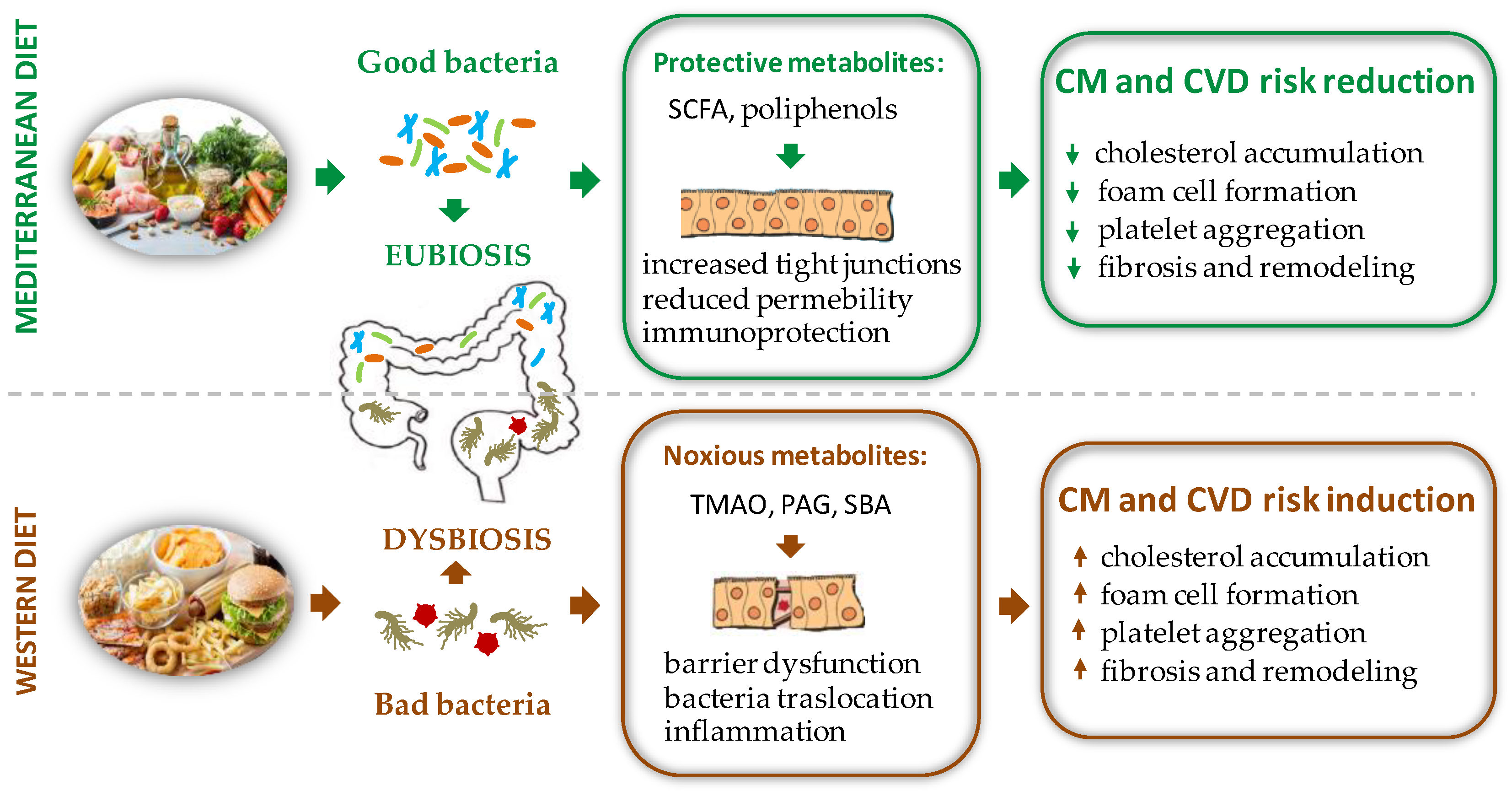
Gut microbiota (GM) is the set of microbe strains colonizing the intestinal tract. Through its genetic heritage, known as the gut microbiome, this complex ecosystem generates bioactive metabolites that impact various physiological processes, far beyond food digestion. Able to communicate with distal districts through multiple pathways, GM is therefore considered the largest endocrine organ of the body and one of the major determinants of humans’ health from infancy through adulthood. Indeed, while a balanced GM facilitates beneficial effects including digestion of macronutrients, synthesis of some vitamins, maintenance of immune homeostasis, and protection against pathogens, detrimental changes in GM composition lead to adverse remodeling of the host phenotype, which predispose to several pathological conditions, such as insulin resistance, atherosclerosis, obesity, and associated disorders, ultimately leading to cardiovascular disease (CVD). Dietary habit and sex hormones (SH) are considered major regulators of the GM variability (6, 7).

  • gut microbiota
  • diet
  • sex hormone

1. Food Intake and Gut Microbiota (GM) -Mediated Cardiovascular Disease (CVD) Risk

Depending on its composition, GM can transform nutrients present in the food into protective or damaging products entailing healthful or noxious effects, respectively [1]. The most representative examples of bioactive GM-derived compounds are: (1) metabolites of cholesterol such as primary bile acids (PBAs), secondary bile acids (SBAs), and coprostanol, (2) trimethylamine (TMA)/trimethylamine N-oxide (TMAO), (3) phenyl-acetyl-glutamine (PAG), (4) short chain fatty acids (SCFAs), and (5) polyphenols [2]. Some of these molecules have been proved to functionally interact with SH, which may explain at least in part, the gender dimorphism in CV risk and CVD outcomes [3].

1.1. Bile Acids Modulation and Cholesterol Metabolism

Bile acids (BAs) are considered critical components of the crosstalk between GM and cardiovascular (CV) health. The GM can directly influence the hepatic regulation of cholesterol metabolism [4] and alter bile acid composition and abundance, which in turn affect systemic cholesterol levels [5]. Originating from cholesterol catabolism, BAs are synthesized in the liver as primary bile acids (PBAs), then conjugated to amino acids taurine or glycine to form bile salts and included in the bile. Next, the bile is secreted in the duodenum to facilitate digestion and absorption of fats, nutrients, and liposoluble vitamins [6]. In the intestine, PBAs are deconjugated by bile salt hydrolase (BSH), an enzyme produced by a broad spectrum of aerobic and anaerobic bacteria (Gram-positive Bifidobacterium, Lactobacillus, Clostridium, and Enterococcus and Gram Negative Bacteroides). Later, a microbial-mediated dehydroxylation converts PBAs into secondary bile acids (SBAs). This reaction is catalyzed by a limited number of bacteria belonging to different genera (Bacteroides, Clostridium, Eubacterium, Lactobacillus, and Escherichia) [7].
PBAs appear to play a role in preventing the development of atherosclerosis through activation of the farnesoid X receptor (FXR) signaling cascade, which improves lipid profile and regulates gluconeogenesis and intestinal barrier function [8]. In addition, PBAs inhibit the NF-kB-dependent activation of Takeda-G protein 5 receptors (TGR5), resulting in a decreased production of proinflammatory cytokines [2][9]. PBAs can also influence CV function and reduce heart rate by regulating channel conductance and calcium dynamics in sinoatrial and ventricular cardiomyocytes. Finally, PBAs modulate vascular tone through a transcriptional regulation of vasoactive molecules as long-lasting effect [10].
Some studies have demonstrated an association between altered plasma or fecal BAs concentrations and CVD risk [11]. In particular, high concentrations of SBAs have been related with atherosclerosis development and CVD through GM-mediated mechanisms [12]. In fact, BSHs of GM are able to hydrolyze glycine and taurine conjugates to liberate free BAs; the resulting SBA overproduction promotes enhanced cholesterol levels and increases foam cell formation and atherosclerotic plaque size [9]. Accordingly, a low SBA excretion represents an independent risk factor for stroke and mortality [11]. Moreover, an alteration of SBAs to PBAs ratio may be implicated in hypercholesterolemia and CAD development. In this regard, Meyerhofer et al. demonstrated that higher plasma levels of SBAs in front of reduced PBAs in heart failure (HF) patients are associated with worse outcomes in univariate analysis [10]. On the other hand, some species of bacteria produce less absorbable SBA molecules that are excreted in the stool. This process stimulates BA neo-synthesis by the liver, leading to a net loss of circulating low-density lipoprotein (LDL) and reduced CV risk [13].
Collectively, the available findings indicate that GM plays a pivotal role in BAs synthesis and in determining the types and amount of PBAs and SBAs, with beneficial or harmful effects on health depending on the composition of the bacteria population.
Another mechanism involving GM in cholesterol metabolism is the conversion of absorbable cholesterol to coprostanol, a reduced non-absorbable sterol [14]. Although further studies are needed to better clarify this pathway, it is conceivable that GM may contribute to lower blood cholesterol and CV risk also by enhancing cholesterol removal through this route. The rate of GM-mediated cholesterol-to-coprostanol conversion in humans is variable: there are high converters showing almost complete cholesterol conversion and low converters with coprostanol representing less than 40% of the fecal neutral sterols content [14]. Strains belonging to the genera of Eubacterium and Bacteroides have been identified as the prevalent cholesterol-reducing strains [15][16]. Interestingly, coprostanol production is sex-dependent, with young women being better converters compared to young males [17].

1.2. Trimethylamine/Trimethylamine N-Oxide (TMAO)

Foods such as meat, eggs, fish, brassica vegetables, peanuts, and soybeans are abundant in choline, betaine, phosphatidylcholine, lecithin, and L-carnitine [18][19][20], which serve as dietary precursors of CM disease-associated compounds. Specifically, these nutrients are converted to trimethylamine (TMA) by particular intestinal bacterial strains endowed with TMA lyases. TMA is subsequently oxidized to trimethylamine N-oxide (TMAO) by the hepatic flavin monooxygenase 3 (FMO3) [21]. TMA producers include: the Firmicutes Anaerococcus, Clostridium, Desulfitobacterium, Enterococcus, and Streptococcus; the Proteobacteria Dseulfovibrio, Enterobacter, Escherichia, Klebsiella, Proteus, Pseudomonas, the Actinobacteria Actinobacter, and Citrobacter [22], while Bacteroidetes are not TMA producers [23]. Additionally, Akkermansia, Sporobacter, Prevotella [23], and Ruminococcus Gnavus, highly represented in GM of patients with atherosclerotic CAD, are great TMAO producers [24].
Noteworthy, the TMAO precursor choline can be also produced directly by GM via phospholipase D (PLD) enzyme, thus confirming the important direct involvement of GM in TMAO production [25].
TMAO represents a risk factor for CAD development as demonstrated by several preclinical and clinical data [20]. In mice, TMAO levels increased upon dietary supplementation of choline and L-carnitine, prompting macrophage foam cell formation and development of atherosclerosis [18][20]. The association of raised TMAO production and adverse CV outcome is also shown in numerous clinical studies [26][27]. In particular, increased TMAO levels have been associated with an elevated risk of fatal and non-fatal myocardial infarction (MI) or stroke [28]. In a large independent clinical cohort (n = 4007), patients in the highest quartile of TMAO plasma levels had a 2.5-fold higher risk of a major adverse CV event than patients in the lowest quartile [19]. Moreover, TMAO circulating levels predict 5-year mortality in patients with stable coronary artery disease [27].
The mechanisms by which TMAO increases GM-mediated CV risk are numerous. TMAO influences lipid composition [28], alters BAs transport, composition, and pool size, induces C-reactive protein production, fosters endothelial dysfunction, and increases serum levels of the proinflammatory LPS endotoxin [29]. Moreover, TMAO enhances platelet responsiveness to multiple distinct agonists (ADP, thrombin, and collagen) by facilitating Ca2+ release from the intracellular stores and inducing a pro-thrombotic effect in vivo [30]. Finally, TMAO promotes platelet aggregation also by activating the toll-like receptor (TLR) pathways [31].

1.3. Phenyl-Acetylglutamine (PAG)

Phenyl-acetylglutamine (PAG) is a recently identified, protein-derived compound produced by GM, and is considered a risk factor for atherosclerosis. It is obtained from the conversion of phenylalanine into phenylacetic acid performed by specific strains of GM. Phenylacetic acid is then conjugated with glutamine in the liver, generating phenylaceticglutamine (PAG) [32]. PAG affects platelet responsiveness inducing thrombosis through adrenergic receptor activation [32]. In the Malmo Offspring Study, Ottoson demonstrated that PAG was correlated with GM composition and associated with an increased risk of future CAD independently of other CV risk factors [33].

1.4. Short Chain Fatty Acid Production (SCFAs)

Besides transforming the compounds introduced with the diet in pro-atherogenic mediators, GM can also produce metabolites with a protective role.
SCFAs are GM-derived metabolites produced by the fermentation of complex carbohydrates, mostly from fermentable fibers such as pectin, beta-glucans, guar-gum, inulin, and phospho-oligosaccharides, largely contained in fruit, vegetables, and whole grains [34]. The most abundant SCFA is acetate (up to 75% of total SCFAs) followed by propionate and butyrate [35]. Acetate and butyrate are produced by members of the Bacteroidetes phylum, whereas butyrate is produced by species of the Firmicutes phylum [36]. In the specific case of Firmicutes, most gut bacteria representing this phylum are gram-positive and are able to produce several SCFAs, contributing to the protective CVD phenotype [7].
SCFAs influence numerous processes such as host-microbe signaling, energy utilization, and control of intestinal pH, entailing effects on the GM composition and gut motility [37]. SCFAs contribute to maintaining the intestinal barrier integrity by stimulating the production of mucin and regulating the expression of tight junction proteins [38]. The efficiency of the intestinal mucosa barrier allows a reduction of the leakage of molecules and cells from the gut into the bloodstream and, in turn, participates in reducing the systemic inflammatory condition. Thus, the SCFAs have anti-inflammatory effects [39].
SCFAs and, especially butyrate, act on specific plasma membrane receptors to mainly transduce inhibitory effects. For example, the interaction of SCFAs with receptors of immune response cells represses the activation of the nuclear factor NFκB and the activity of histone-deacetylases (HDAC) [40][41]. These two mechanisms are responsible for stopping the proliferation of T lymphocytes, and for inducing apoptosis of activated T lymphocyte when SCFA concentration increases further [42]. The same two mechanisms also blunt the release of TNF-alpha by granulocytes, monocytes, and macrophages after contact with bacterial membrane LPS [43]. In addition, SCFAs activate regulatory T (Treg) cells, through HDAC inhibition, playing a regulatory or suppressive activity on inflammatory signaling [40].
Since the SCFA receptors are widespread in many organs and cells such as intestinal epithelium, or adipocytes, the overall action of SCFAs lowers serum lipid levels though several routes: (i) by blocking cholesterol synthesis, (ii) by redirecting lipids towards the liver [44], and (iii) by reducing the triglycerides accumulation in fat cells [45]. Collectively, these SCFA-mediated processes have a protective role against the development of overweight, obesity, and CAD [45]

1.5. Polyphenols

Dietary polyphenols are naturally occurring compounds present in food items such as vegetables, fruits, cereals, tea, coffee, dark chocolate, cocoa powder, and wine [46]. The main groups of dietary polyphenols are phenolic acids, flavonoids, tannins, stilbenes, and diferuloylmethanes. The chemical composition of these molecules is extremely variable but presents common domains, i.e., hydroxylated aromatic rings or phenol rings [47]. A large proportion of polyphenols remain unabsorbed along the small intestine and may accumulate in the large intestine (90–95%), where it is extensively metabolized by GM. In turn, the transformed polyphenols have the ability to modify GM growth and composition in a two-way relationship [46]. For example, berberine, a polyphenol with poor oral bioavailability, significantly reduced atherosclerosis in high-fat-diet fed mice by stimulating the growth of Akkermansia [48]. Along the same line, the ability of resveratrol to counteract the metabolic syndrome-related alterations, including derangement of glucose and lipid homeostasis, increase of fat mass, rise in blood pressure, low-grade inflammation, and oxidative stress, is mainly due to the regulation of GM composition, BAs biosynthesis, and TMAO production [49]. Furthermore, GM converts polyphenols into secondary metabolites with potential health effects. This is the case of the lignin derivatives, such as enterodiol and enterolactone, that exert protective effects including a 30% reduction of all-cause mortality risk, along with decreased CVD mortality and non-fatal myocardial infarction [50].
Therefore, when compared to a western diet, a polyphenol rich nutritional regimen, such as the Mediterranean one, must be preferred to increase the biodiversity of the GM and ensure advantageous effects for the health of the host (Figure 1).
Figure 1. Schematic representation of the effects of different diet regimens on gut microbiota composition with opposite repercussions on CM and CVD risk factors. PAG = phenyl-acetylglutamine, SBA = secondary bile acids, SCFA = short chain fatty acid, TMAO = trimethylamine N-oxide.

2. Sex Difference and GM Diversity: Bidirectional Cross Talk between Intestinal Microbial Composition and Sex Hormones

2.1. Sex Hormones Regulates GM Composition and Function

Like other members of the steroid hormones superfamily, SH act prevalently through the classical genomic mechanism that involves binding to specific nuclear steroid hormone receptors (SHR) transcription factors, including estrogen receptors (ER), progesterone receptors (PR), and androgen receptors (AR) [51]. Besides the classical mechanism of action, sex steroids can act in the cells through the nonclassical or nongenomic mechanism of action, in most cases mediated by membrane receptors.
Through these pathways, SH may influence GM composition: (1) by a direct action on bacteria, or (2) indirectly, by affecting intestinal PH and motility, and by modifying the function of the intestinal epithelium and local immunological response, thus influencing the environment for bacteria growth.

2.1.1. Direct Effects of Sex Hormones on GM Profile

SH directly affect bacterial growth and induction of virulence factors. In particular, SH, by interacting with ER beta, (ERβ) modulate the bacteria metabolism [52]. For instance, it has long been recognized that the uptakes estradiol and progesterone by the anaerobic bacteria Prevotella Intermedius, favors bacterial expansion [53]. In addition, progesterone and estradiol can act as surrogates of vitamin K, an essential growth factor for Prevotella Intermedius [54] (Figure 2).

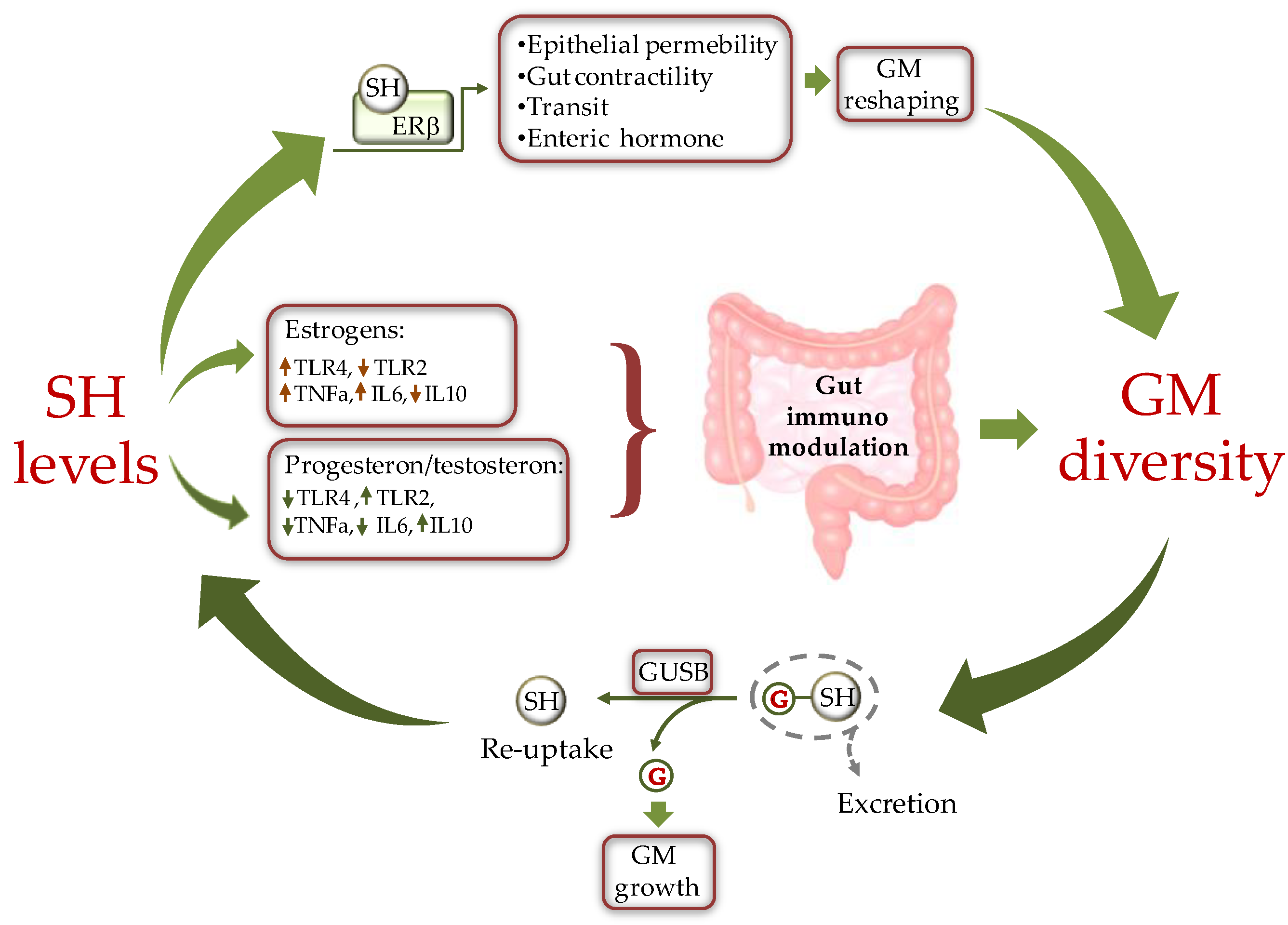
Figure 2. Schematic representation of the cross-talk between sex hormones (SH) and gut microbiota (GM). SH affect GM composition by modulating the intestine milieu via: i) a direct gene expression regulation in epithelial cells; and ii) by modulating the gut immune system through differential activation of TLRs. In turn GM influences SH circulating level by fine-tuning the balance between hormone excretion and reuptake. ERb = estrogen receptor beta, GUSB = β-glucuronidases; G = glucuronic moiety, IL = inteleukin, TLR = toll like receptor.

Sex steroids could also directly affect the composition of the GM by modifying substrate levels and energy production as the result of changes in bacterial beta glucuronidase (GUSB) activity (Figure 2). Many gut bacteria synthesize GUSB enzymes. These proteins catalyze the release of glucuronic acid from host-derived molecules including SH, previously conjugated in the liver. The released carbon sources are then used by the intestinal bacteria as energy substrate to promote their own growth [55].

2.1.2. Indirect Effects of Sex Hormones on GM Profile

Sex steroids may indirectly modulate the GM growth through activation of specific SHR on the intestinal colonic cells of the host [56]. As a consequence, the intestinal abundance and localization of SHR subtypes is expected to greatly impact GM distribution, composition, and function.
ERβ is the most abundant ER in colon epithelial cells, followed by ERα. In accordance with its distribution, ERβ is involved in the organization and architectural maintenance of the colon epithelium [57][58]. Indeed, Erβ-knockout mice exhibit a number of pre-pathogenic phenotypes, including disrupted cell-to-cell tight junctions and altered GM compared with wild-type mice, thus indicating a specific physiological role of ERβ in regulating the permeability of colonic epithelia [57][58].
AR is more expressed in human colon rectal mucosa and in colon stromal cells where it contributes to the maintenance of intestinal homeostasis [59]. The modified microenvironment might, in turn, affect the GM composition.
The classical ligand-dependent regulation of SHR activity by SH in the gastrointestinal cells also influences gut functions such as contractility, transit, or secretion of enteric hormones [60], which in turn induce GM modifications (Figure 2).
Numerous studies have evidenced a close connection between inflammatory state observed in several disease conditions and changes in the composition of GM [61]. SH transcriptional activity influences GM composition by differentially affecting the local intestinal immune environment in a gender-specific way. Estrogens and androgens have opposite effects on the immune response. In particular, estrogens favor a hyperactive immune environment, while androgens (testosterone) have a suppressive effect on T cell proliferation, resulting in an anti-inflammatory condition [62]. On the other hand, progesterone promotes the synthesis of anti-inflammatory cytokines and inhibits the production of their pro-inflammatory counterpart [63].
A major mechanism linking SH, immune response, and GM deals with the integrity of the intestinal epithelial barrier [64]. Decreased cohesion of the gut epithelium is associated with infiltration of gram-negative bacteria into circulation and activation of a peripheral inflammatory response by LPS, which may exasperate existing pathologies such as CM and CV disease [54][65]. The interaction between certain GM strains and estrogens alters the integrity of the intestinal barrier and enhances cell-mediated and humoral immune response, natural killer (NK) cell cytotoxicity, and the production of pro-inflammatory cytokines such as interleukin 1 (IL-1), interleukin 6 (IL-6), and tumor necrosis factor alpha (TNF-α) [64], thus favoring a proinflammatory phenotype.
As concerns the underlying pathway, in females there is an increased expression of genes involved in the pro-inflammatory TLR pathways. As evidenced by experimental studies, estrogenic treatment in mice increases cell membrane content of TLR4 [66]. Once activated, TLRs induce a pro-inflammatory immune environment that compromises gut permeability, causing translocation of gut commensals into the lamina propria where they can amplify pro-inflammatory responses [54] (Figure 2). In contrast, to estrogens, androgens, such as testosterone, decrease TLR4 and TLR2 expression in macrophage and down-regulate NK cells and TNF-α production while enhancing the production of the anti-inflammatory interleukin 10 (IL-10) [67]. Ultimately, the repression of TLR pathways and antigen presentation by testosterone favors the integrity of the intestinal barrier [67]. Similarly to testosterone, progesterone contrasts the activation of LPS-mediated TLR4 signaling [68] and suppresses NK cell activity, thus fostering the integrity of the intestinal epithelium [54]. Additionally, progesterone decreases gut permeability through upregulating occludin in human intestinal tissue [69].
A recently identified pathway involving SHR, intestinal homeostasis, and GM diversity deals with the activity of the orphan nuclear estrogen-related receptor alpha (ESRRA) on mitochondrial function. This factor is critical to maintain mitochondrial biogenesis and macroautophagy/autophagy function in the gut. It has been demonstrated that ESRRA acts as a key regulator of intestinal homeostasis by ameliorating colonic inflammation through activation of autophagic flux and control of host GM composition. Thus, ESRRA contributes to intestinal homeostasis to protect the host from detrimental inflammation and dysfunctional mitochondria [70].
In conclusion, changes in estrogens, progesterone, or androgens signaling could result in altered intestinal functions, such as contractility and transit, and in a local immune unbalance, which may influence the gender-specific GM composition by differently shaping the gut mucosal environment.

2.2. GM Composition Regulates Sex Hormone Levels

Just as GM is influenced by SH, in turn GM may be an important regulator of circulating levels of SH and SH metabolites [71]. In fact, GM is able to produce hormones (e.g., serotonin, dopamine, somatostatine), and to regulate the host’s hormones homeostasis by inhibiting gene transcription (e.g., prolactin) or fostering conversion reactions (e.g., glucocorticoids to androgens) [72].
The set of enteric bacterial genes coding for estrogen-metabolizing enzymes, is defined as “estrobolome” [73]. As above anticipated, the bacterial GUSB influences the systemic estrogen metabolite (EM) profiles. Normally, in the phase I metabolic step, estrogens, and their metabolites are conjugated by the liver and excreted in the bile. Of note, glucuronidation of estrogens serves primarily a classical excretory role given that conjugated EM have a low affinity for the ERs and do not promote appreciable transcription. Once secreted within the intestine, EM can be de-conjugated by the GM GUSB, reabsorbed by the gut, and released into the bloodstream for distal action. Thus, depending on GM composition, the estrobolome favors deconjugation and promotes reabsorption of free estrogens into enterohepatic circulation, which in turn contributes to the host’s total estrogen amount [54][65]. When this process is impaired through dysbiosis, the altered deconjugation process results in variation of circulating SH levels. For example, a reduction of bacterial GUSB activity may contribute to the development of pathological conditions such as obesity, metabolic syndrome, cardiovascular disease (CVD), and decline of cognitive function [3][64]. On the other side, the increased abundance of GUSB-producing bacteria can induce hyperestrogenic pathologies. This condition results in increased free estrogens circulating levels, entailing diseases such as endometriosis and cancer [74][75].
Increasing findings also indicate that male androgen levels are subjected to GM-dependent modulation. In studies on obese male mice, the treatment with lactic acid bacteria isolated from human milk induces growth of testes and increased serum testosterone levels [76]. In addition, feeding the old male mice with purified microbes such as Lactobacillus Reuteri restores the testosterone to the youthful level [77]. Collectively, these results provide evidence that GM is able to affect testosterone production and testicular aging.
Although human androgen biosynthesis and metabolism have been extensively studied in the past, the exact underlying mechanisms still remain elusive. Traditionally, most androgen has been thought to be metabolized by the liver and subsequently excreted by the kidney [78]. However, a recent study by Collden et al. pointed at the GM as a main regulator of androgen metabolism [79]. In effect, similarly to estrogens, conjugated androgens are hydrolyzed in the intestinal tract via bacterial GUSB into free androgens for reabsorption. A reduced concentration of circulating androgen may induce androgen-related disease. As reported by Harada et al., the GM was largely involved in the CV risks associated with male hypogonadism, which could be prevented by antibiotic therapeutic strategies [80][81]. In line with this notion, in a recent post hoc analysis, men with metabolic syndrome and low levels of high-density lipoprotein (HDL)-cholesterol evidenced an association between reduced testosterone concentrations and increased risk of subsequent CV events and death [82]. This finding is confirmed by other epidemiological studies showing that low testosterone levels in men are negatively associated with the degree of carotid atherosclerosis [83]. Finally, it is well-established that male hypogonadism in aging men is closely associated with an increased risk of metabolic syndrome and T2D [84].
Besides the androgen deglucuronidation activity of GM, some specific microbial taxa, including Butyricicoccus Desmolans, Clostridium Cadaveris, Propionimicrobium Lymphophilum, Clostridium Scindens, and Clostridium Innocuum, express classical steroid processing enzymes such as steroid-17, 20-desmolase, 20β-, 20α-, and 3α-hydroxysteroid dehydrogenase, or 5β-reductase, and possess the potential capability to directly metabolize steroid hormones [85][86]. In addition, glucocorticoids can be converted into androgens via side-chain cleaving capacity of bacteria [87][88]. Finally, to make the scenario of interactions between GM and SH even more complex, a recent work has shown the ability of some bacterial strains to retroconvert estrogens into androgens [89].
Overall, the present evidence underlines a critical role of GM in estrogens and androgen metabolism, which deserves to be explored in more detailed studies.

This entry is adapted from the peer-reviewed paper 10.3390/ijms23137154

References

  1. Rodríguez-Morató, J.; Matthan, N.R. Nutrition and Gastrointestinal Microbiota, Microbial-Derived Secondary Bile Acids, and Cardiovascular Disease. Curr. Atheroscler. Rep. 2020, 22, 47.
  2. Witkowski, M.; Weeks, T.L.; Hazen, S.L. Gut Microbiota and Cardiovascular Disease. Circ. Res. 2020, 127, 553–570.
  3. Razavi, A.C.; Potts, K.S.; Kelly, T.N.; Bazzano, L.A. Sex, gut microbiome, and cardiovascular disease risk. Biol. Sex Differ. 2019, 10, 29.
  4. Tang, W.W.; Wang, Z.; Fan, Y.; Levison, B.; Hazen, J.E.; Donahue, L.M.; Wu, Y.; Hazen, S.L. Prognostic Value of Elevated Levels of Intestinal Microbe-Generated Metabolite Trimethylamine-N-Oxide in Patients With Heart Failure. J. Am. Coll. Cardiol. 2014, 64, 1908–1914.
  5. Caesar, R.; Nygren, H.; Oresic, M.; Bäckhed, F. Interaction between dietary lipids and gut microbiota regulates hepatic cholesterol metabolism. J. Lipid Res. 2016, 57, 474–481.
  6. Molinero, N.; Ruiz, L.; Sánchez, B.; Margolles, A.; Delgado, S. Intestinal Bacteria Interplay With Bile and Cholesterol Metabolism: Implications on Host Physiology. Front. Physiol. 2019, 10, 185.
  7. Busnelli, M.; Manzini, S.; Chiesa, G. The Gut Microbiota Affects Host Pathophysiology as an Endocrine Organ: A Focus on Cardiovascular Disease. Nutrients 2020, 12, 79.
  8. Li, Y.; Hou, H.; Wang, X.; Dai, X.; Zhang, W.; Tang, Q.; Dong, Y.; Yan, C.; Wang, B.; Li, Z.; et al. Diammonium Glycyrrhizinate Ameliorates Obesity Through Modulation of Gut Microbiota-Conjugated BAs-FXR Signaling. Front. Pharmacol. 2021, 12, 796590.
  9. Ryan, P.M.; Stanton, C.; Caplice, N.M. Bile acids at the cross-roads of gut microbiome–host cardiometabolic interactions. Diabetol. Metab. Syndr. 2017, 9, 102.
  10. Mayerhofer, C.C.K.; Ueland, T.; Broch, K.; Vincent, R.P.; Cross, G.F.; Dahl, C.P.; Aukrust, P.; Gullestad, L.; Hov, J.R.; Troseid, M. Increased secondary/primary bile acid ratio in chronic heart failure. J Card Fail. 2017, 23, 666–671.
  11. Charach, G.; Karniel, E.; Novikov, I.; Galin, L.; Vons, S.; Grosskopf, I.; Charach, L. Reduced bile acid excretion is an independent risk factor for stroke and mortality: A prospective follow-up study. Atherosclerosis 2020, 293, 79–85.
  12. Li, Y.; Zhang, D.; He, Y.; Chen, C.; Song, C.; Zhao, Y.; Bai, Y.; Wang, Y.; Pu, J.; Chen, J.; et al. Investigation of novel metabolites potentially involved in the pathogenesis of coronary heart disease using a UHPLC-QTOF/MS-based metabolomics approach. Sci. Rep. 2017, 7, 15737.
  13. Pushpass, R.-A.G.; Alzoufairi, S.; Jackson, K.G.; Lovegrove, J.A. Circulating bile acids as a link between the gut microbiota and cardiovascular health: Impact of prebiotics, probiotics and polyphenol-rich foods. Nutr. Res. Rev. 2021, 1–20.
  14. Matysik, S.; Krautbauer, S.; Liebisch, G.; Schött, H.-F.; Kjølbaek, L.; Astrup, A.; Blachier, F.; Beaumont, M.; Nieuwdorp, M.; Hartstra, A.; et al. Short-Chain Fatty Acids and Bile Acids in Human Faeces Are Associated with the Intestinal Cholesterol Conversion Status. Br. J. Pharm. 2021, 178, 3342–3353.
  15. Gérard, P.; Lepercq, P.; Leclerc, M.; Gavini, F.; Raibaud, P.; Juste, C. Bacteroides sp. Strain D8, the First Cholesterol-Reducing Bacterium Isolated from Human Feces. Appl. Environ. Microbiol. 2007, 73, 5742–5749.
  16. Ren, D.; Li, L.; Schwabacher, A.W.; Young, J.W.; Beitz, D.C. Mechanism of cholesterol reduction to coprostanol by Eubacterium coprostanoligenes ATCC 51222. Steroids 1996, 61, 33–40.
  17. Benno, P.; Midtvedt, K.; Alam, M.; Collinder, E.; Norin, E.; Midtvedt, T. Examination of intestinal conversion of cholesterol to coprostanol in 633 healthy subjects reveals an age- and sex-dependent pattern. Microb. Ecol. Health Dis. 2005, 17, 200–204.
  18. Koeth, R.A.; Wang, Z.; Levison, B.S.; Buffa, J.A.; Org, E.; Sheehy, B.T.; Britt, E.B.; Fu, X.; Wu, Y.; Li, L.; et al. Intestinal microbiota metabolism of l-carnitine, a nutrient in red meat, promotes atherosclerosis. Nat. Med. 2013, 19, 576–585.
  19. Tang, W.W.; Wang, Z.; Levison, B.S.; Koeth, R.A.; Britt, E.B.; Fu, X.; Wu, Y.; Hazen, S.L. Intestinal microbial metabolism of phosphatidyl- choline and cardiovascular risk. N. Engl. J. Med. 2013, 368, 1575–1584.
  20. Wang, Z.; Klipfell, E.; Bennett, B.J.; Koeth, R.; Levison, B.S.; DuGar, B.; Feldstein, A.E.; Britt, E.B.; Fu, X.; Chung, Y.-M.; et al. Gut Flora Metabolism of Phosphatidylcholine Promotes Cardiovascular Disease. Nature 2011, 472, 57–63.
  21. Guasti, L.; Galliazzo, S.; Molaro, M.; Visconti, E.; Pennella, B.; Gaudio, G.V.; Lupi, A.; Grandi, A.M.; Squizzato, A. TMAO as a biomarker of cardiovascular events: A systematic review and meta-analysis. Intern. Emerg. Med. 2021, 16, 201–207.
  22. Al-Obaide, M.A.I.; Singh, R.; Datta, P.; Rewers-Felkins, K.A.; Salguero, M.V.; Al-Obaidi, I.; Kottapalli, K.R.; Vasylyeva, T.L. Gut Microbiota-Dependent Trimethylamine-N-oxide and Serum Biomarkers in Patients with T2DM and Advanced CKD. J. Clin. Med. 2017, 6, 86.
  23. Falony, G.; Vieira-Silva, S.; Raes, J. Microbiology Meets Big Data: The Case of Gut Microbiota–Derived Trimethylamine. Annu. Rev. Microbiol. 2015, 69, 305–321.
  24. Wang, Z.; Roberts, A.B.; Buffa, J.A.; Levison, B.S.; Zhu, W.; Org, E.; Gu, X.; Huang, Y.; Zamanian-Daryoush, M.; Culley, M.K.; et al. Non-lethal Inhibition of Gut Microbial Trimethylamine Production for the Treatment of Atherosclerosis. Cell 2015, 163, 1585–1595.
  25. Chittim, C.L.; Del Campo, A.M.; Balskus, E.P. Gut bacterial phospholipase Ds support disease-associated metabolism by generating choline. Nat. Microbiol. 2019, 4, 155–163.
  26. Tang, W.W.; Wang, Z.; Shrestha, K.; Borowski, A.G.; Wu, Y.; Troughton, R.W.; Klein, A.L.; Hazen, S.L. Intestinal Microbiota-Dependent Phosphatidylcholine Metabolites, Diastolic Dysfunction, and Adverse Clinical Outcomes in Chronic Systolic Heart Failure. J. Card. Fail. 2015, 21, 91–96.
  27. Senthong, V.; Wang, Z.; Li, X.S.; Fan, Y.; Wu, Y.; Wilson Tang, W.H.; Hazen, S.L. Intestinal microbiota-generated metabolite trimethyl-amine-N-oxide and 5-year mortality risk in stable coronary artery disease: The contributory role of intestinal microbiota in a COURAGE-like patient cohort. J. Am. Heart Assoc. 2016, 5, e002816.
  28. Allayee, H.; Hazen, S.L. Contribution of Gut Bacteria to Lipid Levels. Circ. Res. 2015, 117, 750–754.
  29. Kanitsoraphan, C.; Rattanawong, P.; Charoensri, S.; Senthong, V. Trimethylamine N-Oxide and Risk of Cardiovascular Disease and Mortality. Curr. Nutr. Rep. 2018, 7, 207–213.
  30. Zhu, W.; Gregory, J.C.; Org, E.; Buffa, J.A.; Gupta, N.; Wang, Z.; Li, L.; Fu, X.; Wu, Y.; Mehrabian, M.; et al. Gut Microbial Metabolite TMAO Enhances Platelet Hyperreactivity and Thrombosis Risk. Cell 2016, 165, 111–124.
  31. Koupenova, M.; Mick, E.; Mikhalev, E.; Benjamin, E.J.; Tanriverdi, K.; Freedman, J.E. Sex Differences in Platelet Toll-Like Receptors and Their Association With Cardiovascular Risk Factors. Arter. Thromb. Vasc. Biol. 2015, 35, 1030–1037.
  32. Nemet, I.; Saha, P.P.; Gupta, N.; Zhu, W.; Romano, K.A.; Skye, S.M.; Cajka, T.; Mohan, M.L.; Li, L.; Wu, Y.; et al. A Cardiovascular Disease-Linked Gut Microbial Metabolite Acts via Adrenergic Receptors. Cell 2020, 180, 862–877.e22.
  33. Ottosson, F.; Brunkwall, L.; Smith, E.; Orho-Melander, M.; Nilsson, P.M.; Fernandez, C.; Melander, O. The gut microbiota-related metabolite phenylacetylglutamine associates with increased risk of incident coronary artery disease. J. Hypertens. 2020, 38, 2427–2434.
  34. Richards, L.B.; Li, M.; van Esch, B.C.; Garssen, J.; Folkerts, G. The effects of short-chain fatty acids on the cardiovascular system. PharmaNutrition 2016, 4, 68–111.
  35. Macfarlane, G.T.; Macfarlane, S. Fermentation in the human large intestine: Its physiologic consequences and the potential contribution of prebiotics. J. Clin. Gastroenterol. 2011, 45, S120–S127.
  36. Macfarlane, S.; Macfarlane, G.T. Regulation of short-chain fatty acid production. Proc. Nutr. Soc. 2003, 62, 67–72.
  37. Musso, G.; Gambino, R.; Cassader, M. Interactions Between Gut Microbiota and Host Metabolism Predisposing to Obesity and Diabetes. Annu. Rev. Med. 2011, 62, 361–380.
  38. Krishnan, S.; Alden, N.; Lee, K. Pathways and functions of gut microbiota metabolism impacting host physiology. Curr. Opin. Biotechnol. 2015, 36, 137–145.
  39. Wang, Z.; Zhao, Y. Gut microbiota derived metabolites in cardiovascular health and disease. Protein Cell 2018, 9, 416–431.
  40. Silva, J.P.B.; Navegantes-Lima, K.C.; Oliveira, A.L.B.; Rodrigues, D.V.S.; Gaspar, S.L.F.; Monteiro, V.V.S.; Moura, D.P.; Monteiro, M.C. Protective mechanisms of butyrate on inflammatory bowel disease. Curr. Pharm. Des. 2018, 24, 4154–4166.
  41. Amiri, P.; Hosseini, S.A.; Ghaffari, S.; Tutunchi, H.; Ghaffari, S.; Mosharkesh, E.; Asghari, S.; Roshanravan, N. Role of Butyrate, a Gut Microbiota Derived Metabolite, in Cardiovascular Diseases: A comprehensive narrative review. Front. Pharmacol. 2022, 12, 837509.
  42. Arpaia, N.; Campbell, C.; Fan, X.; Dikiy, S.; van der Veeken, J.; Deroos, P.; Liu, H.; Cross, J.R.; Pfeffer, K.K.; Coffer, P.J.; et al. Metabolites produced by com- mensal bacteria promote peripheral regulatory T-cell generation. Nature 2013, 504, 451–455.
  43. Gibson, R.; Eriksen, R.; Chambers, E.; Gao, H.; Aresu, M.; Heard, A.; Chan, Q.; Elliott, P.; Frost, G. Intakes and Food Sources of Dietary Fibre and Their Associations with Measures of Body Composition and Inflammation in UK Adults: Cross-Sectional Analysis of the Airwave Health Monitoring Study. Nutrients 2019, 11, 1839.
  44. Deng, M.; Qu, F.; Chen, L.; Liu, C.; Zhang, M.; Ren, F.; Guo, H.; Zhang, H.; Ge, S.; Wu, C.; et al. SCFAs alleviated steatosis and inflammation in mice with NASH induced by MCD. J. Endocrinol. 2020, 245, 425–437.
  45. Reynolds, A.; Mann, J.; Cummings, J.; Winter, N.; Mete, E.; Te Morenga, L. Carbohydrate quality and human health: A series of systematic reviews and meta-analyses. Lancet 2019, 393, 434–445.
  46. Singh, A.K.; Cabral, C.; Kumar, R.; Ganguly, R.; Rana, H.K.; Gupta, A.; Flávio Reis, F.; Pandey, A.K. Beneficial Effects of Dietary Polyphenols on Gut Microbiota and Strategies to Improve Delivery Efficiency. Nutrients 2019, 11, 2216.
  47. Kinger, M.; Kumar, S.; Kumar, V. Some Important Dietary Polyphenolic Compounds: An Anti-inflammatory and Immunoregulatory Perspective. Mini-Reviews Med. Chem. 2018, 18, 1270–1282.
  48. Zhu, L.; Zhang, D.; Zhu, H.; Zhu, J.; Weng, S.; Dong, L.; Liu, T.; Hu, Y.; Shen, X. Berberine treatment increases Akkermansia in the gut and improves high-fat diet-induced atherosclerosis in Apoe−/− mice. Atherosclerosis 2018, 268, 117–126.
  49. Chaplin, A.; Carpéné, C.; Mercader, J. Resveratrol, Metabolic Syndrome, and Gut Microbiota. Nutrients 2018, 10, 1651.
  50. Rienks, J.; Barbaresko, J.; Nöthlings, U. Association of Polyphenol Biomarkers with Cardiovascular Disease and Mortality Risk: A Systematic Review and Meta-Analysis of Observational Studies. Nutrients 2017, 9, 415.
  51. Beato, M. Steroid hormone receptors: An update. Hum. Reprod. Updat. 2000, 6, 225–236.
  52. Yoon, K.; Kim, N. Roles of Sex Hormones and Gender in the Gut Microbiota. J. Neurogastroenterol. Motil. 2021, 27, 314–325.
  53. Hussain, T.; Murtaza, G.; Kalhoro, D.H.; Kalhoro, M.S.; Metwally, E.; Chughtai, M.I.; Mazhar, M.U.; Khan, S.A. Relationship between gut microbiota and host-metabolism: Emphasis on hormones related to reproductive function. Anim. Nutr. 2021, 7, 1–10.
  54. García-Gómez, E.; González-Pedrajo, B.; Camacho-Arroyo, I. Role of Sex Steroid Hormones in Bacterial-Host Interactions. BioMed Res. Int. 2013, 2013, 928290.
  55. Walsh, J.; Olavarria-Ramirez, L.; Lach, G.; Boehme, M.; Dinan, T.G.; Cryan, J.F.; Griffin, B.; Hyland, N.P.; Clarke, G. Impact of host and environmental factors on β-glucuronidase enzymatic activity: Implications for gastrointestinal serotonin. Am. J. Physiol. Liver Physiol. 2020, 318, G816–G826.
  56. Nie, X.; Xie, R.; Tuo, B. Effects of Estrogen on the Gastrointestinal Tract. Am. J. Dig. Dis. 2018, 63, 583–596.
  57. Looijer-van Langen, M.; Hotte, N.; Dieleman, L.A.; Albert, E.; Mulder, C.; Madsen, K.L. Estrogen receptor signaling modulates epithelial barrier function. Am. J. Physiol. Gastroenterol. Liver Physiol. 2011, 300, G621–G626.
  58. Wada-Hiraike, O.; Imamov, O.; Hiraike, H.; Hultenby, K.; Schwend, T.; Omoto, Y.; Warner, M.; Gustafsson, J. Role of estrogen receptor β in colonic epithelium. Proc. Natl. Acad. Sci. USA 2006, 103, 2959–2964.
  59. Yu, X.; Li, S.; Xu, Y.; Zhang, Y.; Ma, W.; Liang, C.; Lu, H.; Ji, Y.; Liu, C.; Chen, D.; et al. Androgen Maintains Intestinal Homeostasis by Inhibiting BMP Signaling via Intestinal Stromal Cells. Stem Cell Rep. 2020, 15, 912–925.
  60. Falony, G.; Joossens, M.; Vieira-Silva, S.; Wang, J.; Darzi, Y.; Faust, K.; Kurilshikov, A.; Bonder, M.J.; Valles-Colomer, M.; Vandeputte, D.; et al. Population-level analysis of gut microbiome variation. Science 2016, 352, 560–564.
  61. Wang, J.; Chen, W.-D.; Wang, Y.-D. The Relationship between Gut Microbiota and Inflammatory Diseases: The Role of Macrophages. Front. Microbiol. 2020, 11, 1065.
  62. Markle, J.G.M.; Frank, D.N.; Mortin-Toth, S.; Robertson, C.E.; Feazel, L.M.; Rolle-Kampczyk, U.; von Bergen, M.; McCoy, K.D.; Macpherson, A.J.; Danska, J.S. Sex Differences in the Gut Microbiome Drive Hormone-Dependent Regulation of Autoimmunity. Science 2013, 339, 1084–1088.
  63. Polikarpova, A.; Levina, I.; Sigai, N.; Zavarzin, I.; Morozov, I.; Rubtsov, P.; Guseva, A.; Smirnova, O.; Shchelkunova, T. Immunomodulatory effects of progesterone and selective ligands of membrane progesterone receptors. Steroids 2019, 145, 5–18.
  64. Vemuri, R.; Sylvia, K.E.; Klein, S.L.; Forster, S.C.; Plebanski, M.; Eri, R.; Flanagan, K.L. The microgenderome revealed: Sex differences in bidirectional interactions between the microbiota, hormones, immunity and disease susceptibility. Semin. Immunopathol. 2019, 41, 265–275.
  65. Thackray, V.G. Sex, Microbes, and Polycystic Ovary Syndrome. Trends Endocrinol. Metab. 2019, 30, 54–65.
  66. Rettew, J.A.; Huet, Y.; Marriott, I. Estrogens Augment Cell Surface TLR4 Expression on Murine Macrophages and Regulate Sepsis Susceptibility in Vivo. Endocrinology 2009, 150, 3877–3884.
  67. Rettew, J.A.; Huet-Hudson, Y.M.; Marriott, I. Testosterone Reduces Macrophage Expression in the Mouse of Toll-Like Receptor 4, a Trigger for Inflammation and Innate Immunity. Biol. Reprod. 2008, 78, 432–437.
  68. Lei, B.; Mace, B.; Dawson, H.N.; Warner, D.S.; Laskowitz, D.T.; James, M.L. Anti-Inflammatory Effects of Progesterone in Lipopolysaccharide-Stimulated BV-2 Microglia. PLoS ONE 2014, 9, e103969.
  69. Zhou, Z.; Bian, C.; Luo, Z.; Guille, C.; Ogunrinde, E.; Wu, J.; Zhao, M.; Fitting, S.; Kamen, D.L.; Oates, J.C.; et al. Progesterone decreases gut permeability through upregulating occludin expression in primary human gut tissues and Caco-2 cells. Sci. Rep. 2019, 9, 8367.
  70. Kim, S.; Lee, J.-Y.; Shin, S.G.; Kim, J.K.; Silwal, P.; Kim, Y.J.; Shin, N.-R.; Kim, P.S.; Won, M.; Lee, S.-H.; et al. ESRRA (estrogen related receptor alpha) is a critical regulator of intestinal homeostasis through activation of autophagic flux via gut microbiota. Autophagy 2021, 17, 2856–2875.
  71. He, S.; Li, H.; Yu, Z.; Zhang, F.; Liang, S.; Liu, H.; Chen, H.; Lü, M. The Gut Microbiome and Sex Hormone-Related Diseases. Front. Microbiol. 2021, 12, 2699.
  72. Neuman, H.; Debelius, J.W.; Knight, R.; Koren, O. Microbial endocrinology: The interplay between the microbiota and the endocrine system. FEMS Microbiol. Rev. 2015, 39, 509–521.
  73. Osadchiy, V.; Martin, C.R.; Mayer, E.A. The gut-brain axis and the microbiome: Mechanisms and clinical implications. Clin. Gastroenterol. Hepatol. 2019, 17, 322–332.
  74. Gomez, A.; Luckey, D.; Taneja, V. The gut microbiome in autoimmunity: Sex matters. Clin. Immunol. 2015, 159, 154–162.
  75. Rubtsova, K.; Marrack, P.; Rubtsov, A.V. Sexual dimorphism in autoimmunity. J. Clin. Investig. 2015, 125, 2187–2193.
  76. Levkovich, T.; Poutahidis, T.; Smillie, C.; Varian, B.J.; Ibrahim, Y.M.; Lakritz, J.R.; Alm, E.J.; Erdman, S.E. Probiotic bacteria induce a ‘glow of health’. PLoS ONE. 2013, 8, e53867.
  77. Poutahidis, T.; Springer, A.D.; Levkovich, T.; Qi, P.; Varian, B.J.; Lakritz, J.; Ibrahim, Y.M.; Chatzigiagkos, A.; Alm, E.J.; Erdman, S.E. Probiotic Microbes Sustain Youthful Serum Testosterone Levels and Testicular Size in Aging Mice. PLoS ONE 2014, 9, e84877.
  78. Schiffer, L.; Arlt, W.; Storbeck, K.-H. Intracrine androgen biosynthesis, metabolism and action revisited. Mol. Cell. Endocrinol. 2018, 465, 4–26.
  79. Colldén, H.; Landin, A.; Wallenius, V.; Elebring, E.; Fändriks, L.; Nilsson, M.E.; Ryberg, H.; Poutanen, M.; Sjögren, K.; Vandenput, L.; et al. The gut microbiota is a major regulator of androgen metabolism in intestinal contents. Am. J. Physiol. Endocrinol. Metab. 2019, 317, E1182–E1192.
  80. Harada, N.; Hanaoka, R.; Horiuchi, H.; Kitakaze, T.; Mitani, T.; Inui, H.; Yamaji, R. Castration influences intestinal microflora and induces abdominal obesity in high-fat diet-fed mice. Sci. Rep. 2016, 6, 23001.
  81. Harada, N.; Hanaoka, R.; Hanada, K.; Izawa, T.; Inui, H.; Yamaji, R. Hypogonadism alters cecal and fecal microbiota in male mice. Gut Microbes 2016, 7, 533–539.
  82. Boden, W.E.; Miller, M.G.; McBride, R.; Harvey, C.; Snabes, M.C.; Schmidt, J.; McGovern, M.E.; Fleg, J.L.; Desvigne-Nickens, P.; Anderson, T.; et al. Testosterone concentrations and risk of cardiovascular events in androgen-deficient men with atherosclerotic cardiovascular disease. Am. Heart J. 2020, 224, 65–76.
  83. Li, X.; Cheng, W.; Shang, H.; Wei, H.; Deng, C. The Interplay between Androgen and Gut Microbiota: Is There a Microbiota-Gut-Testis Axis. Reprod. Sci. 2022, 29, 1674–1684.
  84. Org, E.; Mehrabian, M.; Parks, B.W.; Shipkova, P.; Liu, X.; Drake, T.A.; Lusis, A.J. Sex differences and hormonal effects on gut microbiota composition in mice. Gut Microbes 2016, 7, 313–322.
  85. Devendran, S.; Méndez-García, C.; Ridlon, J.M. Identification and characterization of a 20β-HSDH from the anaerobic gut bacterium Butyricicoccus desmolans ATCC 43058. J. Lipid Res. 2017, 58, 916–925.
  86. Devendran, S.; Mythen, S.M.; Ridlon, J.M. The desA and desB genes from Clostridium scindens ATCC 35704 encode steroid-17,20-desmolase. J. Lipid Res. 2018, 59, 1005–1014.
  87. Mueller, S.; Saunier, K.; Hanisch, C.; Norin, E.; Alm, L.; Midtvedt, T.; Cresci, A.; Silvi, S.; Orpianesi, C.; Verdenelli, M.C.; et al. Differences in fecal microbiota in different European study populations in relation to age, gender, and country: A cross-sectional study. Appl. Environ. Microbiol. 2006, 72, 1027–1033.
  88. Schnorr, S.L.; Candela, M.; Rampelli, S.; Centanni, M.; Consolandi, C.; Basaglia, G.; Turroni, S.; Biagi, E.; Peano, C.; Severgnini, M.; et al. Gut microbiome of the Hadza hunter-gatherers. Nat. Commun. 2014, 5, 3654.
  89. Wang, P.-H.; Chen, Y.-L.; Wei, S.T.-S.; Wu, K.; Lee, T.-H.; Wu, T.-Y.; Chiang, Y.-R. Retroconversion of estrogens into androgens by bacteria via a cobalamin-mediated methylation. Proc. Natl. Acad. Sci. USA 2019, 117, 1395–1403.
More
This entry is offline, you can click here to edit this entry!
Video Production Service