Recently, dental pulp stem cells (DPSCs) and stem cells from human exfoliated deciduous teeth (SHEDs) have attracted substantial attention as promising cell sources for tissue regeneration. Here, we summarized the features of DPSCs and SHEDs such as high growth capacity, multipotency, expression of cell markers, immunomodulatory effects, and their potential to regenerate various somatic tissues.
- dental pulp stem cells
- stem cells from human exfoliated deciduous teeth
- regenerative therapy
- regenerative medicine
1. Content
Mesenchymal stem cells (MSCs) have the capacity for self-renewal and multilineage differentiation potential, and are considered a promising cell population for cell-based therapy and tissue regeneration. MSCs are isolated from various organs including dental pulp, which originates from cranial neural crest-derived ectomesenchyme. Recently, dental pulp stem cells (DPSCs) and stem cells from human exfoliated deciduous teeth (SHEDs) have been isolated from dental pulp tissue of adult permanent teeth and deciduous teeth, respectively. Because of their MSC-like characteristics such as high growth capacity, multipotency, expression of MSC-related markers, and immunomodulatory effects, they are suggested to be an important cell source for tissue regeneration. Here, authors review the features of these cells, their potential to regenerate damaged tissues, and the recently acquired understanding of their potential for clinical application in regenerative medicine.
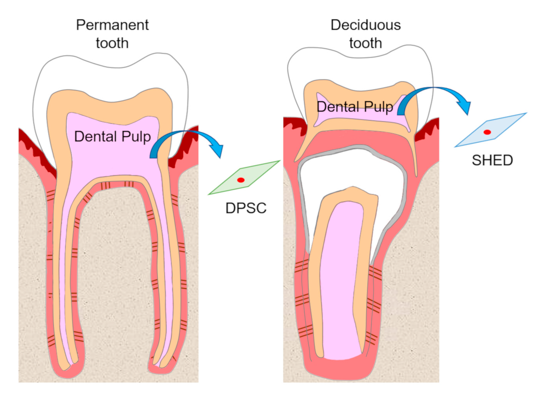
 Figure 1. Isolation of dental pulp stem cells (DPSCs) and stem cells from human exfoliated deciduous teeth (SHEDs). DPSCs and SHEDs isolated from pulp tissue of permanent teeth and deciduous teeth, respectively, have the potential to be cell sources in regenerative medicine.
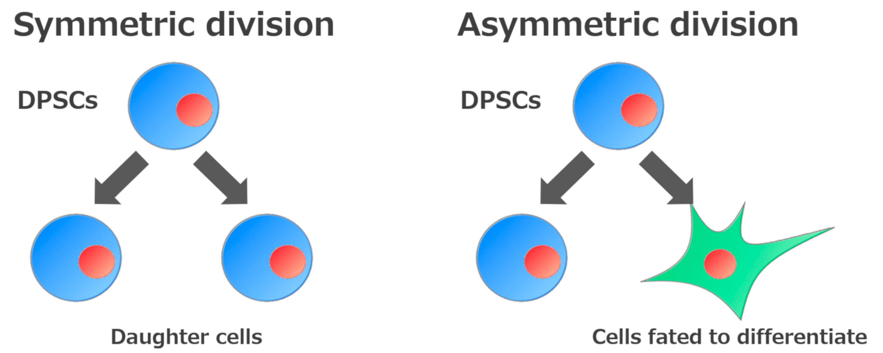
 Figure 2. Self-renewal mechanisms of stem cells. Stem cells are defined by their ability to make new stem cells (self-renewal). Self-renewal of stem cells is divided into two processes: Generation of daughter cells or of cells fated to differentiate.
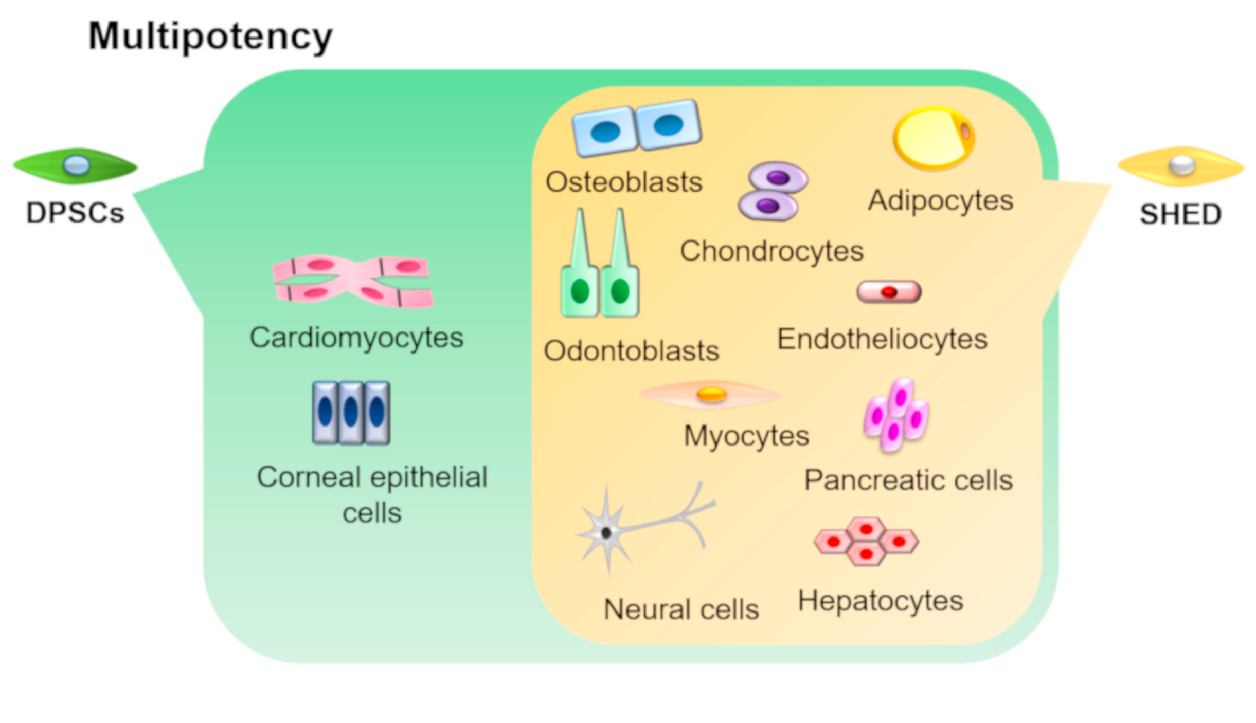
 Figure 3. Multipotency of DPSCs and SHEDs. DPSCs and SHEDs can differentiate into multiple lineages such as osteoblasts, odontoblasts, adipocytes, chondrocytes, neural cells, endotheliocytes, myocytes, hepatocytes, and pancreatic cells under appropriate culture conditions. In addition, DPSCs can also differentiate into corneal epithelial cells and cardiomyocytes.
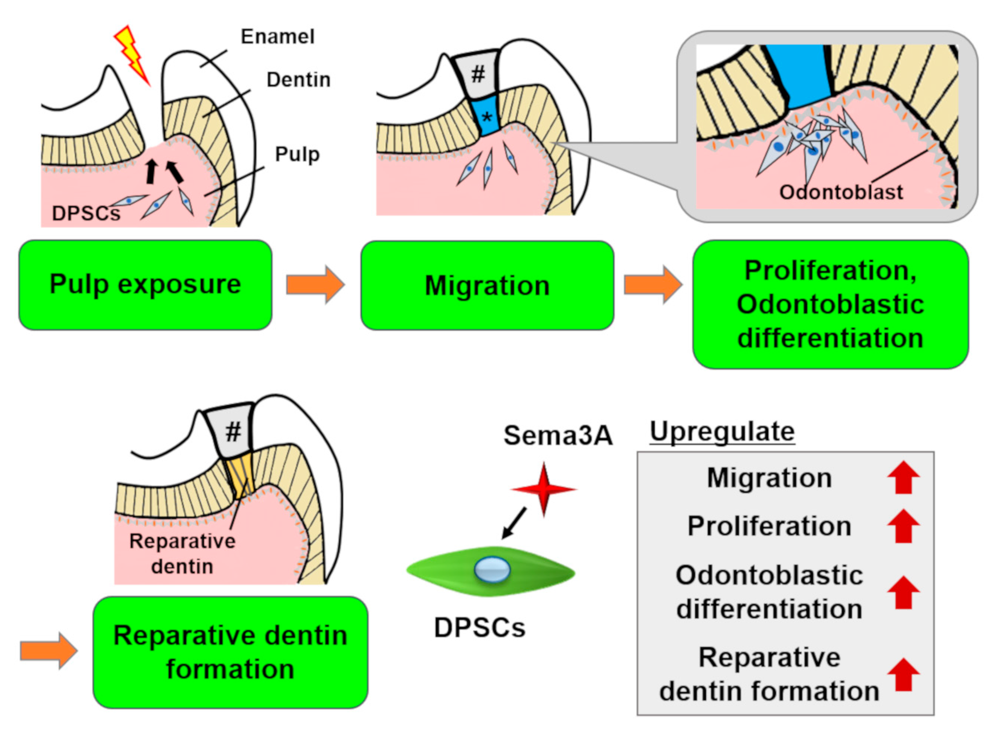
 Figure 4. Schema of the effects of Semaphorin 3A (Sema3A) on DPSCs. In case of pulp exposure, DPSCs migrate toward the exposure site, then they proliferate and differentiate into odontoblasts to form reparative dentin. Sema3A can act as a bioactive agent to facilitate these regenerative processes. #, sealing material; *, Sema3A.
This entry is adapted from the peer-reviewed paper 10.3390/biology9070160
