Your browser does not fully support modern features. Please upgrade for a smoother experience.
Please note this is an old version of this entry, which may differ significantly from the current revision.
Autoimmune liver diseases (AILDs) include autoimmune hepatitis, primary biliary cholangitis and primary sclerosing cholangitis. The etiologies of AILD are not well understood but appear to involve a combination of genetic and environmental factors. AILDs commonly affect young individuals and are characterized by a highly variable clinical course. These diseases significantly influence quality of life and can progress toward liver decompensation or the onset of hepatocellular or cholangiocarcinoma; a significant number of patients eventually progress to end-stage liver disease, requiring liver transplantation.
- gender
- autoimmune liver diseases
- autoimmune hepatitis primary biliary cirrhosis
- primary sclerosing cholangitis
- overlap syndromes
- liver transplant
1. Autoimmune Hepatitis
AIH affects the female sex more than the male sex across all ethnicities and ages (children, 60–76%; adults, 71–95%) [1][2][3][4][5][6][7][8][9][10][11][12]. The male-to-female ratio in the population with AIH is considered to have changed over time, indicating a relative increase in the number of male patients. In Japan, the male-to-female ratio was 1:7 in 2004 and 1:4 in 2016 [13]. It should also be stressed that cases with “acute” presentation (transaminases higher than 10 times the upper limit and bilirubin higher than 5 mg/mL) are increasing worldwide. In an Italian multicenter study, among 479 patients diagnosed as AIH, 202 (43%) met the criteria for “acute” onset, and no significant differences were observed between the sexes [14].
Factors responsible for gender differences in immune response are illustrated in Figure 1.
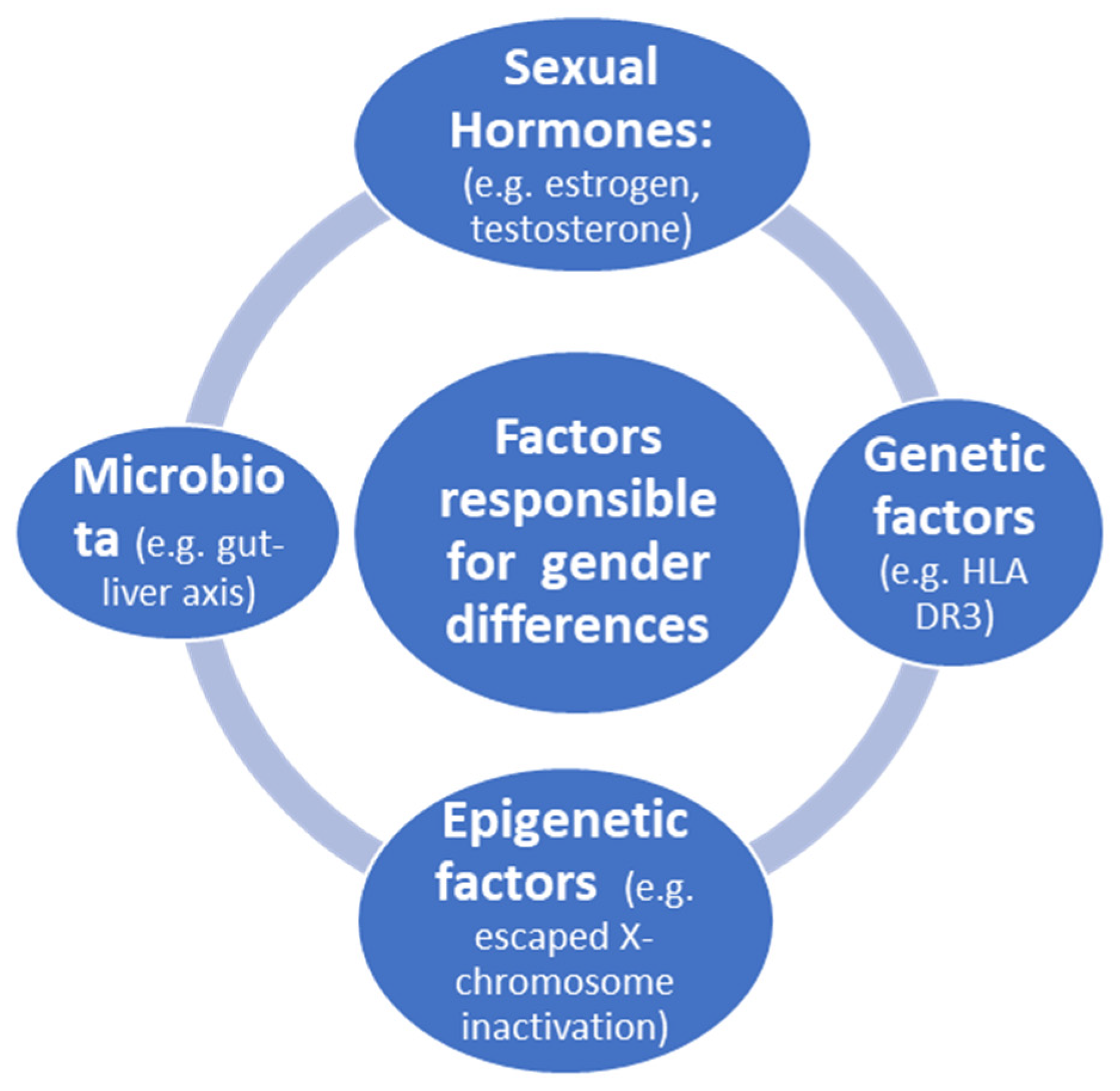
Figure 1. Factors responsible for gender differences in immune response.
Sexual hormones influence innate immunity. High levels of estrogen reduce the synthesis of interleukin-1β (IL-1β), IL-6 and tumor necrosis factor (TNF) by macrophages and monocytes. On the other hand, low levels of estrogen increase the same cytokines [15]. However, in cases in which cytotoxic T cells target liver cells, estrogen may shift the onset of AIH to the late reproductive phase. This is the case of AIH with onset after menopausal state. Moreover, depending on the reproductive status, before puberty, B-cell-dependent autoimmunity plays a key role in AIH, with the production of autoantibodies [15]. Indeed, estrogen also plays a role in natural killer (NK) cells; low estrogen levels increase the activity of NK cells, as well as that of dendritic cells. Estrogen treatment (compared to pregnancy levels) was found to decrease TNF and IL-12 production in mature mouse dendritic cells [16]. These changes also explain the beneficial effect of pregnancy for AIH. During pregnancy, high levels of estrogen and progesterone exert a tremendous inhibitory influence on most inflammatory pathways, but females might be prone to disease flare thereafter in the vulnerable postpartum phase.
Genetic factors linked to X chromosomes have been extensively studied [17][18][19][20]. During embryonic development, one of the two X chromosomes is randomly inactivated in females. This process is initiated by long non-coding RNA X inactivation and results in a cellular mosaicism, where about one-half of the cells in a given tissue express either the maternal X chromosome or the paternal X chromosome. However, X chromosome inactivation is not complete, with 15 to 23% of genes escaping inactivation, thereby contributing to the emergence of a female-specific heterogeneous population of cells with biallelic expression of some X-linked genes. Although X chromosomes provide clues as to the cause of PBC [17], few studies have been published on AIH.
Epigenetic factors were extensively reviewed by Liu et al. [18]. An important issue is the role of X-chromosome-located microRNAs in immunity, which has been hypothesized to contribute to the enhanced immune response of females [19]. The human X chromosome contains 10% of all microRNAs detected to date. According to recent studies, in several mammalian species, including humans and mice, the X chromosome has a higher density of miRNAs, whereas the Y chromosome has only four miRNA sequences (although not experimentally validated) with two shared by both sex chromosomes [20]. Indeed, miRNAs on the X chromosome may influence sex-specific responses. It has been shown that bone-marrow-derived mesenchymal stem-cell-secreted miR-223-containing exosomes prevent liver injury in an autoimmune hepatitis mouse model by suppressing hepatic NLRP3 and caspase-1 [21]. Moreover, modification of miR-223 further improves its therapeutic efficacy against AIH [21]. In general, miR-223 is involved in the pathogenesis of various liver diseases by influencing immune cell differentiation, neutrophil infiltration, macrophage polarization and inflammasome activation by both metabolic and inflammatory signaling pathways [22]. However, a few studies have been conducted on the epigenetics of AIH utilizing miRNAs, but altogether, these studies suggest the possibility that miRNAs could be used as biomarkers for diagnosis and prognosis of AIH [23].
The microbiome can influence a number of physiological aspects of the host, including the immune response, and several studies have attempted to unravel the role of the microbiome in the pathogenesis of autoimmune diseases [24][25]. Moreover, the sex difference in this setting is currently not sufficiently explored, representing a gap that needs to be filled.
Neoplastic risk for hepatocellular carcinoma (HCC) in AIH is lower than other types of liver disease [26]. HCC develops in patients with AIH and cirrhosis in 1–9% of cases, and the annual incidence in patients with cirrhosis is 1.1–1.9% [27], with the same proportion in females and males [28]. Besides cirrhosis, other risk factors linked to HCC are older age, increased frequency of relapses, concurrent alcohol consumption and a trend for male sex [26].
The long-term outcome of patients with AIH was evaluated in 238 patient (51 men) at a single center from 1971 to 2005 [29], showing increased survival in males; however, the reason was unclear. Moreover, the age at death in women with liver-related causes compared to non-liver causes was significantly lower (39 years. vs. 70.5 years., p = 0.001). Interestingly, HLA a1-B8-DR3 (associated with increased susceptibility to AIH) was more than twice as prevalent in males compared to females. When considering a large cohort of patients including all ages, patients diagnosed under the age of 18 years were found to have a significantly reduced life expectancy [30] and were at high risk of relapses and liver transplantation (LT), with no significant differences observed between the sexes [30].
2. Primary Biliary Cholangitis
A significant female preponderance is a well-known clinical feature of PBC, whereas differences between sexes in the clinical presentation at PBC diagnosis are not so well-defined. Twelve studies including 51,290 PBC patients worldwide were analyzed to evaluate sex-related differences at PBC diagnosis [31][32][33][34][35][36][37][38][39][40][41]. Seven studies showed that male sex is associated with delayed diagnosis and, consequently, older age at PBC identification [31][34][35][36][38][40][41]. Six studies showed that male patients received PBC diagnosis at more advanced and severe liver disease with cirrhosis and its decompensation events, as well as portal hypertension signs [32][33][35][36][39][42]. Three studies found that male PBC patients presented with worse liver biochemistries [34][35][42]. Finally, six studies showed that fatigue is more associated with female sex at PBC presentation [32][33][35][36][39][42]. Furthermore, Marzioni et al. analyzed data from electronic medical records of patients from 900 general practitioners in Italy, identifying 412 PBC patients and showing that osteoporosis, inflammatory arthritis and other connective tissue diseases were significantly more common in women, whereas inflammatory bowel diseases were significantly more common in men (p < 0.01) [43].
The clinical impact of PBC is highly variable, and one of the most important factors contributing to this variability is the response to primary therapy with ursodeoxycholic acid (UDCA) [44]. Several studies have demonstrated that the efficacy of UDCA therapy strongly determines long-term outcomes [44][45][46]. Moreover, many studies have evaluated the role of sex in the response to UDCA therapy, although results are conflicting because of small sample size, retrospectivity and different criteria used to determine response. Eleven studies including 9748 patients with PBC were analyzed [31][39][46][47][48][49][50][51][52][53][54]. All of these studies, except one [50], evaluated the impact of sex on UDCA therapy response. In particular, four of studies showed that no response to UDCA is more frequent in the male than female sex [31][47][48][49]. However, although these studies presented large sample sizes (7677 PBC patients), they evaluated response to UDCA with four different criteria, and this heterogeneity makes the results not comparable. Particularly, Carbone et al. [31] used the UK-PBC cohort to evaluate UDCA response with Paris I criteria and demonstrated that sex is an independent predictor of therapy failure. Instead, Cheung et al. [47] analyzed the largest cohort of PBC patients utilizing the GLOBE score criteria. Finally, Lammert et al. [48] used the Toronto criteria, and Tian et al. [49] utilized a combination of the Barcelona and Paris I criteria. A similar heterogeneity in the evaluation of UDCA response was observed in six, studies showing that sex has no impact on response to therapy [39][46][51][52][53][54]. In 2016, obeticholic acid (OCA) was approved as a second-line therapy in PBC patients with inadequate response or intolerant to UDCA [55]. Four studies evaluated the response to OCA therapy according to POISE criteria [56][57][58][59]. Only D’Amato et al. [59] considered the role of sex in OCA therapy and showed that sex has no impact on inadequate response to OCA.
There is currently little information available regarding the exact magnitude of HCC risk in PBC patients according to sex. A recent meta-analysis evaluating 18 studies examining the incidence of HCC in PBC patients according to sex showed a pooled HCC incidence rate of 9.82 per 1000 person-years (95% CI 5.92–16.28) in men and 3.82 per 1000 person-years (95% CI 2.85–5.11) in women, with moderate-to-high between-study heterogeneity [60]. Additionally, Trivedi et al. showed that HCC incidence was higher in male UDCA non-responders versus responders (HR 4.44, 95% CI 1.29 to 10.20; p < 0.001) in a cohort of 4565 PBC patients [61]. Moreover, Harada et al. found that the cumulative incidence of HCC was 6.5% in males and 2.0% in females (p < 0.0001) during the 10 years after PBC diagnosis, indicating that male PBC patients had a 3.3-fold higher risk of HCC compared with female PBC patients [62]. Nonetheless, although PBC primarily affects females, the authors postulated that HCC might be more common in male PBC patients because of a lack of estrogen-mediated prevention. In females, the HCC incidence gradually increased according to histological stage, indicating that the terminal stage of PBC, which is a cirrhotic state, may be a risk factor for HCC development in females, whereas males are likely to develop HCC at any stage [62].
The role of sex in the prognosis of PBC patients has been widely evaluated. Seven studies analyzing the presence of ACLD were considered [33][36][39][42][46][47][63]. All of them showed that the presence of ACLD was more frequent in male than in female patients. In particular, Cheung et al. [47] and Marschall et al. [33] demonstrated that male sex had a higher prevalence of portal hypertension and liver decompensation. Moreover, Adejumo et al. [63] showed that male sex had higher risk of jaundice, spontaneous bacterial peritonitis and acute liver failure, whereas female sex had a higher risk of hospitalizations. Furthermore, 11 studies evaluated the role of sex in mortality for PBC patients [33][38][39][41][44][46][47][64][65][66][67]. Eight of these studies demonstrated that males have a higher risk of mortality. In particular, John et al. [46] showed that male sex is a risk factor for death, liver-related mortality and liver decompensation. Lleo et al. [65] demonstrated that male sex was associated to an increased risk of all-cause mortality.
Primary Sclerosing Cholangitis
PSC is considered an immune-mediated disease with atypical features, including prevalence in men, the absence of disease-specific autoantibodies and poor response to immunosuppression. Regarding gender distribution, PSC affects men prevalently; in a large regional population study from Sweden, the mean crude annual incidence of PSC was 1.22 per 100,000 in the total population aged ≥18 years in the period from 1992 to 2005; among men and women, the incidence was 1.8 and 0.7, respectively. The point prevalence of PSC in the same population was 16 (24 among men and 9 among women) per 100,000, and the proportion of men was 71% [68]. In other nations, proportion of men ranged from 51% in New Zealand [69] to 71% in the USA [70] and in Norway [8]. Similarly, in a recent data collection from the National Rare Diseases Registry (RNMR) and the National Mortality Database (NMD) in Italy, 60% of new PSC diagnoses were in male patients, with a male-to-female ratio of 1.5:1 [71]. Mean age at disease onset was 33 years (SD = 17), and mean age at diagnosis was 37 years. There were no statistically significant differences in age at diagnosis, age at onset and diagnostic delay between male and female patients. In other studies, the median age at PSC onset was generally higher in women than men; a large cohort study from Germany published in 2018 evaluated patients with late disease onset (defined as first diagnosis after 50 years), revealing that the proportion of females was significantly higher in the late-onset group compared with the earlier-onset group (50/183 (27%) vs. 15/32 (47%), p = 0.02) [72]. A study population from Sweden was reported to have a time-trend increase in the incidence of large-duct PSC among women but not among men; conversely, the incidence of small-duct PSC increased significantly among men but not among women. Diverging trends were also observed for the incidence of PSC related to IBD, with a significant increase in the incidence of PSC-IBD in women, whereas in men, an increase in PSC without IBD was observed [68].
Patients with PSC are at increased risk of developing several hepatobiliary cancers, mainly cholangiocarcinoma, gallbladder and colon cancer—and hepatocellular carcinoma to a lesser degree. Cholangiocarcinoma (CCA) is a model of malignancies occurring in the inflammatory background. Chronic inflammation of the biliary tree induced by PSC promotes oncogenesis and predisposes to development of CCA through DNA damage, cellular proliferation and oxidative stress [73][74]. The annual risk for CCA in PSC is approximately 2%, with a 10- and 30-year cumulative incidence of 6–11% and 20%, respectively, and an increase of 400-fold when compared with the general population [75][76]. Regarding the sex-specific risk of CCA development, female sex seems to be associated with a lower risk of CCA (HR,0.68; p < 0.001, respectively), as reported in the data from a large international PSC cohort, which included 7121 patients encompassing >30 years of clinical observation. According to multivariate analysis, advancing age at diagnosis is an independent risk factor of CCA development, whereas female sex and having small-duct disease or CD at the time of PSC diagnosis are protective factors against CCA development [77].
Inflammatory bowel disease occurs in 70–80% of patients with PSC, and PSC seems to confer additional risk of developing colorectal cancer (CRC) when compared with the risk in patients with IBD alone [78][79]. CRC can appear in up to 20–30% of PSC-IBD patients, and an annual colonoscopy is recommended [80].
In 2020, a nationwide population-based study from national healthcare registries in England identified incident cases of IBD with and without PSC over ten years; the study showed that patients with PSC-IDB younger than 40 years old had a fourfold higher risk of CRC, whereas there was no difference between groups for patients in which the IBD diagnosis was made in patients older than 60 years. Regarding sex differences, the risk for CRC was significantly lower among women than men (HR 0.46 p < 0.001) [81]. The oncologic additive risk of association between PSC and IBD was confirmed in a Spanish multicenter retrospective cohort study. The risk of CRC was increased four- to fivefold in PSC-IBD patients compared to IBD controls, including patients submitted to annual colonoscopic surveillance [82].
Data related to sex differences in PSC clinical presentation and evolution are scarce. From the Italian registry, including 502 PSC patients in population-based data, the survival rate was 92% at 10 years from diagnosis and 82% at 20 years, considering all causes of deaths. The Kaplan–Meier curves show no significant difference between male and female patients with respect to estimated survival times from diagnosis [71]. The International PSC Study Group published a multicenter outcome study in 2017 to describe the natural history of the disease, including 7121 patients across 17 countries and encompassing ≥30 years of clinical observation from 1980 through 2010. The study registered sex-specific variations in clinical phenotype and correlations with liver disease progression and neoplastic complications. Men comprised the majority of the cohort (66%) and were younger than women (average age of 37 years vs. 40 years). Women more commonly exhibited small-duct PSC phenotype, ulcerative colitis (UC) was less common in women than men (48% vs. 61%, respectively p < 0.001) and small-duct PSC was characterized by a low-risk phenotype in both sexes (adjusted HR for men, 0.23; p < 0.001 and adjusted HR for women, 0.48; p = 0.003). Female sex was an independent protective factor against liver progression; in particular, females maintained a significantly higher transplant-free survival than males matched for age and PSC phenotype. Moreover, the lower prevalence of UC in women may partially account for differences in liver disease progression between the sexes.
3. Overlap Syndromes and Gender
The term overlap syndrome (OS) describes a subtype of clinical syndromes that share common features relating to AIH and PBC or AIH and PSC. Very rarely, an overlap syndrome between PBC and PSC has been described. However, the term remains controversial, and it is not known whether overlaps are situations occurring simultaneously or represent a different development during the natural course of the disease.
PBC-AIH is the most common form of overlap syndromes [83][84]. A systematic review included 17 studies of PBC-AIH comprising a total of 402 patients [85]. Female gender was present in 87–100% of either retrospective or prospective studies.
AIH-PSC overlap syndrome has been described in both children and adults. In children, the syndrome is particularly important, reported in up to 40% of patients with AIH [86]. Adults diagnosed with AIH-PSC overlap are significantly younger at the time of diagnosis than those with classical PSC (24–27 years vs. 39–46 years, respectively) [87][88][89]. The proportion of adult males with AIH-PSC overlap is 69–81%, which is higher than in AIH [90]. However, the proportion of male gender in AIH-PSC undergoing liver transplantation was slightly lower (49%) than the number reported in classical PSC [91].
4. Conclusions
The mechanisms behind the sex differences observed in autoimmune liver diseases, specifically the female predominance in AIH and PBC; the worse disease course in male PBC; male predominance in PSC patients with a lower risk of UC; and cholangiocarcinoma among female patients remain largely unknown. Understanding the effects of sex-related genes and intestinal microbiota underlying AILD immune dysregulation, as well as the role of sex hormones in immune cells, may pave the way for novel treatment strategies for AILD.
In PBC patients, the frequent delay in diagnosis plays an important role among the male sex, leading to more advanced liver disease at PBC presentation; consequently, the risk of ACLD, liver decompensation, HCC development and mortality is higher in male than in female patients, with worse biochemical response rates. Delayed diagnosis could be partially explained by the lower incidence of PBC in male patients, leading clinicians to not consider the disease as a first choice, was well the minor presence of PBC-related symptoms. Despite its rarity, the diagnosis of PBC should be considered in men with elevated cholestatic parameters maintaining a high index of suspicion for PBC to prevent diagnostic delays.
This entry is adapted from the peer-reviewed paper 10.3390/jpm12060925
References
- Coss Adame, E.; Granados, J.; Uribe, M.; Torre, A. Does HLA-DR7 differentiate the overlap syndrome of auto-immune hepatitis-primary biliary cirrhosis (AIH-PBC) from those with auto-immune hepatitis type 1? Ann. Hepatol. 2011, 10, 28–32.
- Manns, M.P.; Czaja, A.J.; Gorham, J.D.; Krawitt, E.L.; Mieli-Vergani, G.; Vergani, D.; Vierling, J.M. Diagnosis and management of autoimmune hepatitis. Hepatology 2010, 51, 2193–2219.
- Gregorio, G.V.; Portmann, B.; Reid, F.; Donaldson, P.T.; Doherty, D.G.; McCartney, M.; Mowat, A.P.; Vergani, D.; Milei-Vergani, G. Autoimmune hepatitis in childhood: A 20-year experience. Hepatology 1997, 25, 541–547.
- Radhakrishnan, K.R.; Alkhouri, N.; Worley, S.; Arrigain, S.; Hupertz, V.; Kay, M.; Yerian, L.; Wyllie, R.; Feldstein, A.E. Autoimmune hepatitis in children—Impact of cirrhosis at presentation on natural history and long-term outcome. Dig. Liver Dis. 2010, 42, 724–728.
- Deneau, M.; Jensen, M.K.; Holmen, J.; Williams, M.S.; Book, L.S.; Guthery, S.L. Primary sclerosing cholangitis, autoimmune hepatitis, and overlap in Utah children: Epidemiology and natural history. Hepatology 2013, 58, 1392–1400.
- Jimenez-Rivera, C.; Ling, S.C.; Ahmed, N.; Yap, S.; Aglipay, M.; Borrowman, N.; Graitson, S.; Critch, J.; Rashid, M.; Ng, V.L.; et al. Incidence and characteristics of autoimmune hepatitis. Pediatrics 2015, 136, e1237–e1248.
- Czaja, A.J.; Carpenter, H.A.; Santrach, P.J.; Moore, S.B.; Taswell, H.F.; Homburger, H.A. Evidence against hepatitis viruses as important causes of severe autoimmune hepatitis in the United States. J. Hepatol. 1993, 18, 342–352.
- Boberg, K.M.; Aadland, E.; Jahnsen, J.; Raknerud, N.; Stiris, M.; Bell, H. Incidence and prevalence of primary biliary cirrhosis, primary sclerosing cholangitis, and autoimmune hepatitis in a Norwegian population. Scand. J. Gastroenterol. 1998, 33, 99–103.
- Werner, M.; Prytz, H.; Ohlsson, B.; Almer, S.; Bjornsson, E.; Berquist, A.; Wallerstedt, S.; Sandberg-Gertzén, H.; Hultcrantz, R.; Sangfelt, P.; et al. Epidemiology and the initial presentation of autoimmune hepatitis in Sweden: A nationwide study. Scand. J. Gastroenterol. 2008, 43, 1232–1240.
- Ngu, J.H.; Bechly, K.; Chapman, B.A.; Burt, M.J.; Barclay, M.L.; Gearry, R.B.; Stedman, C.A.M. Population-based epidemiology study of autoimmune hepatitis: A disease of older women? J. Gastroenterol. Hepatol. 2010, 25, 1681–1686.
- Gronbaek, L.; Vilstrup, H.; Jepsen, P. Autoimmune hepatitis in Denmark: Incidence, prevalence, prognosis, and causes of death. A nationwide registry-based cohort study. J. Hepatol. 2014, 60, 612–617.
- Van Gerven, N.M.; Vermer, N.J.; Witte, B.I.; van Erpecum, K.J.; van Buuren, H.R.; Maijers, I.; Visscher, A.P.; Verschuren, E.C.; van Hoek, B.; Coenraad, M.J.; et al. Epidemiology and clinical characteristics of autoimmune hepatitis in the Netherlands. Scand. J. Gastroenterol. 2014, 49, 1245–1254.
- Tanaka, A.; Mori, M.; Matsumoto, K.; Ohiza, H.; Takazuma, S.; Takikawa, H. Increase trend in the prevalence and to male-to-female ratio of primary biliary cholangitis, autoimmune hepatitis and primary sclerosing cholangitis in Japan. Hepatol. Res. 2019, 49, 881–889.
- Muratori, P.; Carbone, M.; Stangos, G.; Perini, L.; Lalanne, C.; Ronca, V.; Cazzagon, N.; Bianchi, G.; Lenzi, M.; Floreani, A.; et al. Clinical and prognostic implications of acute onset of autoimmune hepatitis: An Italian multicentre study. Dig. Liver Dis. 2018, 50, 698–702.
- Straub, R.H. The complex role of estrogens in inflammation. Endocr. Rev. 2007, 28, 521–574.
- Liu, H.Y.; Buenafe, A.C.; Matejuk, A.; Ito, A.; Zamora, A.; Dwyer, J.; Vandenbark, A.A.; Offner, H. Estrogens inhibition of EAE involves effects on dendritic cell function. J. Neurosci. Res. 2002, 70, 238–248.
- Asselta, R.; Paraboschi, E.M.; Gerussi, A.; Cordell, H.J.; Mells, G.F.; Sandford, R.N.; Jones, D.E.; Nakamura, M.; Ueno, K.; Hitomi, Y. X chromosome contribution to the genetic architecture of primary biliary cholangitis. Gastroenterology 2021, 160, 2483–2495.
- Liu, Q.; Li, Y.; Ma, X.; Tang, R. Epigenetics of autoimmune liver diseases: Current progress and future directions. J. Bio-X Res. 2019, 2, 46–55.
- Pinheiro, I.; Dejager, L.; Libert, C. X-chromosome-located microRNAs in immunity: Might they explain male/female differences? Bioassays 2011, 33, 791–802.
- Di Palo, A.; Siniscalchi, C.; Salerno, M.; Russo, A.; Gravholt, C.H.; Potenza, N. What microRNAs could tell us about the X chromosome. Cell Mol. Life Sci. 2020, 77, 4069–4080.
- Chen, L.; Lu, F.-B.; Chen, D.-Z.; Wu, J.-L.; Hu, E.; Xu, L.M.; Zheng, M.-H.; Li, H.; Huang, Y.; Jin, X.-Y. BMSCs-derived miR-223-containing exosomes contribute to liver protection in experimental autoimmune hepatitis. Mol. Immunol. 2018, 93, 38–46.
- Ye, D.; Zhang, T.; Lou, G.; Liu, Y. Role of miR-223 in the pathophysiology of liver diseases. Exp. Mol. Med. 2018, 50, 128.
- Huang, C.; Xing, X.; Xiang, X.; Fan, X.; Men, R.; Ye, T.; Yang, L. MicroRNAs in autoimmune liver diseases: From diagnosis to potential therapeutic targets. Biomed. Pharmacother. 2020, 13, 110558.
- De Luca, F.; Shoenfeld, Y. The microbiome in autoimmune diseases. Clin. Exp. Immunol. 2019, 195, 74–85.
- Zheng, D.; Liwinski, T.; Elinav, E. Interaction between microbiota and immunity in health and disease. Cell Res. 2020, 30, 492–506.
- Tansel, A.; Katz, L.H.; El-Serag, H.B.; Thrift, A.P.; Parepally, M.; Shakhatreh, M.H.; Kanwal, F. Incidence and determinants of hepatocellular carcinoma in autoimmune hepatitis: A systematic review and meta-analysis. Clin. Gastroenterol. Hepatol. 2017, 15, 1207–1217.
- Czaja, A.J. Hepatocellular carcinoma and other malignancies in autoimmune hepatitis. Dig. Dis. Sci. 2013, 58, 1459–1476.
- Yeoman, A.D.; Al-Chalabi, T.; Karani, J.B.; Quaglia, A.; Devlin, J.; Mieli-Vergani, G.; Bomford, A.; O’Grady, J.G.; Harrison, P.M.; Heneghan, M.A. Evaluation of risk factors in the development of hepatocellular carcinoma in autoimmune hepatitis: Implication for follow-up and screening. Hepatology 2008, 48, 863–870.
- Al-Chalabi, T.; Underhill, J.A.; Portmann, B.C.; McFarlane, I.G.; Heneghan, M.A. Impact of gender on the long-term survival of patients with autoimmune hepatitis. J. Hepatol. 2008, 48, 140–147.
- Kirstein, M.M.; Metzler, F.; Geiger, E.; Einrich, E.; Hallensleben, M.; Manns, M.P.; Vogel, A. Prediction of short- and long-term outcome in patients with autoimmune hepatitis. Hepatology 2015, 62, 1524–1535.
- Carbone, M.; Mells, G.F.; Pells, G.; Dawwas, M.F.; Newton, J.L.; Heneghan, M.A.; Neuberger, J.M.; Day, D.B.; Ducker, S.J.; UK PBC Consortium; et al. Sex and age are determinants of the clinical phenotype of primary biliary cirrhosis and response to ursodeoxycholic acid. Gastroenterology 2013, 144, 560–569.e7.
- Ali, A.H.; Sinakos, E.; Silveira, M.G.; Jorgensen, R.A.; Angulo, P.; Lindor, K.D. Varices in early histological stage primary biliary cirrhosis. J. Clin. Gastroenterol. 2011, 45, e66–e71.
- Marschall, H.U.; Henriksson, I.; Lindberg, S.; Söderdahl, F.; Thuresson, M.; Wahlin, S.; Ludvigsson, J.F. Incidence, prevalence, and outcome of primary biliary cholangitis in a nationwide Swedish population-based cohort. Sci. Rep. 2019, 9, 11525.
- Drazilova, S.; Babinska, I.; Gazda, J.; Halanova, M.; Janicko, M.; Kucinsky, B.; Safcak, D.; Martinkova, D.; Tarbajova, L.; Cekanova, A.; et al. Epidemiology and clinical course of primary biliary cholangitis in Eastern Slovakia. Int. J. Public Health 2020, 65, 683–691.
- Chen, S.; Duan, W.; Li, M.; Li, S.; Lv, T.; Tian, Q.; Wang, Q.; Wu, X.; Zhao, X.; Wang, X.; et al. Prognosis of 732 ursodeoxycholic acid-treated patients with primary biliary cholangitis: A single center follow-up study from China. J. Gastroenterol. Hepatol. 2019, 34, 1236–1241.
- Abdulkarim, M.; Zenouzi, R.; Sebode, M.; Schulz, L.; Quaas, A.; Lohse, A.W.; Schramm, C.; Weiler-Normann, C. Sex differences in clinical presentation and prognosis in patients with primary biliary cholangitis. Scand. J. Gastroenterol. 2019, 54, 1391–1396.
- Yagi, M.; Tanaka, A.; Abe, M.; Namisaki, T.; Yoshiji, H.; Takahashi, A.; Ohira, H.; Komori, A.; Yamagiwa, S.; Kikuchi, K.; et al. Symptoms and health-related quality of life in Japanese patients with primary biliary cholangitis. Sci. Rep. 2018, 8, 12542.
- Cheung, K.S.; Seto, W.K.; Fung, J.; Lai, C.L.; Yuen, M.F. Epidemiology and Natural History of Primary Biliary Cholangitis in the Chinese: A Territory-Based Study in Hong Kong between 2000 and 2015. Clin. Transl. Gastroenterol. 2017, 8, e116.
- Gatselis, N.K.; Zachou, K.; Lygoura, V.; Azariadis, K.; Arvaniti, P.; Spyrou, E.; Papadamou, G.; Koukoulis, G.K.; Dalekos, G.N.; Rigopoulou, E.I. Geoepidemiology, clinical manifestations and outcome of primary biliary cholangitis in Greece. Eur. J. Intern. Med. 2017, 42, 81–88.
- Kim, K.A.; Ki, M.; Choi, H.Y.; Kim, B.H.; Jang, E.S.; Jeong, S.H. Population-based epidemiology of primary biliary cirrhosis in South Korea. Aliment. Pharmacol. Ther. 2016, 43, 154–162.
- Boonstra, K.; Bokelaar, R.; Stadhouders, P.H.; Tuynman, H.A.; Poen, A.C.; van Nieuwkerk, K.M.; Witteman, E.M.; Hamann, D.; Witteman, B.J.; Beuers, U.; et al. Increased cancer risk in a large population-based cohort of patients with primary biliary cirrhosis: Follow-up for up to 36 years. Hepatol. Int. 2014, 8, 266–274.
- Younossi, Z.M.; Stepanova, M.; Golabi, P.; Epstein, R.S.; Strauss, M.E.; Nader, F.; Racila, A. Factors Associated with Potential Progressive Course of Primary Biliary Cholangitis: Data from Real-world US Database. J. Clin. Gastroenterol. 2019, 53, 693–698.
- Marzioni, M.; Bassanelli, C.; Ripellino, C.; Urbinati, D.; Alvaro, D. Epidemiology of primary biliary cholangitis in Italy: Evidence from a real-world database. Dig. Liver Dis. 2019, 51, 724–729.
- Lammers, W.J.; Hirschfield, G.M.; Corpechot, C.; Nevens, F.; Lindor, K.D.; Janssen, H.L.; Floreani, A.; Ponsioen, C.Y.; Mayo, M.J.; Invernizzi, P.; et al. Development and Validation of a Scoring System to Predict Outcomes of Patients with Primary Biliary Cirrhosis Receiving Ursodeoxycholic Acid Therapy. Gastroenterology 2015, 149, 1804–1812.e4.
- Zhang, L.N.; Shi, T.Y.; Shi, X.H.; Wang, L.; Yang, Y.J.; Liu, B.; Gao, L.X.; Shuai, Z.W.; Kong, F.; Chen, H.; et al. Early biochemical response to ursodeoxycholic acid and long-term prognosis of primary biliary cirrhosis: Results of a 14-year cohort study. Hepatology 2013, 58, 264–272.
- John, B.V.; Aitcheson, G.; Schwartz, K.B.; Khakoo, N.S.; Dahman, B.; Deng, Y.; Goldberg, D.; Martin, P.; Taddei, T.H.; Levy, C.; et al. Male Sex Is Associated with Higher Rates of Liver-Related Mortality in Primary Biliary Cholangitis and Cirrhosis. Hepatology 2021, 74, 879–891.
- Cheung, A.C.; Lammers, W.J.; Murillo Perez, C.F.; van Buuren, H.R.; Gulamhusein, A.; Trivedi, P.J.; Lazaridis, K.N.; Ponsioen, C.Y.; Floreani, A.; Hirschfield, G.M.; et al. Effects of Age and Sex of Response to Ursodeoxycholic Acid and Transplant-free Survival in Patients with Primary Biliary Cholangitis. Clin. Gastroenterol. Hepatol. 2019, 17, 2076–2084.e2.
- Lammert, C.; Juran, B.D.; Schlicht, E.; Chan, L.L.; Atkinson, E.J.; de Andrade, M.; Lazaridis, K.N. Biochemical response to ursodeoxycholic acid predicts survival in a North American cohort of primary biliary cirrhosis patients. J. Gastroenterol. 2014, 49, 1414–1420.
- Tian, S.; Liu, Y.; Sun, K.; Zhou, X.; Ma, S.; Zhang, M.; Zhou, X.; Wang, L.; Han, Y. A nomogram based on pretreatment clinical parameters for the prediction of inadequate biochemical response in primary biliary cholangitis. J. Clin. Lab. Anal. 2020, 34, e23501.
- Melchor-Mendoza, Y.K.; Martínez-Benítez, B.; Mina-Hawat, A.; Rodríguez-Leal, G.; Duque, X.; Moran-Villota, S. Ursodeoxycholic Acid Therapy in Patients with Primary Biliary Cholangitis with Limited Liver Transplantation Availability. Ann. Hepatol. 2017, 16, 430–435.
- Delgado, J.S.; Vodonos, A.; Delgado, B.; Jotkowitz, A.; Rosenthal, A.; Fich, A.; Novack, V. Primary biliary cirrhosis in Southern Israel: A 20 year follow up study. Eur. J. Intern. Med. 2012, 23, e193–e198, Erratum in Lancet 2015, 386, 1536.
- Chen, J.; Xue, D.; Gao, F.; Tao, L.; Li, Y.; Zhang, Q.; Wang, R.; Sun, L.; Yang, X.; Liu, Y.; et al. Influence factors and a predictive scoring model for measuring the biochemical response of primary biliary cholangitis to ursodeoxycholic acid treatment. Eur. J. Gastroenterol. Hepatol. 2018, 30, 1352–1360.
- Madir, A.; Božin, T.; Mikolašević, I.; Milić, S.; Štimac, D.; Mijić, M.; Filipec Kanižaj, T.; Biloglav, Z.; Lucijanić, M.; Lucijanić, I.; et al. Epidemiological and clinical features of primary biliary cholangitis in two Croatian regions: A retrospective study. Croat. Med. J. 2019, 60, 494–502.
- Cortez-Pinto, H.; Liberal, R.; Lopes, S.; Machado, M.V.; Carvalho, J.; Dias, T.; Santos, A.; Agostinho, C.; Figueiredo, P.; Loureiro, R.; et al. Predictors for incomplete response to ursodeoxycholic acid in primary biliary cholangitis. Data from a national registry of liver disease. United Eur. Gastroenterol. J. 2021, 9, 699–706.
- European Association for the Study of the Liver. Clinical Practice Guidelines: The Diagnosis and Management of Patients with Primary Biliary Cholangitis. J. Hepatol. 2017, 67, 145–172.
- Roberts, S.B.; Ismail, M.; Kanagalingam, G.; Mason, A.L.; Swain, M.G.; Vincent, C.; Yoshida, E.M.; Tsien, C.; Flemming, J.A.; Janssen, H.L.A.; et al. Real-World Effectiveness of Obeticholic Acid in Patients with Primary Biliary Cholangitis. Hepatol. Commun. 2020, 4, 1332–1345.
- Gomez, E.; Garcia Buey, L.; Molina, E.; Casado, M.; Conde, I.; Berenguer, M.; Jorquera, F.; Simón, M.A.; Olveira, A.; Hernández-Guerra, M.; et al. Effectiveness and safety of obeticholic acid in a Southern European multicentre cohort of patients with primary biliary cholangitis and suboptimal response to ursodeoxycholic acid. Aliment Pharmacol. Ther. 2021, 53, 519–530.
- Harms, M.H.; Hirschfield, G.M.; Floreani, A.; Mayo, M.J.; Parés, A.; Liberman, A.; Malecha, E.S.; Pencek, R.; MacConell, L.; Hansen, B.E. Obeticholic acid is associated with improvements in AST-to-platelet ratio index and GLOBE score in patients with primary biliary cholangitis. JHEP Rep. 2020, 3, 100191.
- D’Amato, D.; De Vincentis, A.; Malinverno, F.; Viganò, M.; Alvaro, D.; Pompili, M.; Picciotto, A.; Palitti, V.P.; Russello, M.; Storato, S.; et al. Real-world experience with obeticholic acid in patients with primary biliary cholangitis. JHEP Rep. 2021, 3, 100248.
- Natarajan, Y.; Tansel, A.; Patel, P.; Emologu, K.; Shukla, R.; Qureshi, Z.; El-Serag, H.B.; Thrift, A.P.; Kanwal, F. Incidence of Hepatocellular Carcinoma in Primary Biliary Cholangitis: A Systematic Review and Meta-Analysis. Dig. Dis. Sci. 2021, 66, 2439–2451.
- Trivedi, P.J.; Lammers, W.J.; van Buuren, H.R.; Parés, A.; Floreani, A.; Janssen, H.L.; Invernizzi, P.; Battezzati, P.M.; Ponsioen, C.Y.; Corpechot, C.; et al. Stratification of hepatocellular carcinoma risk in primary biliary cirrhosis: A multicentre international study. Gut 2016, 65, 321–329.
- Harada, K.; Hirohara, J.; Ueno, Y.; Nakano, T.; Kakuda, Y.; Tsubouchi, H.; Ichida, T.; Nakanuma, Y. Incidence of and risk factors for hepatocellular carcinoma in primary biliary cirrhosis: National data from Japan. Hepatology 2013, 57, 1942–1949.
- Adejumo, A.C.; Akhtar, D.H.; Dennis, B.B.; Cholankeril, G.; Alayo, Q.; Ogundipe, O.A.; Kim, D.; Ahmed, A. Gender and Racial Differences in Hospitalizations for Primary Biliary Cholangitis in the USA. Dig. Dis. Sci. 2021, 66, 1461–1476.
- Fan, X.; Wang, T.; Shen, Y.; Xi, X.; Yang, L. Underestimated Male Prevalence of Primary Biliary Cholangitis in China: Results of a 16-yr cohort study involving 769 patients. Sci. Rep. 2017, 7, 6560.
- Lleo, A.; Jepsen, P.; Morenghi, E.; Carbone, M.; Moroni, L.; Battezzati, P.M.; Podda, M.; Mackay, I.R.; Gershwin, M.E.; Invernizzi, P. Evolving Trends in Female to Male Incidence and Male Mortality of Primary Biliary Cholangitis. Sci. Rep. 2016, 6, 25906.
- Lu, M.; Zhou, Y.; Haller, I.V.; Romanelli, R.J.; VanWormer, J.J.; Rodriguez, C.V.; Anderson, H.; Boscarino, J.A.; Schmidt, M.A.; Daida, Y.G.; et al. Increasing Prevalence of Primary Biliary Cholangitis and Reduced Mortality with Treatment. Clin. Gastroenterol. Hepatol. 2018, 16, 1342–1350.e1.
- Sayiner, M.; Golabi, P.; Stepanova, M.; Younossi, I.; Nader, F.; Racila, A.; Younossi, Z.M. Primary Biliary Cholangitis in Medicare Population: The Impact on Mortality and Resource Use. Hepatology 2019, 69, 237–244.
- Lindkvist, B.; Benito de Valle, M.; Gullberg, B.; Bjornsson, E. Incidence and Prevalence of Primary SclerosingCholangitis in a Defined Adult Population in Sweden. Hepatology 2010, 52, 571–577.
- Lamba, M.; Hieng Ngu, J.; Stedman, C.A.M. Trends in Incidence of Autoimmune Liver Diseases and Increasing Incidence of Autoimmune Hepatitis. Clin. Gastroenterol. Hepatol. 2020, 19, 573–579.e1.
- Bakhshi, Z.; Hilscher, M.B.; Gores, G.J.; Harmsen, W.S.; Viehman, J.K.; LaRusso, N.F.; Gossard, A.A.; Lazaridis, K.N.; Lindor, K.D.; Eaton, J.E. An update on primary sclerosing cholangitis epidemiology, outcomes and quantification of alkaline phosphatase variability in a population-based cohort. J. Gastroenterol. 2020, 55, 523–532.
- Carbone, M.; Kodra, Y.; Rocchetti, A.; Manno, V.; Minelli, G.; Gerussi, A.; Ronca, V.; Malinverno, F.; Cristoferi, L.; Floreani, A.; et al. Primary Sclerosing Cholangitis: Burden of Disease and Mortality Using Data from the National Rare Diseases Registry in Italy. Int. J. Environ. Res. Public Health 2020, 17, 3095.
- Trivedi, P.J.; Crothers, H.; Mytton, J.; Bosch, S.; Iqbal, T.; Ferguson, J.; Hirschfield, G.M. Effects of Primary Sclerosing Cholangitis on Risks of Cancer and Death in People with Inflammatory Bowel Disease, Based on Sex, Race, and Age. Gastroenterology 2020, 159, 915–928.
- Rizvi, S.; Gores, G.J. Pathogenesis, diagnosis, and management of cholangiocarcinoma. Gastroenterology. 2013, 145, 1215–1229.
- Jaiswal, M.; La Russo, N.F.; Burgart, L.J.; Gores, G.J. Inflammatory cytokines induce DNA damage and inhibit DNA repair in cholangiocarcinoma cells by a nitric oxide-dependent mechanism. Cancer Res. 2000, 60, 184–190.
- Boonstra, K.; Weersma, R.K.; van Erpecum, K.J.; Rauws, E.A.; Spanier, B.W.; Poen, A.C.; van Nieuwkerk, K.M.; Drenth, J.P.; Witteman, B.J.; Tuynman, H.A.; et al. Population-based epidemiology, malignancy risk, and outcome of primary sclerosing cholangitis. Hepatology 2013, 58, 2045–2055.
- Kornfeld, D.; Ekbom, A.; Ihre, T. Survival and risk of cholangiocarcinoma in patients with primary sclerosing cholangitis. A population-based study. Scand. J. Gastroenterol. 1997, 32, 1042–1045.
- Weismüller, T.J.; Trivedi, P.J.; Bergquist, A.; Imam, M.; Lenzen, H.; Ponsioen, C.Y.; Holm, K.; Gotthardt, D.; Färkkilä, M.A.; Marschall, H.U.; et al. Patient Age, Sex, and Inflammatory Bowel Disease Phenotype Associate with Course of Primary Sclerosing Cholangitis. Gastroenterology 2017, 152, 1975–1984.e8.
- Soetikno, R.M.; Lin, O.S.; Heidenreich, P.A.; Young, H.S.; Blackstone, M.O. Increased risk of colorectal neoplasia in patients with primary sclerosing cholangitis and ulcerative colitis: A meta-analysis. Gastrointest. Endosc. 2002, 56, 48–54.
- Zheng, H.H.; Jiang, X.L. Increased risk of colorectal neoplasia in patients with primary sclerosing cholangitis and inflammatory bowel disease: A meta-analysis of 16 observational studies. Eur. J. Gastroenterol. Hepatol. 2016, 28, 383–390.
- Karlsen, T.H. Primary sclerosing cholangitis: 50 years of a gut-liver relationship and still no love? Gut 2016, 65, 1579–1581.
- Rupp, C.; Rössler, A.; Zhou, T.; Rauber, C.; Friedrich, K.; Wannhoff, A.; Weiss, K.H.; Sauer, P.; Schirmacher, P.; Süsal, C.; et al. Impact of age at diagnosis on disease progression in patients with primary sclerosing cholangitis. United Eur. Gastroenterol. J. 2018, 6, 255–262.
- Guerra, I.; Bujanda, L.; Castro, J.; Merino, O.; Tosca, J.; Camps, B.; Gutiérrez, A.; Gordillo Ábalos, J.; de Castro, L.; Iborra, M.; et al. Clinical Characteristics, Associated Malignancies and Management of Primary Sclerosing Cholangitis in Inflammatory Bowel Disease Patients: A Multicentre Retrospective Cohort Study. J. Crohns Colitis 2019, 13, 1492–1500.
- Carey, E.J.; Ali, A.H.; Lindor, K.D. Primary biliary cirrhosis. Lancet 2015, 386, 1565–1575.
- Floreani, A.; Franceschet, I.; Cazzagon, N. Primary biliary cirrhosis: Overlaps with other autoimmune disorders. Semin. Liver Dis. 2014, 34, 352–360.
- Freedman, B.L.; Danford, C.J.; Patwardhan, V.; Bonder, A. Treatment of overlap syndromes in autoimmune liver disease: A systematic review and meta-analysis. J. Clin. Med. 2020, 9, 1449.
- Gregorio, G.V.; Portmann, B.; Karani, J.; Harrison, P.; Donaldson, P.T.; Vergani, D.; Mieli-Vergani, G. Autoimmune hepatitis/sclerosing cholangitis overlap syndrome in childhood: A 16-year prospective study. Hepatology 2001, 33, 544–553.
- Abdalian, R.; Dhar, P.; Jhaveri, K.; Haider, M.; Guindi, M.; Heathcote, E.J. Prevalence of sclerosing cholangitis in adults with autoimmune hepatitis: Evaluating the role of routine magnetic resonance imaging. Hepatology 2008, 47, 949–957.
- Al-Chalabi, T.; Portmann, B.C.; Bernal, W.; McFarlane, I.G.; Heneghan, M.A. Autoimmune hepatitis overlap syndromes: An evaluation of treatment response, long-term outcome and survival. Aliment Pharmacol. Ther. 2008, 28, 209–220.
- Gheorghe, L.; Iacob, S.; Gheorghe, C.; Iacob, R.; Simionov, I.; Vadan, R.; Becheanu, G.; Parvulescu, I.; Toader, C. Frequency and predictive factors for overlap syndrome between autoimmune hepatitis and primary cholestatic liver disease. Eur. J. Gastroenterol. Hepatol. 2004, 16, 585–592.
- Lüth, S.; Kanzler, S.; Frenzel, C.; Kasper, H.U.; Dienes, H.P.; Schramm, C.; Galle, P.R.; Herkel, J.; Lohse, A.W. Characteristics and long-term prognosis of the autoimmune hepatitis/primary sclerosing cholangitis overlap syndrome. J. Clin. Gastroenterol. 2009, 43, 75–80.
- Chayanupatkul, M.; Fiel, M.I.; Schiano, T.D. The clinical characteristics, pre- and post-liver transplantation outcomes in patients having autoimmune overlap syndromes. Clin. Transplant. 2020, 34, e13841.
This entry is offline, you can click here to edit this entry!