The HIF-1α and Gastric Cancer: History
Please note this is an old version of this entry, which may differ significantly from the current revision.
Contributor: ,

Gastric cancer is one of the most aggressive tumors in the clinic that is resistant to chemotherapy. Gastric tumors are rich in hypoxic niches, and high expression of hypoxia-inducible factor-1α is associated with poor prognosis. Hypoxia is the principal architect of the topographic heterogeneity in tumors. Hypoxia-inducible factor-1α (HIF-1α) reinforces all hallmarks of cancer and donates cancer cells with more aggressive characteristics at hypoxic niches. HIF-1α potently induces sustained growth factor signaling, angiogenesis, epithelial–mesenchymal transition, and replicative immortality. Hypoxia leads to the selection of cancer cells that evade growth suppressors or apoptotic triggers and deregulates cellular energetics. HIF-1α is also associated with genetic instability, tumor-promoting inflammation, and escape from immunity. 

  • gastric cancer
  • hypoxia
  • hypoxia-inducible factor-1α

1. HIF-1α and Hallmarks in Gastric Cancer

1.1. HIF-1α and Sustained Growth Factor Signaling in Gastric Cancer

Growth factor (GF) signaling is tightly regulated in normal tissues to control the cell number and maintain homeostasis. Cancer cells circumvent this regulation by producing GFs themselves, upregulating the GF receptors (GFRs), or employing constitutive active signaling proteins downstream to the GF receptors. This way, dependence on GFs and GFRs for cellular growth is eliminated [1]. Hypoxia potently induces these mechanisms leading to the sustained proliferation of cancer cells (Figure 1).
Figure 1. Induction of cancer hallmarks by hypoxia and HIF-1α in gastric cancer. Hypoxia induces sustained growth factor signaling and evasion from growth suppressors, resistance to apoptosis, replicative immortality, angiogenesis, and epithelial–mesenchymal transition in gastric cancer. Processes for which there is specific evidence in gastric cancer are shown in blue. Processes common in different cancers are shown in light pink. Upregulation or downregulation of specific proteins is shown with an upward or downward arrow, respectively. The figures were drawn in Inkscape 1.1.2.
In a hypoxic microenvironment, HIFs can induce the synthesis of various GFs, including epidermal growth factor (EGF), transforming growth factor-α (TGF- α), insulin-like growth factors 1 and 2 (IGF1 and IGF2), platelet-derived growth factor (PDGF), endothelin1 (EDN1), adrenomedullin (ADM) and erythropoietin (EPO) in renal cell carcinoma, colorectal carcinoma, pancreatic cancer, breast cancer, prostate cancer, melanoma, and ovarian cancer cells [2][3]. Among the GFRs, hypoxia increased the expression of fibroblast growth factor receptor 1 (FGFR1) via the MAPK signaling pathway in lung cancer cell lines and xenograft models [4]. Both vascular endothelial growth factor (VEGF) and VEGF receptor 1 (VEGFR1) are upregulated by HIF-1α and boost the proliferation of endothelial cells leading to angiogenesis, as shown in endothelial cells isolated from the human umbilical vein and pig aorta [5]. Moreover, GFRs can be constitutively active in a hypoxic environment and stimulate cell proliferation regardless of the presence of GFs. For instance, HIF-induced upregulation of the caveolin increased EGFR dimerization and phosphorylation, leading to EGF-independent activation of EGFR signaling in renal cell carcinoma cell lines [6]. In addition to the regulatory role of HIF-1α on GF signaling pathways, downstream signaling molecules in GF pathways also seem to have control over HIF-1α, suggesting the possibility of a positive feedback interaction between these two. The overactivity of RAS/MAPK and PI3K/AKT/mTOR pathways upregulates HIF-1α [7]. Findings in vulvar squamous adenocarcinoma, colorectal carcinoma, and non-small cell lung cancer cell lines suggest that EGFR can induce HIF-1α expression by the PI3K/AKT pathway [8].
In GC, it requires a laborious effort to find studies that demonstrate a direct relationship between hypoxia and increased expression of GFs or GFRs. Except for VEGFR. On the other hand, a reasonable number of studies in GC suggest that hypoxia regulates GF signaling through the activation of signaling mechanisms downstream of the GFRs (Figure 1). Recepteur d’origine nantais (RON) is a member of the c-met family of receptor tyrosine kinases that induce cell proliferation, migration, and invasion via activating oncogenic signaling pathways. RON was reported to be upregulated in GC tissues. It is not clear whether hypoxia controls RON expression. Nonetheless, it was observed that binding of HIF-1α to the RON/β-catenin complex increases under hypoxic conditions and enhances the proliferation of GC cell lines [9].

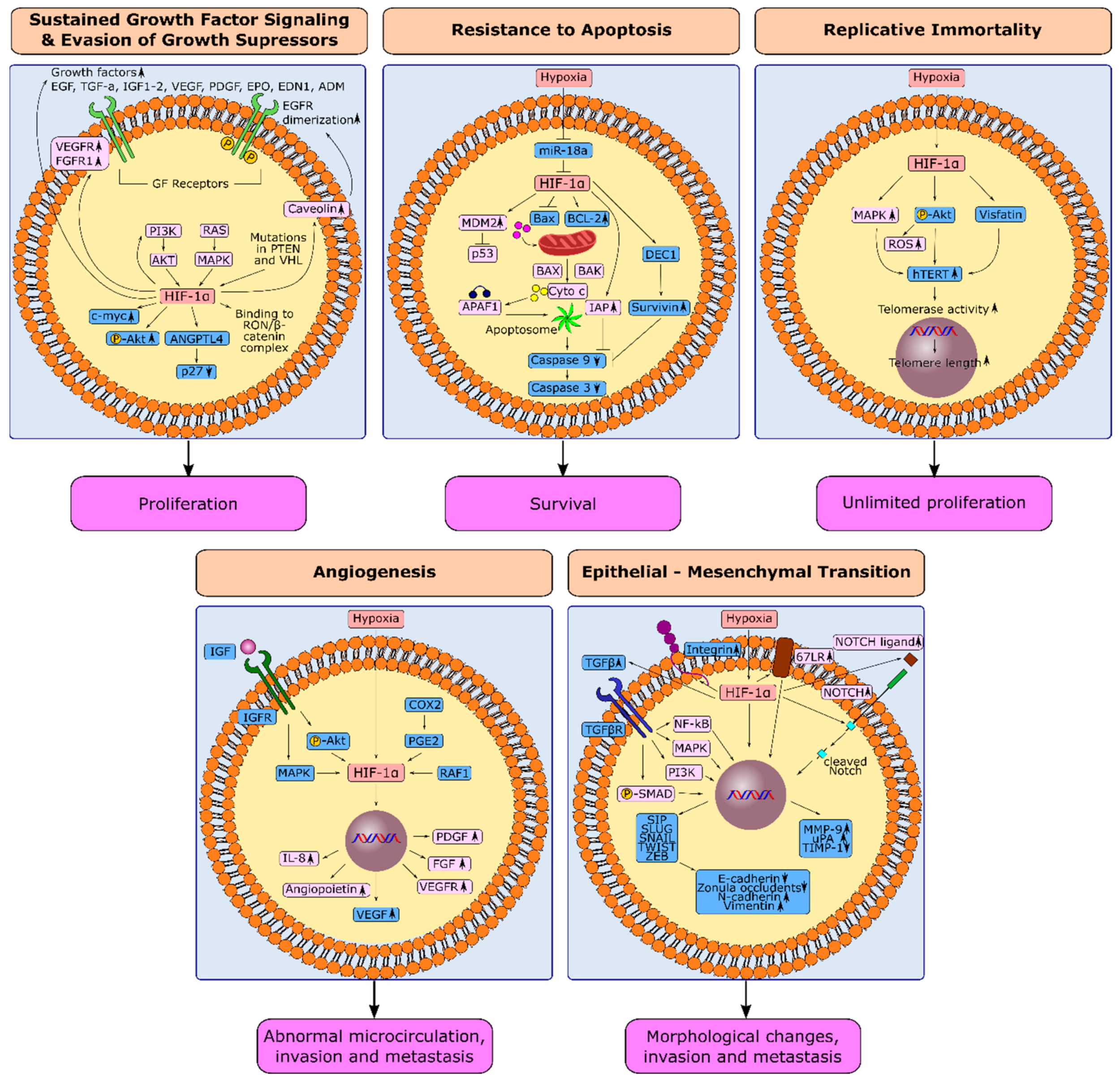
1.2. HIF-1α and Evasion from Growth Suppressors in Gastric Cancer

Cancer cells breach the suppressive actions of tumor suppressor genes on cellular growth through genetic and epigenetic alterations [1][10]. Decreased expression of tumor suppressors PTEN and pVHL is associated with poor prognosis in GC [11][12][13]. The mutations in tumor suppressor genes PTEN and pVHL are associated with the upregulation of HIF-1α which may contribute to poor prognosis in PTEN or pVHL-deficient cancers [7][14] (Figure 1). Hypoxia may also pose a selective pressure leading to the selection of GC cells with dysfunctional tumor suppressor genes. Notably, concomitant overexpression of tumor suppressor protein p53 with HIF-1α was correlated with a dismal prognosis in GC patients, where HIF-1α(+)/p53(+) primary gastric tumors were more frequently associated with an undifferentiated, infiltrative, and metastatic phenotype compared to HIF-1α(−)/p53(−) tumors [15]. The authors did not investigate whether p53(+) tumor samples expressed wild-type or mutant p53 in the study. However, extensive literature suggests that selective pressure posed by hypoxia may lead to enrichment of cells with loss of function mutations in p53 and could explain the dismal prognosis in HIF-1α(+)/p53(+) tumors [16]. This may explain the failure of HIF-1α inhibitors since selected cancer cells will be inherently resistant to apoptosis in these circumstances. Therefore, p53 expression and mutation status of hypoxic cells in gastric tumors should be elucidated as a prospect for the possible use of HIF-1α inhibitors in GC.

1.3. HIF-1α and Resistance to Apoptosis in Gastric Cancer

The evasion of cancer cells from apoptotic triggers is a hallmark of cancer that is crucial for tumor progression and resistance to cancer therapy [1]. Hypoxia triggers apoptosis through a HIF-1α-mediated increase in mitochondrial membrane permeability. As a result, cytochrome c is released from the mitochondria and induces p53-dependent apoptosis where APAF1 and caspases are involved [17]. Additionally, HIF-1α induces caspase-independent cell death through upregulation of BNIP3 and NIX, pro-apoptotic members of the BCL-2 family [18]. Activated JNK is also involved in hypoxia-induced apoptosis [19].
Although hypoxia itself is an apoptotic trigger, cancer cells that can escape from the apoptotic action of hypoxia are selected under hypoxic pressure, generating tumors with a more aggressive and resistant clonal composition [17][20]. Under recurrent hypoxia, p53-mutant and apoptosis-deficient subpopulations are selected remarkably and expand rapidly [20]. Hypoxia was shown to induce anti-apoptotic mechanisms simultaneously with pro-apoptotic processes in ovarian cancer cell lines. Hypoxia conferred resistance to apoptosis by promoting the expression of anti-apoptotic protein BCL-2 in ovarian cancer cell lines, apoptosis inhibitory protein IAP-2 in proximal tubule cells from rat kidney, and MDM2, which is the negative regulator of p53, in murine fibrosarcoma and squamous cell carcinoma cell lines [21][22][23] (Figure 1). The activation of the PI3K/AKT pathway by hypoxia led to survival under hypoxic conditions and resistance to apoptosis in prostate cancer cell lines and rat pheochromocytoma cell lines [24][25]. Additionally, hypoxia inhibited the apoptotic effect of the tumor necrosis factor-related apoptosis-inducing ligand (TRAIL) by blocking the translocation of the pro-apoptotic protein BAX from the cytosol to the mitochondria in colon and lung cancer cell lines [26]. Whether hypoxia will induce apoptosis or confer resistance to apoptosis is possibly dependent on the extent of hypoxia, the cell type, and the phosphorylation status of HIF-1α, which warrants further investigation to be clarified [17][27].
In GC, human differentiated embryonic chondrocyte-expressed gene 1 (DEC1), a hypoxia-induced gene, was reported to prevent apoptosis through upregulation of Survivin in MKN45 and BGC823 GC cells. Survivin expression was higher in mouse models established from DEC1 overexpressing cells compared with the DEC1 knock-down models. Expression of DEC1 and survivin was also positively correlated in GC specimens and associated with a dismal prognosis [28]. Suppression of miR-18a was proposed as a mechanism for hypoxia-induced resistance to apoptosis in MGC-803 and HGC-27 GC cells. Hypoxia suppressed the expression of miR-18a significantly in these cell models. miR-18a mimics increased apoptosis and decreased the invasion in hypoxic conditions. Further investigation demonstrated that miR-18a leads to transcriptional suppression of HIF-1α, downregulation of BCL-2 protein, and upregulation of BAX, caspase-3, and caspase-9 proteins (Figure 1) [29].
In GC, hypoxia may also induce resistance to anoikis, a specific type of programmed cell death following the detachment of anchorage-dependent cells from the extracellular matrix. In AGS and MKN-28 GC cells, silencing or pharmacological inhibition of HIF-1α induced the expression of integrin-5 and led to anoikis [30]. In scirrhous GC cell models, activation of the FAK/SRC/PI3K/AKT and ERK signaling pathways by ANGPTL4 was proposed as a mechanism of resistance to anoikis under hypoxia. In xenograft models established from ANGPTL4 knock-down GC cells, tumorigenicity declined, and metastasis to the peritoneum diminished. Hypoxia could induce anoikis in ANGPTL4 knock-down GC cells [31]. These observations suggest a possible role of HIF-1α in facilitating the survival of metastatic GC cells in the circulation or peritoneal cavity and increasing the metastatic potential in gastric tumors.

1.4. HIF-1α and Replicative Immortality in Gastric Cancer

The number of times that normal cells divide is limited due to the shortening of telomeres [32]. Cancer cells can overcome this limit by activating telomere maintenance mechanisms and gaining replicative immortality. This process differs from sustained growth factor signaling. The total number of times that a cell divide may still be limited, although the proliferation may occur without exogenous growth stimuli in the case of sustained growth factor signaling. Therefore, sustained growth factor signaling may not guarantee replicative immortality, which is highly dependent on the maintenance of the telomere length [1]. Telomerase maintains the telomere length in 85% of human tumors [33]. Human telomerase reverse transcriptase (hTERT), which is the catalytic subunit of telomerase, adds hexameric telomere repeat sequences to the 3′ ends of chromosomal DNA using the intrinsic RNA moiety human telomerase RNA (hTERC) as a template [34].
Hypoxia upregulated telomerase activity in ovarian cancer, colon cancer, cervical cancer, and renal cancer cell lines through transcriptional activation of hTERT by HIF-1α [35][36][37]. In rat gastric mucosa, telomere length increased with the increased duration of exposure to hypoxia, supported by a positive correlation between HIF-1α and hTERT expression. These observations suggested that HIF-1α may protect the gastric mucosa from lethal damage through increased telomerase activity [38].
The MAPK signaling pathway is a pivotal mediator of hypoxia-induced telomerase activity in some cancers, as shown in ovarian cancer and colon cancer cell lines [35]. Despite the limited number of studies investigating the role of hypoxia/HIF-1α in the replicative immortality of GC, the AKT pathway comes forward as the primary mediator for hypoxia-induced telomerase activity in GC (Figure 1). In AGS GC cells, treatment with the VEGFR inhibitor bevacizumab increased the expression of hTERT through the PI3K/AKT/mTOR pathway and HIF-1α [39]. In MKN28 GC cells, AKT activation upregulated hTERT. Inhibition of AKT downregulated hTERT and telomerase activity in these cells. AKT phosphorylation positively correlated with hTERT positivity and telomere length in tumor samples from 40 GC patients [40]. Additionally, visfatin, a HIF-1α-induced proinflammatory cytokine [41][42], was demonstrated to be overexpressed in AGS cells and increase hTERT expression [43]. A high perioperative plasma visfatin level was associated with poor prognosis in a study where 262 GC patients were enrolled [44]. Based on this knowledge, AKT and visfatin may be potential targets in the future to prevent HIF-1α induced replicative immortality in GC cells.

1.5. Induction of Angiogenesis through HIF-1α in Gastric Cancer

When tumors reach a critical size, the hypoxic microenvironment and HIFs turn on the angiogenic switch by inducing the expression of proangiogenic factors such as VEGF, VEGFRs, angiopoietins, matrix metalloproteinases (MMPs), interleukin-8, FGF, and PDGF [45] (Figure 1). Among these factors, VEGF plays a crucial role in angiogenesis. High expression of VEGF leads to tumor progression, metastasis, and poor prognosis [46]. HIF-1α is a master regulator of VEGF and angiogenesis in almost all tumors, including GC. Immunohistochemical investigation of patient specimens [47] in in vitro and in vivo studies also showed the association between HIF-1α, VEGF, and angiogenesis in GC. In TMK-1 GC cells transfected with a dominant-negative form of HIF-1α, the secretion of VEGF decreased substantially both in normoxic and hypoxic conditions. Subcutaneous or orthotopic tumor models established from TMK-1 cells with a dominant-negative form of HIF-1α were significantly smaller. The vessel maturation and density were lower compared with the HIF-1α naïve control groups [48].
Though hypoxia is the primary stimulator for HIF-1α-mediated angiogenesis, studies suggest that inflammatory mediators can also induce angiogenesis through HIF-1α. A correlation was observed between the expression of cyclooxygenase-2 (COX-2) and VEGF in GC specimens. Subsequent studies in AGS cells showed that COX-2 activity and exogenous prostaglandin E2 (PGE2) lead to stimulation of HIF-1α and VEGF expression concomitantly, suggesting the involvement of the COX-2/PGE2/HIF-1α/VEGF pathway in the induction of angiogenesis [49].
Growth factor signaling pathways also play a role in the hypoxia-independent induction of HIF-1α and angiogenesis. In a study, the expression of phosphorylated AKT (pAKT) positively correlated with HIF-1α and VEGF in 268 GC specimens. In SNU-216 and SNU-668 GC cells, constitutively active AKT (CA-AKT) induced the expression of HIF-1 protein and VEGF mRNA in a normoxic environment. However, HIF-1α and VEGF were downregulated in cells with a kinase-dead mutant of AKT. Xenograft models established from CA-AKT-expressing GC cells exhibited a higher incidence of tumor formation with larger volumes, higher micro-vessel density, and HIF-1α expression [50]. RAF1 was also reported as a stimulator of angiogenesis through HIF-1α in GC. The silencing of RAF1 down-regulated the expression of HIF-1α and VEGF in SGC7901 GC cells [51].
Growth factor receptors such as insulin-like growth factor receptors (IGFRs) also emerge as hypoxia-independent regulators of angiogenesis [52]. The blockage of IGF-1R reduced tumor angiogenesis in GC xenograft models [53]. The expression of HIF-1α and IGF2 mRNA correlated in gastric tumor samples [54]. Findings suggest that the association between IGFs and HIF-1α induction is mediated through PI3K/AKT and MAPK activation [52]. Besides these, a novel hypoxamiR, miR-382, was defined recently as an inducer of angiogenesis with possible importance as a predictive marker for progression in GC patients [55].

1.6. Hypoxia-Induced Epithelial-Mesenchymal Transition in Gastric Cancer

Epithelial–mesenchymal transition (EMT) is a physiological process where polarized epithelial cells adhere to the basal membrane or neighboring cells lose their polarity and gain migratory characteristics [56]. Through EMT, epithelial cancer cells transform into mesenchymal cancer cells that invade surrounding tissues and metastasize to distant sites to form new tumors [57][58]. HIF-1α induces EMT through activation of EMT-associated transcription factors TWIST, SNAIL, SLUG, SIP1, ZEB1, and MMPs, the key players in invasion and metastasis, as shown in several in vitro cancer models, including head and neck squamous carcinoma, hepatocellular carcinoma, colorectal cancer, ovarian cancer, and renal cell carcinoma cell lines [59][60][61] (Figure 1). Additionally, an increase in the expression of mesenchymal markers vimentin, fibronectin, and N-cadherin in parallel with a decrease in E-cadherin and destruction of cadherin-mediated cell–cell adhesions are critical alterations, especially for the early stages of metastasis [61].
Transforming growth factor β (TGFβ) is a critical mediator for hypoxia-induced EMT. Prolonged hypoxia (exposure to the hypoxic microenvironment for more than 10 days) induced the production of TGFβ at much higher levels compared to normoxic conditions in Lewis lung carcinoma cell lines [62]. TGFβ activates serine/threonine kinase receptors that lead to the phosphorylation of SMAD proteins in the cytoplasm. Phosphorylated SMADs activate SNAIL, ZEB1, and SIP1 in the nucleus, altering the transcription of several genes responsible for cell proliferation, differentiation, migration, and EMT. TGFβ can also induce EMT-associated transcription factors via SMAD-independent mechanisms such as MAPK, PI3K/AKT, and NF-κB signaling, as observed in in vitro models of murine mammary epithelial cells, human cervical carcinoma cells, human breast cancer cells, human kidney cells, and human salivary gland epithelial cells [63][64][65][66]. Moreover, TGFβ may cooperate with various oncogenic pathways such as NOTCH and WNT/β-catenin to trigger EMT and moderate hypoxia-induced tumor invasion and migration, as shown in prostate cancer and breast cancer cell lines [67][68][69].
The NOTCH signaling pathway also mediates hypoxia-induced EMT, besides its regulatory role in stemness, embryonic development, and cell fate. HIF-1α activated NOTCH signaling, and NOTCH1 increased HIF-1α expression in ovarian and breast cancer cell lines. Thus, once activated, NOTCH can imitate hypoxia to stimulate EMT [59]. After ligand-stimulated cleavage, intracellular domains of NOTCH translocate to the nucleus and directly increase SNAIL1 expression by binding to the SNAIL1 promoter together with other genes critical for tumor progression, as demonstrated in cervical cancer, colon cancer, ovarian cancer, and glioma cell lines. NOTCH also stabilizes SNAIL1 through increased expression of lysyl-oxidase (LOX) which protects SNAIL1 from degradation [68]. Moreover, hypoxia can increase the expression of NOTCH receptors and ligands, which stimulate a higher expression of NOTCH target genes by the accumulation of HIF-1α. Suppression of the NOTCH pathway in breast cancer cell lines decreased SLUG and SNAIL expression and blocked cellular migration, supporting the role of NOTCH signaling in hypoxia-induced EMT [69].
In GC, evidence that supports the involvement of NOTCH signaling in hypoxia-induced EMT is almost lacking. However, there is strong evidence for the involvement of TGFβ signaling. Both TGFβ1 and TGFβR increased under hypoxic conditions in OCUM-2MD3 and OCUM-12 cells. TGFβ inhibitors repressed the induction of EMT in these cells, demonstrating the mediator role of TGFβ in hypoxia-induced EMT in diffuse-type GC. Although the HIF-1α expression increased with hypoxia in the cells, the authors stated that knocking down HIF-1α did not decrease the expression of TGFβ. Therefore, the role of HIF-1α is not clear in the process [70]. Hypoxia increased integrin expression, potentiating the implantation ability of diffuse-type GC cells to the peritoneum [71]. In SGC7901 and MKN45 (poorly differentiated) GC cell lines, the hypoxic microenvironment increased the expression of MMP-9 and urokinase-type plasminogen activator and decreased the expression of tissue inhibitor of matrix metalloproteinase (TIMP)-1, through HIF-1α-induced upregulation of 67 kDa Laminin receptor (67LR). Thus, HIF-1α increased the invasion ability of GC cells [72]. Moreover, HIF-1α upregulated chemokine receptor 4 (CXCR4), increasing the migration and invasion of KATO III GC cells [73].

2. HIF-1α and Next-Generation Hallmarks in Gastric Cancer

2.1. Hypoxia and Genetic Instability in Gastric Cancer

Hypoxia increases the mutation rate in mammalian cells. Mutation frequency increases further with subsequent exposures [74]. Hypoxia can induce genomic instability by gene amplification, chromosome rearrangement, and suppression of DNA mismatch repair (MMR) genes and gives rise to chemotherapy-resistant clones through microsatellite instability in several cancers, as demonstrated in breast cancer, lung cancer, and rectal cancer in vitro cell models [75][76][77][78].
ROS produced during hypoxia/reoxygenation cycles play a pivotal role in genetic instability. ROS damage DNA and generate many genomic aberrations such as base modifications, nucleotide transversions, DNA slippage mutations at microsatellites, and chromosomal-fragile sites [79]. Moreover, hypoxia suppresses DNA repair mechanisms. RAD51 and BRCA1, key actors in homologous recombination (HR), were shown to be downregulated by hypoxia, leading to impaired HR in hypoxic and post-hypoxic conditions in breast cancer, lung cancer, cervical cancer, prostate cancer, and colorectal cancer cell lines (Figure 2) [80]. Hypoxia can also suppress nucleotide excision repair through downregulation of RAD23, and MMR through downregulation of MSH2, MSH6, MSH3, MLH1, and PMS2, leading to microsatellite slippage mutations, as evidenced in lung cancer and colon cancer in vitro models [79]. Furthermore, hypoxia was proposed as an inducer of polyploidy in studies conducted with melanoma and colon cancer cell lines [81][82].
Figure 2. Induction of next-generation cancer hallmarks by hypoxia and HIF-1α in gastric cancer. Hypoxia induces genetic instability, deregulation in cellular energetics, escape from immune surveillance, and tumor-promoting inflammation in gastric cancer. Processes for which there is specific evidence in gastric cancer are shown in blue. Processes common in different cancers are shown in light pink. Upregulation or downregulation of specific proteins is shown with an upward or downward arrow, respectively. The figures were drawn in Inkscape 1.1.2.

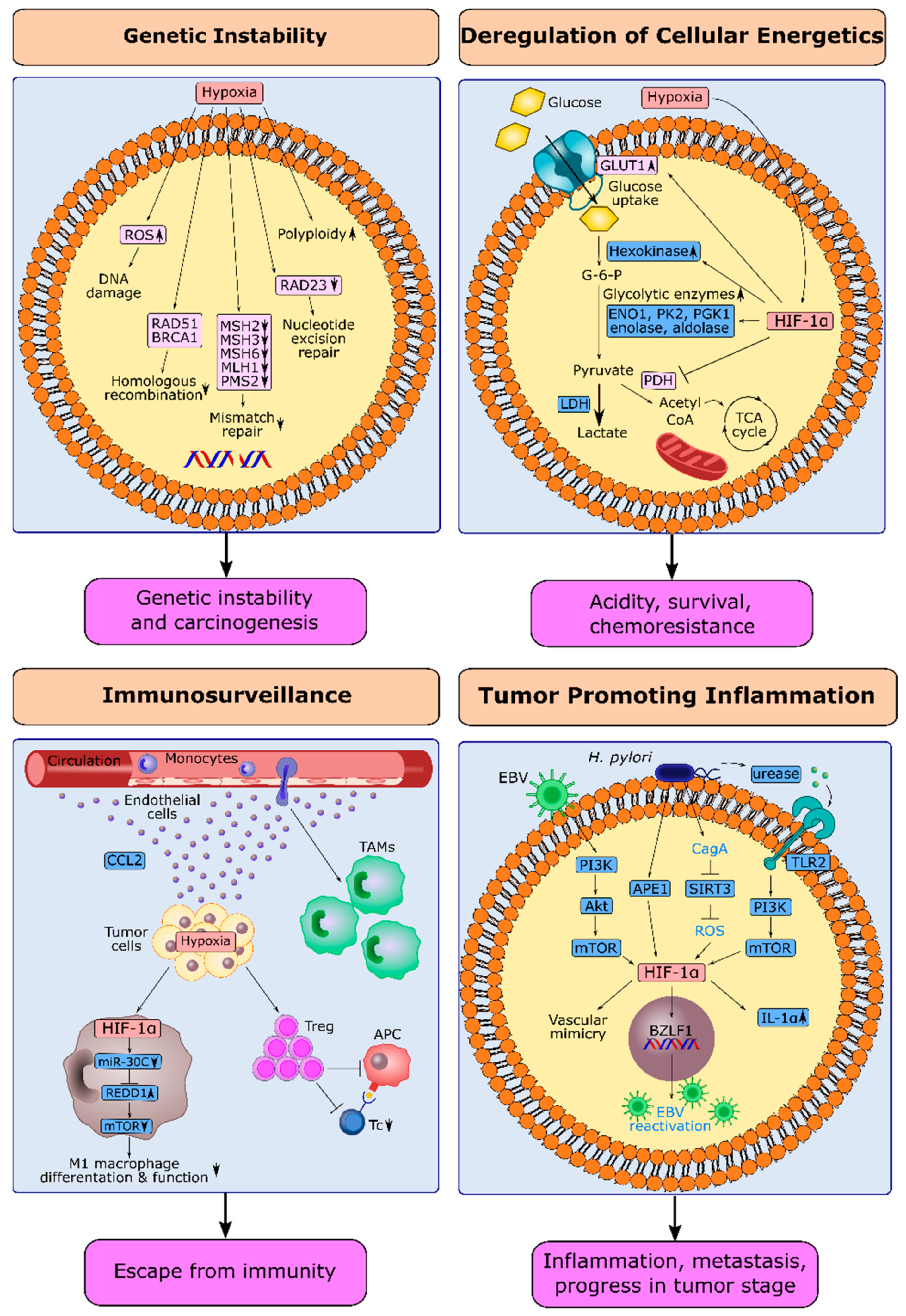
2.2. HIF-1α and Deregulation of Cellular Energetics in Gastric Cancer

Cellular energy metabolism is regulated dynamically, based on the energy requirements of the cell and changes in the microenvironment. Oxidative phosphorylation of glucose is the major source of energy in normal cells under normoxic conditions. Cancer cells switch from oxidative phosphorylation to oxygen-independent glycolysis and convert glucose into lactate instead of directing it to the tricarboxylic acid (TCA) cycle. This switch in cancer cells is known as the Warburg effect [83]. Though oxygen-independent glycolysis is a less efficient process in terms of ATP production than oxidative phosphorylation, it provides some advantages for the survival of cancer cells and their rapid adaptation to the microenvironment. Via glycolysis, cellular ATP requirements are met even under hypoxic conditions, and cancer cells protect themselves against the excess production of ROS that would occur during the TCA and electron transport chain (ETC) [84]. Additionally, it is a way to spare glycolysis intermediates to synthesize fatty acids, amino acids, and nucleotide precursors essential in rapidly proliferating cells [85][86].
HIF-1α has a central role in the Warburg effect [87]. HIF-1α increases the uptake of glucose into cancer cells via the transcriptional upregulation of the glucose transporter GLUT1. Thereby it provides cancer cells with higher glucose needed to keep up with cellular ATP requirements [88]. HIF-1α-induced GLUT-1 was significantly correlated with the depth of invasion, advanced stage, and shorter overall survival in GC patients [89][90]. HIF-1α increases the expression of enzymes involved in oxygen-independent glycolysis, such as enolase and aldolase [91] and inhibits the activity of pyruvate dehydrogenase required for the entry into the TCA (Figure 2) [92]. In GC cell lines, HIF-1α increased the expression of ENO1, pyruvate kinase 2, phosphoglycerate kinase 1, and lactate dehydrogenase A (LDHA), critical enzymes in the glycolytic pathway [93]. HIF-1 transcriptionally up-regulates hexokinase (HK), the catalyzer of the first reaction in glycolysis. HK2 was overexpressed in 16.7% of the GC specimens. HK2 expression was inversely related to BCL-2 expression and associated with poor survival [94]. HIF-1α-induced glycolysis was also suggested as a mechanism for hypoxia-induced 5-fluorouracil resistance in GC cells [95].

2.3. HIF-1α and Escape from Immune Surveillance in Gastric Cancer

Cancer cells are in continuous interaction with the immune system and utilize several strategies to evade anti-tumor immunity. Hypoxia plays a reinforcing role in these strategies [96]. Hypoxic tumor niches are enriched in tumor-associated macrophages (TAMs), which suppress anti-tumor immunity and induce tumor progression [97]. Monocytes recruited to tumors differentiate into TAMs and secrete growth factors, proangiogenic factors, and immunosuppressive mediators under the influence of HIFs [98]. In GC specimens, the expression of HIF-1α was correlated with monocyte chemoattractant protein-1, which plays a critical role in the recruitment of monocytes and macrophages to tumors (Figure 2) [99]
In GC-infiltrating macrophages, HIF-1α suppressed miR-30c, increasing the expression of REDD1, an inhibitor of mTOR. Consequently, the concomitant decreases in mTOR activity and glycolysis led to the inhibition of M1 macrophage differentiation and function. Since M1 macrophages have cytotoxic activity on cancer cells, the HIF-1α/miR-30c/REDD1/mTOR axis was suggested as a mechanism for hypoxia-induced suppression of anti-tumor immunity in GC (Figure 2) [100]. Dysregulation of cellular energetics may also be a mechanism by which hypoxia impairs anti-tumor immunity. Hypoxia-induced down-regulation of miR-34a increased the expression of LDHA, which increases lactate production in GC tumor-infiltrating lymphocytes. A high lactate concentration was negatively correlated with the number of pro-inflammatory Th1 cells and cytotoxic T lymphocytes (CTLs) in GC specimens [101]. Suppression of CTLs and antigen-presenting cells via regulatory T (Treg) cells is another important strategy for tumor cells to evade anti-tumor immunity [102]. HIF-1α expression was correlated with the number of Treg cells in GC specimens. This correlation was stronger in metastatic tumors compared to non-metastatic tumors. Hypoxia-induced upregulation of TGF-β was suggested as a mechanism for the expansion of Treg cells under hypoxia in GC [103].

2.4. HIF-1α and Tumor-Promoting Inflammation in Gastric Cancer

The inflammatory tumor microenvironment is a critical hallmark for carcinogenesis and tumor progression that facilitates the acquisition of other hallmark capabilities via the release of GFs, proangiogenic mediators, proteases, ROS, and EMT-activating signals [1][104]. Pro-inflammatory cytokine interleukin-1α (IL-1α) increases in several malignancies, including GC. Increased IL-1α expression is significantly associated with liver metastasis, lymph node metastasis, increased tumor stage, and decreased survival in GC patients [105][106]. Hypoxia increased IL-1α expression via HIF-1α in GC cell lines [106].
Bacterial or viral infections precede chronic inflammation in approximately 20% of all cancers [104]. GC associated with H. pylori or Epstein–Barr virus (EBV) is among the most prominent examples of tumor-promoting inflammation in cancer [104][107]. Chronic inflammation induced by H. pylori in gastric epithelium promotes a sequence of pathologies from gastritis to intestinal-type GC [108]. Additionally, H. pylori infection is associated with MALT lymphoma [104]. Based on the molecular classification of GC, 9% of GCs are associated with EBV infection, which constitutes a distinct molecular subtype [109].
Investigation of H. pylori-positive gastritis specimens demonstrated that HIF-1α expression particularly increases in areas infiltrated by macrophages. Bone marrow-derived macrophages also exhibited high HIF-1α expression in mice models infected
HIF-1α is also involved in the natural life cycle of EBV and lytic infection that induce tumorigenesis. HIF-1α stabilizers increased the EBV lytic proteins and reactivated EBV infection in EBV-positive GC cell lines. HIF-1α induced this response via the direct binding and activation of the EBV primary latent-lytic switch BZLF1 gene, Zp (Figure 2) [110]. Additionally, the PI3K/AKT/mTOR/HIF-1α axis has been put forth as a mediator of EBV-induced vascular mimicry (VM). VM defines channel-like structures established by tumor cells that mimic the vasculature and contribute substantially to tumor progression and metastasis under hypoxic conditions. VM formation is observed in EBV-positive GC cells, while EBV-negative GC cells do not exhibit VM. Moreover, HIF-1α expression and AKT phosphorylation were correlated with VM formation in tumor samples from EBV-associated GC patients [111].

This entry is adapted from the peer-reviewed paper 10.3390/cancers14112711

References

  1. Hanahan, D.; Weinberg, R.A. Hallmarks of cancer: The next generation. Cell 2011, 144, 646–674.
  2. Powis, G.; Kirkpatrick, L. Hypoxia inducible factor-1alpha as a cancer drug target. Mol Cancer Ther. 2014, 3, 647–654.
  3. Semenza, G.L. Hypoxia-inducible factors: Mediators of cancer progression and targets for cancer therapy. Trends Pharmacol. Sci. 2012, 33, 207–214.
  4. Lu, Y.; Liu, Y.; Oeck, S.; Zhang, G.J.; Schramm, A.; Glazer, P.M. Hypoxia Induces Resistance to EGFR Inhibitors in Lung Cancer Cells via Upregulation of FGFR1 and the MAPK Pathway. Cancer Res. 2020, 80, 4655–4667.
  5. Waltenberger, J.; Mayr, U.; Pentz, S.; Hombach, V. Functional Upregulation of the Vascular Endothelial Growth Factor Receptor KDR by Hypoxia. Circulation 1996, 94, 1647–1654.
  6. Wang, Y.; Roche, O.; Xu, C.; Moriyama, E.H.; Heir, P.; Chung, J.; Roos, F.C.; Chen, Y.; Finak, G.; Milosevic, M.; et al. Hypoxia promotes ligand-independent EGF receptor signaling via hypoxia-inducible factor–mediated upregulation of caveolin-1. Proc. Natl. Acad. Sci. USA 2012, 109, 4892–4897.
  7. Kitajima, Y.; Miyazaki, K. The Critical Impact of HIF-1a on Gastric Cancer Biology. Cancers 2013, 5, 15–26.
  8. Li, X.; Lu, Y.; Liang, K.; Pan, T.; Mendelsohn, J.; Fan, Z. Requirement of hypoxia-inducible factor-1α down-regulation in mediating the antitumor activity of the anti–epidermal growth factor receptor monoclonal antibody cetuximab. Mol. Cancer Ther. 2008, 7, 1207–1217.
  9. Zhou, D.; Huang, L.; Zhou, Y.; Wei, T.; Yang, L.; Li, C. RON and RONΔ160 promote gastric cancer cell proliferation, migration, and adaption to hypoxia via interaction with β-catenin. Aging 2019, 11, 2735–2748.
  10. Tamura, G. Alterations of tumor suppressor and tumor-related genes in the development and progression of gastric cancer. World J. Gastroenterol. 2006, 12, 192–198.
  11. Yang, L.; Kuang, L.G.; Zheng, H.C.; Li, J.Y.; Wu, D.Y.; Zhang, S.M.; Xin, Y.; Chen, Y.; Yang, S. PTEN encoding product: A marker for tumorigenesis and progression of gastric carcinoma. World J. Gastroenterol. 2003, 9, 35–39.
  12. Zheng, H.-C.; Qiu, Y.-H.; Zhao, S. The roles of PTEN expression in gastric cancer: A bibliometric, meta and bioinformatics analysis. Oncotarget 2018, 9 (Suppl. 1), s1074–s1089.
  13. Lee, H.S.; Lee, H.K.; Kim, H.S.; Yang, H.K.; Kim, W.H. Tumour suppressor gene expression correlates with gastric cancer prognosis. J. Pathol. 2003, 200, 39–46.
  14. Song, M.S.; Salmena, L.; Pandolfi, P.P. The functions and regulation of the PTEN tumour suppressor. Nat. Rev. Mol. Cell Biol. 2012, 13, 283–296.
  15. Sumiyoshi, Y.; Kakeji, Y.; Egashira, A.; Mizokami, K.; Orita, H.; Maehara, Y. Overexpression of Hypoxia-Inducible Factor 1α and p53 Is a Marker for an Unfavorable Prognosis in Gastric Cancer. Clin. Cancer Res. 2006, 12, 5112–5117.
  16. Hammond, E.M.; Giaccia, A.J. Hypoxia-Inducible Factor-1 and p53: Friends, Acquaintances, or Strangers? Clin. Cancer Res. 2006, 12, 5007–5009.
  17. Ruan, K.; Song, G.; Ouyang, G. Role of hypoxia in the hallmarks of human cancer. J. Cell. Biochem. 2009, 107, 1053–1062.
  18. Sowter, H.M.; Ratcliffe, P.J.; Watson, P.; Greenberg, A.H.; Harris, A.L. HIF-1-dependent regulation of hypoxic induction of the cell death factors BNIP3 and NIX in human tumors. Cancer Res. 2001, 61, 6669–6673.
  19. Kunz, M.; Ibrahim, S.; Koczan, D.; Thiesen, H.J.; Köhler, H.J.; Acker, T.; Plate, K.H.; Ludwig, S.; Rapp, U.R.; Bröcker, E.B.; et al. Activation of c-Jun NH2-terminal kinase/stress-activated protein kinase (JNK/SAPK) is critical for hypoxia-induced apoptosis of human malignant melanoma. Cell Growth Differ. Mol. Biol. J. Am. Assoc. Cancer Res. 2001, 12, 137–145.
  20. Graeber, T.G.; Osmanian, C.; Jacks, T.; Housman, D.E.; Koch, C.J.; Lowe, S.W.; Giaccia, A.J. Hypoxia-mediated selection of cells with diminished apoptotic potential in solid tumours. Nature 1996, 379, 88–91.
  21. Wang, Y.; Pakunlu, R.I.; Tsao, W.; Pozharov, V.; Minko, T. Bimodal Effect of Hypoxia in Cancer: Role of Hypoxia Inducible Factor in Apoptosis. Mol. Pharm. 2004, 1, 156–165.
  22. Dong, Z.; Venkatachalam, M.A.; Wang, J.; Patel, Y.; Saikumar, P.; Semenza, G.L.; Force, T.; Nishiyama, J. Up-regulation of Apoptosis Inhibitory Protein IAP-2 by Hypoxia: HIF-1-independent mechanisms. J. Biol. Chem. 2001, 276, 18702–18709.
  23. Zhang, L.; Hill, R.P. Hypoxia Enhances Metastatic Efficiency by Up-Regulating Mdm2 in KHT Cells and Increasing Resistance to Apoptosis. Cancer Res. 2004, 64, 4180–4189.
  24. Alvarez-Tejado, M.; Naranjo-Suárez, S.; Jiménez, C.; Carrera, A.C.; Landázuri, M.O.; del Peso, L. Hypoxia Induces the Activation of the Phosphatidylinositol 3-Kinase/Akt Cell Survival Pathway in PC12 Cells: Protective Role in Apoptosis. J. Biol. Chem. 2001, 276, 22368–22374.
  25. Chae, Y.C.; Vaira, V.; Caino, M.C.; Tang, H.-Y.; Seo, J.H.; Kossenkov, A.V.; Ottobrini, L.; Martelli, C.; Lucignani, G.; Bertolini, I.; et al. Mitochondrial Akt Regulation of Hypoxic Tumor Reprogramming. Cancer Cell 2016, 30, 257–272.
  26. Kim, M.; Park, S.Y.; Pai, H.S.; Kim, T.H.; Billiar, T.R.; Seol, D.W. Hypoxia Inhibits Tumor Necrosis Factor-Related Apoptosis-Inducing Ligand-Induced Apoptosis by Blocking Bax Translocation. Cancer Res. 2004, 64, 4078–4081.
  27. Greijer, A.E.; van der Wall, E. The role of hypoxia inducible factor 1 (HIF-1) in hypoxia induced apoptosis. J. Clin. Pathol. 2004, 57, 1009–1014.
  28. Jia, Y.; Hu, R.; Li, P.; Zheng, Y.; Wang, Y.; Ma, X. DEC1 is required for anti-apoptotic activity of gastric cancer cells under hypoxia by promoting Survivin expression. Gastric Cancer 2018, 21, 632–642.
  29. Wu, F.; Huang, W.; Wang, X. microRNA-18a regulates gastric carcinoma cell apoptosis and invasion by suppressing hypoxia-inducible factor-1α expression. Exp. Ther. Med. 2015, 10, 717–722.
  30. Rohwer, N.; Welzel, M.; Daskalow, K.; Pfander, D.; Wiedenmann, B.; Detjen, K.; Cramer, T. Hypoxia-Inducible Factor 1 Mediates Anoikis Resistance via Suppression of 5 Integrin. Cancer Res. 2008, 68, 10113–10120.
  31. Baba, K.; Kitajima, Y.; Miyake, S.; Nakamura, J.; Wakiyama, K.; Sato, H.; Okuyama, K.; Kitagawa, H.; Tanaka, T.; Hiraki, M.; et al. Hypoxia-induced ANGPTL4 sustains tumour growth and anoikis resistance through different mechanisms in scirrhous gastric cancer cell lines. Sci. Rep. 2017, 7, 11127.
  32. Shay, J.W.; Wright, W.E. Hayflick, his limit, and cellular ageing. Nat. Rev. Mol. Cell Biol. 2000, 1, 72–76.
  33. Heeg, S. Variations in telomere maintenance and the role of telomerase inhibition in gastrointestinal cancer. Pharmacogenomics Pers. Med. 2015, 8, 171–180.
  34. Yuan, X.; Larsson, C.; Xu, D. Mechanisms underlying the activation of TERT transcription and telomerase activity in human cancer: Old actors and new players. Oncogene 2019, 38, 6172–6183.
  35. Seimiya, H.; Tanji, M.; Oh-Hara, T.; Tomida, A.; Naasani, I.; Tsuruo, T. Hypoxia Up-Regulates Telomerase Activity via Mitogen-Activated Protein Kinase Signaling in Human Solid Tumor Cells. Biochem. Biophys. Res. Commun. 1999, 260, 365–370.
  36. Yatabe, N.; Kyo, S.; Maida, Y.; Nishi, H.; Nakamura, M.; Kanaya, T.; Tanaka, M.; Isaka, K.; Ogawa, S.; Inoue, M. HIF-1-mediated activation of telomerase in cervical cancer cells. Oncogene 2004, 23, 3708–3715.
  37. Lou, F.; Chen, X.; Jalink, M.; Zhu, Q.; Ge, N.; Zhao, S.; Fang, X.; Fan, Y.; Björkholm, M.; Liu, Z.; et al. The Opposing Effect of Hypox-ia-Inducible Factor-2α on Expression of Telomerase Reverse Transcriptase. Mol. Cell. Biol. 2007, 5, 793–800.
  38. Wang, Y.-P.; Zhu, Z.-Y.; Tang, Y.; Ma, Y.-C. Effects of acute hypoxia on telomere length of rat gastric mucosa tissue and underlying mechanism. Sheng Li Xue Bao 2017, 69, 429–436.
  39. Mahfouz, N.; Tahtouh, R.; Alaaeddine, N.; EL Hajj, J.; Sarkis, R.; Hachem, R.; Raad, I.; Hilal, G. Gastrointestinal cancer cells treatment with bevacizumab activates a VEGF autoregulatory mechanism involving telomerase catalytic subunit hTERT via PI3K-AKT, HIF-1α and VEGF receptors. PLoS ONE 2017, 12, e0179202.
  40. Sasaki, T.; Kuniyasu, H.; Luo, Y.; Kitayoshi, M.; Tanabe, E.; Kato, D.; Shinya, S.; Fujii, K.; Ohmori, H.; Yamashita, Y. AKT Activation and Telomerase Reverse Transcriptase Expression are Concurrently Associated with Prognosis of Gastric Cancer. Pathobiology 2014, 81, 36–41.
  41. Bae, S.-K.; Kim, S.-R.; Kim, J.G.; Kim, J.Y.; Koo, T.H.; Jang, H.-O.; Yun, I.; Yoo, M.-A.; Bae, M.-K. Hypoxic induction of human visfatin gene is directly mediated by hypoxia-inducible factor-1. FEBS Lett. 2006, 580, 4105–4113.
  42. Segawa, K.; Fukuhara, A.; Hosogai, N.; Morita, K.; Okuno, Y.; Tanaka, M.; Nakagawa, Y.; Kihara, S.; Funahashi, T.; Komuro, R.; et al. Visfatin in adipocytes is upregulated by hypoxia through HIF1α-dependent mechanism. Biochem. Biophys. Res. Commun. 2006, 349, 875–882.
  43. Mohammadi, M.; Zarghami, N.; Hedayati, M.; Ghaemmaghami, S.; Yamchi, R.M.; Mohaddes, M. Visfatin effects on telomerase gene expression in AGS gastric cancer cell line. Indian J. Cancer 2015, 52, 32–35.
  44. Lu, G.-W.; Wang, Q.-J.; Xia, M.-M.; Qian, J. Elevated plasma visfatin levels correlate with poor prognosis of gastric cancer patients. Peptides 2014, 58, 60–64.
  45. Krock, B.L.; Skuli, N.; Simon, M.C. Hypoxia-Induced Angiogenesis: Good and Evil. Genes Cancer 2011, 2, 1117–1133.
  46. Lv, X.; Li, J.; Zhang, C.; Hu, T.; Li, S.; He, S.; Yan, H.; Tan, Y.; Lei, M.; Wen, M.; et al. The role of hypoxia-inducible factors in tumor angiogenesis and cell metabolism. Genes Dis. 2017, 4, 19–24.
  47. Urano, N.; Fujiwara, Y.; Doki, Y.; Tsujie, M.; Yamamoto, H.; Miyata, H.; Takiguchi, S.; Yasuda, T.; Yano, M.; Monden, M. Overexpression of hypoxia-inducible factor-1 alpha in gastric adenocarcinoma. Gastric Cancer 2006, 9, 44–49.
  48. Stoeltzing, O.; McCarty, M.F.; Wey, J.S.; Fan, F.; Liu, W.; Belcheva, A.; Bucana, C.D.; Semenza, G.L.; Ellis, L.M. Role of Hypoxia-Inducible Factor 1α in Gastric Cancer Cell Growth, Angiogenesis, and Vessel Maturation. J. Natl. Cancer Inst. 2004, 96, 946–956.
  49. Huang, S.-P.; Wu, M.-S.; Shun, C.-T.; Wang, H.-P.; Hsieh, C.-Y.; Kuo, M.-L.; Lin, J.-T. Cyclooxygenase-2 increases hypoxia-inducible factor-1 and vascular endothelial growth factor to promote angiogenesis in gastric carcinoma. J. Biomed. Sci. 2005, 12, 229–241.
  50. Lee, B.L.; Kim, W.H.; Jung, J.; Cho, S.J.; Park, J.-W.; Kim, J.; Chung, H.-Y.; Chang, M.S.; Nam, S.Y. A hypoxia-independent up-regulation of hypoxia-inducible factor-1 by AKT contributes to angiogenesis in human gastric cancer. Carcinogenesis 2008, 29, 44–51.
  51. Meng, F.; Ding, J.; Liu, N.; Zhang, J.; Shao, X.; Shen, H.; Xue, Y.; Xie, H.; Fan, D. Inhibition of gastric cancer angiogenesis by vector-based RNA interference for Raf-1. Cancer Biol. Ther. 2005, 4, 120–124.
  52. Van Beijnum, J.R.; Pieters, W.; Nowak-Sliwinska, P.; Griffioen, A.W. Insulin-like growth factor axis targeting in cancer and tumour angiogenesis-The missing link. Biol. Rev. 2017, 92, 1755–1768.
  53. Li, H.; Adachi, Y.; Yamamoto, H.; Min, Y.; Ohashi, H.; Ii, M.; Arimura, Y.; Endo, T.; Lee, C.-T.; Carbone, D.P.; et al. Insulin-like growth factor-I receptor blockade reduces tumor angiogenesis and enhances the effects of bevacizumab for a human gastric cancer cell line, MKN45. Cancer 2011, 117, 3135–3147.
  54. Ru, G.-Q.; Zhao, Z.-S.; Tang, Q.-L.; Xu, W.-J. Expressions of hypoxia inducible factor-1alpha and insulin-like growth factor-II in gastric carcinoma: Correlation with angiogenesis and prognosis. Zhonghua Wai Ke Za Zhi Chin. J. Surg. 2007, 45, 905–908.
  55. Na Seo, A.; Jung, Y.; Jang, H.; Lee, E.; Bae, H.-I.; Son, T.; Kwon, O.; Chung, H.Y.; Yu, W.; Lee, Y.M. Clinical significance and prognostic role of hypoxia-induced microRNA 382 in gastric adenocarcinoma. PLoS ONE 2019, 14, e0223608.
  56. Lamouille, S.; Xu, J.; Derynck, R. Molecular mechanisms of epithelial–mesenchymal transition. Nat. Rev. Mol. Cell Biol. 2014, 15, 178–196.
  57. Roche, J. The Epithelial-to-Mesenchymal Transition in Cancer. Cancers 2018, 10, 52.
  58. Lambert, A.W.; Pattabiraman, D.R.; Weinberg, R.A. Emerging Biological Principles of Metastasis. Cell 2017, 168, 670–691.
  59. Jiang, J.; Tang, Y.-L.; Liang, X.-H. EMT: A new vision of hypoxia promoting cancer progression. Cancer Biol. Ther. 2011, 11, 714–723.
  60. Peng, Z.; Wang, C.X.; Fang, E.H.; Wang, G.B.; Tong, Q. Role of epithelial-mesenchymal transition in gastric cancer initiation and progression. World J. Gastroenterol. 2014, 20, 5403–5410.
  61. Kalluri, R.; Weinberg, R.A. The basics of epithelial-mesenchymal transition. J. Clin. Investig. 2009, 119, 1420–1428.
  62. Furuta, C.; Miyamoto, T.; Takagi, T.; Noguchi, Y.; Kaneko, J.; Itoh, S.; Watanabe, T.; Itoh, F. Transforming growth factor-β signaling enhancement by long-term exposure to hypoxia in a tumor microenvironment composed of Lewis lung carcinoma cells. Cancer Sci. 2015, 106, 1524–1533.
  63. Wendt, M.K.; Allington, T.M.; Schiemann, W.P. Mechanisms of the epithelial–mesenchymal transition by TGF-β. Future Oncol. 2009, 5, 1145–1168.
  64. Bakin, A.; Rinehart, C.; Tomlinson, A.K.; Arteaga, C.L. p38 mitogen-activated protein kinase is required for TGFβ-mediated fibroblastic transdifferentiation and cell migration. J. Cell Sci. 2002, 115, 3193–3206.
  65. Lamouille, S.; Derynck, R. Cell size and invasion in TGF-β–induced epithelial to mesenchymal transition is regulated by activation of the mTOR pathway. J. Cell Biol. 2007, 178, 437–451.
  66. Azuma, M.; Motegi, K.; Aota, K.; Yamashita, T.; Yoshida, H.; Sato, M. TGF-β1 Inhibits NF-κB Activity through Induction of IκB-α Expression in Human Salivary Gland Cells: A Possible Mechanism of Growth Suppression by TGF-β1. Exp. Cell Res. 1999, 250, 213–222.
  67. Jiang, Y.G.; Luo, Y.; He, D.L.; Li, X.; Zhang, L.L.; Peng, T.; Li, M.C.; Lin, Y.H. Role of Wnt/β-catenin signaling pathway in epithelial-mesenchymal transition of human prostate cancer induced by hypoxia-inducible factor-1α. Int. J. Urol. 2007, 14, 1034–1039.
  68. Sahlgren, C.; Gustafsson, M.V.; Jin, S.; Poellinger, L.; Lendahl, U. Notch signaling mediates hypoxia-induced tumor cell migration and invasion. Proc. Natl. Acad. Sci USA 2008, 105, 6392–6397.
  69. Chen, J.; Imanaka, N.; Chen, J.; Griffin, J.D. Hypoxia potentiates Notch signaling in breast cancer leading to decreased E-cadherin expression and increased cell migration and invasion. Br. J. Cancer 2010, 102, 351–360.
  70. Matsuoka, J.; Yashiro, M.; Doi, Y.; Fuyuhiro, Y.; Kato, Y.; Shinto, O.; Noda, S.; Kashiwagi, S.; Aomatsu, N.; Hirakawa, T.; et al. Hypoxia Stimulates the EMT of Gastric Cancer Cells through Autocrine TGFβ Signaling. PLoS ONE 2013, 8, e62310.
  71. Noda, S.; Yashiro, M.; Nshii, T.; Hirakawa, K. Hypoxia upregulates adhesion ability to peritoneum through a transforming growth factor-β-dependent mechanism in diffuse-type gastric cancer cells. Eur. J. Cancer 2010, 46, 995–1005.
  72. Liu, L.; Sun, L.; Zhao, P.; Yao, L.; Jin, H.; Liang, S.; Wang, Y.; Zhang, D.; Pang, Y.; Shi, Y.; et al. Hypoxia promotes metastasis in human gastric cancer by up-regulating the 67-kDa laminin receptor. Cancer Sci. 2010, 101, 1653–1660.
  73. Oh, Y.S.; Kim, H.Y.; Song, I.-C.; Yun, H.-J.; Jo, D.-Y.; Kim, S.; Lee, H.J. Hypoxia induces CXCR4 expression and biological activity in gastric cancer cells through activation of hypoxia-inducible factor-1α. Oncol. Rep. 2012, 28, 2239–2246.
  74. Reynolds, T.Y.; Rockwell, S.; Glazer, P. Genetic instability induced by the tumor microenvironment. Cancer Res. 1996, 56, 5754–5757.
  75. Bindra, R.S.; Glazer, P.M. Co-repression of mismatch repair gene expression by hypoxia in cancer cells: Role of the Myc/Max network. Cancer Lett. 2007, 252, 93–103.
  76. Rodríguez-Jiménez, F.J.; Moreno-Manzano, V.; Lucas-Dominguez, R.; Sánchez-Puelles, J.M. Hypoxia Causes Downregulation of Mismatch Repair System and Genomic Instability in Stem Cells. Stem Cells 2008, 26, 2052–2062.
  77. Yuan, J.; Narayanan, L.; Rockwell, S.; Glazer, P.M. Diminished DNA repair and elevated mutagenesis in mammalian cells exposed to hypoxia and low pH. Cancer Res. 2000, 60, 4372–4376.
  78. Bindra, R.S.; Glazer, P.M. Genetic instability and the tumor microenvironment: Towards the concept of microenvironment-induced mutagenesis. Mutat. Res. Mol. Mech. Mutagen. 2005, 569, 75–85.
  79. Koi, M.; Boland, C.R. Tumor hypoxia and genetic alterations in sporadic cancers. J. Obstet. Gynaecol. Res. 2011, 37, 85–98.
  80. Bindra, R.S.; Schaffer, P.J.; Meng, A.; Woo, J.; Måseide, K.; Roth, M.E.; Lizardi, P.; Hedley, D.W.; Bristow, R.G.; Glazer, P.M. Down-Regulation of Rad51 and Decreased Homologous Recombination in Hypoxic Cancer Cells. Mol. Cell. Biol. 2004, 24, 8504–8518.
  81. Rofstad, E.K.; Johnsen, N.M.; Lyng, H. Hypoxia-induced tetraploidisation of a diploid human melanoma cell line in vitro. Br. J. Cancer Suppl. 1996, 27, S136–S139.
  82. Lopez-Sánchez, L.M.; Jimenez, C.; Valverde, A.; Hernández, V.; Peñarando, J.; Martinez, A.; López-Pedrera, C.; Muñoz-Castañeda, J.R.; De la Haba-Rodríguez, J.R.; Aranda, E.; et al. CoCl2, a Mimic of Hypoxia, Induces Formation of Polyploid Giant Cells with Stem Characteristics in Colon Cancer. PLoS ONE 2014, 9, e99143.
  83. Vander Heiden, M.G.; Cantley, L.C.; Thompson, C.B. Understanding the Warburg Effect: The Metabolic Requirements of Cell Proliferation. Science 2009, 324, 1029–1033.
  84. Ghanbari Movahed, Z.G.; Rastegari-Pouyani, M.; Mohammadi, M.; Mansouri, K. Cancer cells change their glucose metabolism to overcome increased ROS: One step from cancer cell to cancer stem cell? Biomed. Pharmacother. 2019, 112, 108690.
  85. DeBerardinis, R.J.; Mancuso, A.; Daikhin, E.; Nissim, I.; Yudkoff, M.; Wehrli, S.; Thompson, C.B. Beyond aerobic glycolysis: Transformed cells can engage in glutamine metabolism that exceeds the requirement for protein and nucleotide synthesis. Proc. Natl. Acad. Sci. USA 2007, 104, 19345–19350.
  86. Liberti, M.V.; Locasale, J.W. The Warburg Effect: How Does it Benefit Cancer Cells? Trends Biochem. Sci. 2016, 41, 211–218.
  87. Semenza, G.L. HIF-1: Upstream and downstream of cancer metabolism. Curr. Opin. Genet. Dev. 2010, 20, 51–56.
  88. Airley, R.E.; Mobasheri, A. Hypoxic Regulation of Glucose Transport, Anaerobic Metabolism and Angiogenesis in Cancer: Novel Pathways and Targets for Anticancer Therapeutics. Chemotherapy 2007, 53, 233–256.
  89. Hao, L.S.; Liu, Q.; Tian, C.; Zhang, D.X.; Wang, B.; Zhou, D.X.; Li, Z.P.; Yuan, Z.X. Correlation and expression analysis of hypoxia-inducible factor 1α, glucose transporter 1 and lactate dehydrogenase 5 in human gastric cancer. Oncol. Lett. 2019, 18, 1431–1441.
  90. Jung, J.-H.; Im, S.; Jung, E.S.; Kang, C.S. Clinicopathological Implications of the Expression of Hypoxia-related Proteins in Gastric Cancer. Int. J. Med. Sci. 2013, 10, 1217–1223.
  91. Semenza, G.L.; Jiang, B.-H.; Leung, S.W.; Passantino, R.; Concordet, J.-P.; Maire, P.; Giallongo, A. Hypoxia Response Elements in the Aldolase A, Enolase 1, and Lactate Dehydrogenase A Gene Promoters Contain Essential Binding Sites for Hypoxia-inducible Factor 1. J. Biol. Chem. 1996, 271, 32529–32537.
  92. Kim, J.-W.; Tchernyshyov, I.; Semenza, G.L.; Dang, C.V. HIF-1-mediated expression of pyruvate dehydrogenase kinase: A metabolic switch required for cellular adaptation to hypoxia. Cell Metab. 2006, 3, 177–185.
  93. Song, I.-S.; Wang, A.-G.; Yoon, S.Y.; Kim, J.-M.; Kim, J.H.; Lee, D.-S.; Kim, N.-S. Regulation of glucose metabolism-related genes and VEGF by HIF-1α and HIF-1β, but not HIF-2α, in gastric cancer. Exp. Mol. Med. 2009, 41, 51–58.
  94. Rho, M.; Kim, J.; Jee, C.D.; Lee, Y.M.; Lee, H.E.; Kim, M.A.; Lee, H.S.; Kim, W.H. Expression of type 2 hexokinase and mitochondria-related genes in gastric carcinoma tissues and cell lines. Anticancer Res. 2007, 27, 251–258.
  95. Xu, G.; Li, M.; Wu, J.; Qin, C.; Tao, Y.; He, H. Circular RNA circNRIP1 Sponges microRNA-138-5p to Maintain Hypoxia-Induced Resistance to 5-Fluorouracil Through HIF-1α-Dependent Glucose Metabolism in Gastric Carcinoma. Cancer Manag. Res. 2020, 12, 2789–2802.
  96. Noman, M.Z.; Hasmim, M.; Messai, Y.; Terry, S.; Kieda, C.; Janji, B.; Chouaib, S. Hypoxia: A key player in antitumor immune response. A Review in the Theme: Cellular Responses to Hypoxia. Am. J. Physiol. Cell Physiol. 2015, 309, C569–C579.
  97. Zhou, J.; Tang, Z.; Gao, S.; Li, C.; Feng, Y.; Zhou, X. Tumor-Associated Macrophages: Recent Insights and Therapies. Front. Oncol. 2020, 10, 188.
  98. Lewis, C.; Murdoch, C. Macrophage Responses to Hypoxia: Implications for Tumor Progression and Anti-Cancer Therapies. Am. J. Pathol. 2005, 167, 627–635.
  99. Tao, L.-L.; Shi, S.-J.; Chen, L.-B.; Huang, G.-C. Expression of monocyte chemotactic protein-1/CCL2 in gastric cancer and its relationship with tumor hypoxia. World J. Gastroenterol. 2014, 20, 4421–4427.
  100. Zhihua, Y.; Yulin, T.; Yibo, W.; Wei, D.; Yin, C.; Jiahao, X.; Runqiu, J.; Xuezhong, X. Hypoxia decreases macrophage glycolysis and M1 percentage by targeting microRNA-30c and mTOR in human gastric cancer. Cancer Sci. 2019, 110, 2368–2377.
  101. Ping, W.; Senyan, H.; Li, G.; Yan, C.; Long, L. Increased Lactate in Gastric Cancer Tumor-Infiltrating Lymphocytes Is Related to Impaired T Cell Function Due to miR-34a Deregulated Lactate Dehydrogenase A. Cell. Physiol. Biochem. 2018, 49, 828–836.
  102. Ohue, Y.; Nishikawa, H. Regulatory T (Treg) cells in cancer: Can Treg cells be a new therapeutic target? Cancer Sci. 2019, 110, 2080–2089.
  103. Deng, B.; Zhu, J.M.; Wang, Y.; Liu, T.T.; Ding, Y.B.; Xiao, W.M.; Lu, G.-T.; Bo, P.; Shen, X.Z. Intratumor Hypoxia Promotes Immune Tolerance by Inducing Regulatory T Cells via TGF-β1 in Gastric Cancer. PLoS ONE 2013, 8, e63777.
  104. Grivennikov, S.I.; Greten, F.R.; Karin, M. Immunity, inflammation, and cancer. Cell 2010, 140, 883–899.
  105. Baker, K.J.; Houston, A.; Brint, E. IL-1 Family Members in Cancer; Two Sides to Every Story. Front. Immunol. 2019, 10, 1197.
  106. Xuan, Y.; Wang, Y.N. Hypoxia/IL-1α axis promotes gastric cancer progression and drug resistance. J. Dig. Dis. 2017, 18, 511–520.
  107. Nishikawa, J.; Iizasa, H.; Yoshiyama, H.; Shimokuri, K.; Kobayashi, Y.; Sasaki, S.; Nakamura, M.; Yanai, H.; Sakai, K.; Suehiro, Y.; et al. Clinical Importance of Epstein–Barr Virus-Associated Gastric Cancer. Cancers 2018, 10, 167.
  108. Zhang, X.Y.; Zhang, P.Y.; Aboul-Soud, M.A. From inflammation to gastric cancer: Role of Helicobacter pylori. Oncol. Lett. 2017, 13, 543–548.
  109. Bass, A.J.; Thorsson, V.; Shmulevich, I.; Reynolds, S.M.; Miller, M.; Bernard, B.; Hinoue, T.; Laird, P.W.; Curtis, C.; Shen, H.; et al. Comprehensive molecular characterization of gastric adenocarcinoma. Nature 2014, 513, 202–209.
  110. Kraus, R.J.; Yu, X.; Cordes, B.-L.A.; Sathiamoorthi, S.; Iempridee, T.; Nawandar, D.M.; Ma, S.; Romero-Masters, J.C.; McChesney, K.G.; Lin, Z.; et al. Hypoxia-inducible factor-1α plays roles in Epstein-Barr virus’s natural life cycle and tumorigenesis by inducing lytic infection through direct binding to the immediate-early BZLF1 gene promoter. PLOS Pathog. 2017, 13, e1006404.
  111. Xiang, T.; Lin, Y.-X.; Ma, W.; Zhang, H.-J.; Chen, K.-M.; He, G.-P.; Zhang, X.; Xu, M.; Feng, Q.-S.; Chen, M.-Y.; et al. Vasculogenic mimicry formation in EBV-associated epithelial malignancies. Nat. Commun. 2018, 9, 5009.
More
This entry is offline, you can click here to edit this entry!
Video Production Service