Your browser does not fully support modern features. Please upgrade for a smoother experience.
Please note this is an old version of this entry, which may differ significantly from the current revision.
Subjects:
Engineering, Biomedical
Several types of microneedles (MNs) are majorly categorized based on the fabrication method and applications of MNs. Each type of MNs has its merit and pitfalls over others for drug delivery applications.
- MNs
- drug delivery
- nanoparticles
- permeation
- skin
- transdermal
1. Solid Microneedles (MNs)
The drug delivery from solid MNs is based on the “poke-and-patch” approach. The drug delivery mechanism is the disruption of the stratum corneum and the creation of microchannels by the solid MNs. After the formation of microchannels, the drug patch is applied to the skin, through which the drug is efficiently diffused through the microchannels into the skin [12].
The solid MNs were used to transfer the drug through micronized channels formed inside the skin layer and improved drug diffusion [15,16]. In solid MNs, the drug is bound to the channel, and the microchannel is closed to prevent the entry of toxic materials by using termination therapy [17,18,19]. Solid MNs serve as reservoirs for drugs [20,21]. Non-biodegradable metals can be used to fabricate solid MNs. The fabrication is conducted by forming pointed tips at the end, which helps to make micronized pores on the epidermal surface of the skin [22,23]. Materials used to prepare solid MNs are biodegradable and non-biodegradable materials such as silicon, stainless steel, titanium, nickel with polymers methyl vinyl ether polymethylmethacrylate, maleic anhydride, polycarbonate, maltose, Poly(lactide), Poly(lactide-co-glycolide), etc., [24,25]—various parameters affecting solid MN’s performance strip sharpness, insertion force, and density [26,27]. Solid MNs can be fabricated by microfabrication (micro-electromechanical system) and other methods such as using microreactors and micropumps [4,28].
2. Dissolving MNs
“Poke-and-release” is the drug delivery approach seen with dissolving MNs. In this approach, the drug is encapsulated within MNs. After insertion into the skin, these MNs are retained in the skin and not removed. The encapsulated drug is released when these MNs are degraded within the skin [26]. The dissolving MNs are not removed from the skin after the insertion as the biocompatible composition of natural, semisynthetic and synthetic biodegradable polymers such as poly (propylene), dextrin, chondroitin sulphate, polyvinylpyrrolidone (PVP) albumin, polylactic acid, poly (methyl vinyl ether-maleic anhydride) polyvinylpyrrolidone, polyglycolic acid, polylactic-co-glycolic acid, and poly (vinylpyrrolidone methacrylic acid) [4,29]. The advantage of this technology is the easy fabrication, high drug loading and convenient drug delivery.
Furthermore, these MNs do not leave any biologically harmful waste behind after dissolution, so the drug delivery is safe. The first research work on dissolving MNs and their utility were reported by Miyano et al. in 2015 as a pioneering study within this field [30,31]. The important step for the fabrication of dissolving MNs was the selection of the appropriate polymer, considering its effects on the release kinetics [32].
The literature has reported various examples regarding dissolving MNs for its synergistic drug delivery system and other techniques [32,33]. The application of the dissolving MN load cargo for delivery and improved permeation of MN array patches is clear for the vaccine delivery of influenza, adenovirus vector, etc. [34,35]. Various methods for preparing dissolving MNs include solvent casting, droplet-born air blowing, laser machining, hot embossing, microinjection molding, ultrasonic welding, and lithography [36]. The most frequently used method is the solvent casting method for the fabrication of dissolving MNs. In this method, the ultrasonic wedding fuses the polymer without heating [37]. The dissolving MNs were reported to show poor mechanical performance due to the high hygroscopicity nature [38].
3. Coated MNs
The drug delivery through the coated MNs is by a “coat-and-poke approach”. In this approach, the drug coating is applied to the MNs, and then these MNs are inserted into the skin. The drug coating present on inserted MNs gets dissolved into the skin, and after the dissolution of the drug, the MNs are removed. The advantage of this approach is that it only requires one step and has simple delivery, while a disadvantage is that a much smaller amount of the drug is delivered by this technique [39]. Coated MNs surface completely covered with the drug enables sustained release. Coated MNs were successfully studied for DNA, gene, protein, and peptide delivery [40]. These non-invasive MNs comprise steel for siRNA [41]. Important parameters that need to be optimized in these MNs preparations are the homogenous coating, stability, the method used for MN coating (spraying or dip coating) and release from the MN [15]. Gill and Prausnitz et al. showed a reduction in the surface area and a high viscosity could improve the efficiency of these MNs for drug delivery [42]. In the case of the layer coating of MNs, it has been reported that MNs are immersed in oppositely charged solutions for effective coating. The coating of antifungals on MNs was reported using piezoelectric inkjet printing [43].
| Sr. No. | Type of MNs | Material Used for Fabrication | Drug Delivery Approach | Benefits | Limitations |
|---|---|---|---|---|---|
| 1. | Solid | Silicon, stainless steel, acrylic | Poke and Patch | High mechanical strength |
|
| 2. | Coated | Stainless steel, titanium, polymer | Coat and Poke |
|
|
| 3. | Dissolving/ Biodegradable |
polyvinylpyrrolidone (PVP), carboxymethyl cellulose, sugar, dextran, polyvinyl alcohol (PVA), poly(lactic acid), chitosan, poly(glycolic acid), poly (lactide-co-glycolide) (PLGA) |
Poke and Release |
|
|
| 4. | Hollow | Silicon, metal, glass, ceramic and polymers | Poke and Flow |
|
|
| 5. | Hydrogel forming | Chitosan, PVA, PLGA, poly(methyl vinyl ether-co- maleic acid) |
Poke and Release |
|
|
4. Hydrogel Forming MNs
Hydrogel forming MNs fabricated with cross-linking polymers. The drug release approach of hydrogel-forming MNs is “poke-and-release”. The factors affecting MNs fabrication for solution parameters include a swelling index, molecular weight, and concentration of the foaming agent. This strategy was first established by Donnelly et al. for highly swellable polymers [45]. Iontophoresis, along with MN formation, enhances the efficiency of therapy [46]. The array does not contain a drug, but it imbibes through the skin layer during penetration.
This type of MNs can overcome the pitfalls of the conventional microarray technique by reducing drug loading capacity and modifying release [47]. These hydrogel-based MNs prefer sustained-release formulations [48].
5. Hollow MNs
The drug delivery approach used in Hollow MN is the “poke-and-flow”. The drug delivery from Hollow MNs is similar to the hypodermic injection. The micropump is generally used to execute them under pressure drug delivery into the skin. The advantage of hollow MN is the fast drug delivery as compared to other approaches as the drug delivery is pressure-driven. Another advantage of this technology is the painless and precisely controlled drug delivery into the skin [49]. The Hollow MNs are micron-sized hollow needles, unlike other MNs in length and diameter [50]. The usual size of the hollow MN is 30 gauge of the hypodermic needle of 300-micrometer length, and the materials mainly used in the fabrication are silicone, glass, ceramic and polymer, etc. [51]. It delivers drugs more promptly through the passive diffusion technique than the other types of MNs [52]. It is investigated that various parameters, including tip dimension, length, pressure, inner diameter, insertion and retraction of depth, affect the drug flow rate through hollow MNs [53]. Various techniques are available, such as MEMS techniques, deep reactive ion etching of silicon, deep X-ray photolithography, wet chemical etching, an integrated lithographic molding technique, and microfabrication to fabricate hollow MNs [54]. In the current era, hollow MNs are engaged in fabrication through the 3D printing method [55]. Figure 2 shows MNs drug delivery approaches.
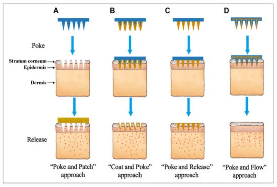
Figure 2. Diagrams showing various microneedle drug delivery approaches. (A) Solid microneedles, for skin pretreatment to create microchannels, followed by the application of transdermal patch; (B) coated microneedles, for deposition of drug formulations into the skin, followed by removal of microneedles; (C) dissolving microneedles, incorporated into the substrate of microneedles, remaining in the skin and dissolving over time to release the drugs; and (D) hollow microneedles, for inserted into the skin and continuous infusion of the drug through the created microchannels [56]. (Adapted from Ref. [56]).
This entry is adapted from the peer-reviewed paper 10.3390/pharmaceutics14051097
This entry is offline, you can click here to edit this entry!