Antigen Presentation and Autophagy in Teleost Adaptive Immunity: History
Please note this is an old version of this entry, which may differ significantly from the current revision.

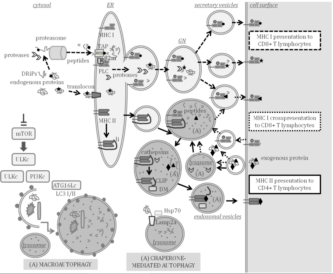
Infectious diseases are a burden for aquaculture. Antigen processing and presentation (APP) to the immune effector cells that fight pathogens is key in the adaptive immune response. At the core of the adaptive immunity that appeared in lower vertebrates during evolution are the variable genes encoding the major histocompatibility complex (MHC). MHC class I molecules mainly present peptides processed in the cytosol by the proteasome and transported to the cell surface of all cells through secretory compartments. Professional antigen-presenting cells (pAPC) also express MHC class II molecules, which normally present peptides processed from exogenous antigens through lysosomal pathways. Autophagy is an intracellular self-degradation process that is conserved in all eukaryotes and is induced by starvation to contribute to cellular homeostasis. Self-digestion during autophagy mainly occurs by the fusion of autophagosomes, which engulf portions of cytosol and fuse with lysosomes (macroautophagy) or assisted by chaperones (chaperone-mediated autophagy, CMA) that deliver proteins to lysosomes. Thus, during self-degradation, antigens can be processed to be presented by the MHC to immune effector cells, thus, linking autophagy to APP.

  • antigen processing
  • antigen-presenting cell
  • bacteria
  • chaperone-mediated autophagy
  • LC3-Associated phagocytosis
  • macroautophagy
  • major histocompatibility complex MHC class I
  • MHC class II
  • vaccine
  • virus
  • teleost

1. Introduction

Antigen Processing and Presentation (APP) in Adaptive Immunity

During the early phases of the development of the adaptive immune system, only T lymphocytes, which are capable of recognizing non-self-peptides are selected. This negative selection allows the activation of the effector functions of surveying T lymphocytes, only when their T cell receptor (TCR) specifically interacts with a foreign peptide presented by a major histocompatibility complex (MHC) protein on an antigen-presenting cell (APC).
Antigen processing and presentation (APP) is the process through which an antigenic protein is processed to peptides, which are loaded and transported to the cell surface on MHC proteins. Autophagy is a self-digestion process that is highly conserved in evolution and involves the degradation of cytoplasmic material delivered to lysosomes by chaperones or by forming double membrane vesicles called autophagosomes [1]. One of the non-canonical functions of autophagy is to participate in APP for the MHC class I and II activation of T lymphocytes [2][3].
The sophisticated adaptive immune system allows the recognition and destruction of pathogens and host cells infected by pathogens. A clear picture of the molecular players in APP has been drawn after almost five decades of research [4]. Although most of the research has been centered in mammals, APP in teleost fish has also been assessed through comparative studies [5].

2. Antigen Processing and Presentation in Teleost Fish

The demonstration of APP occurrence in teleost fish came from studies with the channel catfish (Ictalurus punctatus) with evidence indicating that MHC-like molecules govern APP with a similar restriction as observed in mammals [6] and with exogenous antigens being endocytosed by APC and inducing the proliferation of lymphocytes [7]. There is sufficient evidence supporting that the functions of MHC class I and II molecules in fish and mammals are similar [8]. A scheme of mammal APP indicating the key molecular players that have been studied and characterized in teleost fish is shown in Figure 1, indicating the compartments where APP and autophagy can take place at the same time.
Figure 1. Major histocompatibility complex (MHC) class I and II antigen processing and presentation (APP) and autophagy. MHC class I presents endogenous peptides produced in the cytosol (peptide represented by a circle) or in secretory compartments (star) to CD8 + T lymphocytes through the pathways indicated with the dashed arrows. Endogenously synthesized proteins and defective ribosomal products (DRiPs) are processed to peptides in the cytosol by the proteasome and cytosolic proteases. Peptides are then transported by transporter associated to antigen processing (TAP) to the ER where the peptide loading complex (PLC) is formed by a peptide, TAP, MHC class I, β2-microglobulin (β2m) and other assistant proteins. Endogenous proteins can enter the lumen of the ER through the translocon and be processed by proteases of the ER or the Golgi network (GN), thus, producing peptides that can also be loaded on MHC class I beyond the ER. Loaded MHC class I molecules are transported to the cell surface in secretory vesicles. MHC class I can also cross present peptides and prime CD8+ T lymphocytes through the pathway indicated with the dotted arrows. Cross-presented peptides (triangle) are produced by processing exogenous antigens in the cytosol or in secretory or endocytic compartments in which autophagy (A) may take place. MHC class II presents peptides (diamond) processed from exogenous proteins to CD4 + T lymphocytes through the pathway indicated with the continuous lined arrows. In the ER, MHC class II molecules are assembled and bind to the invariant chain li. The MHC class II is exported to endosomal compartments, in which autophagy can take place and where class II-associated invariant chain peptide (CLIP) remains bound to MHC class II after cleavage of li by lysosomal cathepsins. HLA-DM assists in the exchange of CLIP by a peptide that is exported to the cell surface through exocytic vesicles. Chaperone-mediated autophagy allows the targeting of cytosolic proteins to be degraded in lysosomes and is assisted by Lamp2a, Hsp70 and other chaperones. Macroautophagy is the engulfment of diverse cytosolic material to be degraded, induced by inhibition of the mammalian target of rapamycin (mTOR). Several multiprotein complexes (ULK, PI3K and ATG16L1) are involved in the formation of vesicles and lipidation of LC3-I to LC3-II, thereby, forming autophagosomes that fuse with lysosomes.

2.1. Antigen Processing and Presentation Genes Conserved in Teleost Fish

The emergence of adaptive immunity is related to the highly polymorphic mhc genes encoding MHC, which in most jawed vertebrates are linked, scattered throughout a chromosomal genomic region with varying levels of recombination that also encodes other genes related to APP. Teleost fish are, however, the exception, as the mhc class I gene family is expanded and located on several chromosomes, and the mhc class II genes are lost in some fish species [9][10]. In spite of the loss of key components of the adaptive machinery, teleost fish are capable of mounting a robust immune response to pathogens [5].
Interestingly, the polymorphism in mhc class I observed in many teleosts is proposed to be driven by additional evolutionary pressures apart from pathogen resistance [8]. Initial studies in comparative genomics with medaka (Oryzias latipes) revealed the evolutionarily conserved core in teleost APP is mhc class I and its related genes [11] with the genes encoding MHC class I, proteasome subunits LMP2, LMP7 and TAP2 appearing as linked in linkage analysis [11]. Genes related to MHC class I APP that have been cloned and characterized in teleost fish include several proteasome subunits [12][13][14]β2m [15] or the ERp57 gene pdia3 [16][17] and the tapasin gene tapbp [18].
The mhc class I genes are organized in teleost fish in UZLS or P lineages, although not all species have all lineages. Only the U lineage is predicted to encode the genes that provide APP functions, displaying haplotypic variation and sharing conserved synteny with the core MHC that is observed in mammalian genomes [19]. The mhc class I genes have been cloned and characterized in a wide number of teleost fish [15][20][21][22]. The zebrafish (Danio rerio) shows substantial diversity within genes of proteasome subunits, tap and mhc class I genes [23], indicating teleosts have polymorphisms within the MHC class I APP steps of cleavage, transport and presentation, all predicted to alter peptide specificity.
Vertebrate genome duplication events in evolution have provided cyprinids, salmonids and neoteleosts with duplicates of genes involved in MHC class I APP and therefore with the potential of different PLC [24]. The U lineage in the zebrafish is located on chromosome 19, although two U genes map to chromosome 22 and are predicted to play non classical roles in APP because they lack key residues for peptide binding and display limited polymorphic variation [25]. Further studies are needed to decipher the complexity in the number of functional genes involved in MHC class I APP, as gene duplicates respond differently to stimulation and may not share biological functions [26].
The mhc class II genes in teleost fish are classified in three major groups (DADB and DE), with only DA exhibiting the classical features [19]. The gene coding for the α chain of class II (spau-daa) has been characterized in the gilthead seabream (Sparus aurata), showing conserved important features and a high variability in the α1 domain that forms the peptide-binding domain (PBD) [27]. The mhc class II loci have been lost independently in gadiform fishes, such as Atlantic cod (Gadus morhua) [28] and in some syngnathiform fishes, such as the pipefish (Syngnathus typhle) [29].
HLA-DM, which assists MHC class II in peptide loading, is highly conserved in tetrapod species. The HLA-DM-lineage had only been reported to the level of amphibians until it was recently reported to exist in lungfish [30]. A critical Trp residue required for the HLA-DM system found in mammals is not encoded in classical teleost mhc class II and only found in non-classical mhc class II genes [31]. The invariant chain (li) and cathepsins, which are involved in the cleavage of li have been studied in depth [32][33][34][35][36][37][38][39][40]. Conserved motifs in the CLIP region of li exist throughout jawed vertebrates. Conserved li residues and cathepsin orthologs suggest their long co-evolution in the APP, with sub-functional duplicated cd74 genes [30][41]. In rainbow trout (Oncorhynchus. mykiss), two cd74 genes have been characterized, and their expression is regulated differently [32][33].

2.2. Antigen-Presenting Cells (APC) and Professional APC (pAPC) in Teleost Fish

All cells are APC as they express MHC class I and can perform APP. Indeed, MHC class I molecules was reported to be expressed in teleost fish in similar cell types as in higher vertebrates [42], and there is evidence indicating that the activation of CD8+ T lymphocyte-mediated cytotoxicity is MHC class I restricted [8][43]. MHC class II is, however, only expressed in the professional APC (pAPC), which are in charge of the initial activation or priming of effector T lymphocytes.
These pAPC mainly reside in lymphoid organs, such as the head-kidney, the thymus or the spleen, and are capable of priming T lymphocytes to become cytotoxic CD8+ T lymphocytes or CD4+ T helper lymphocytes. The role of pAPC is thus to act as immune sentries that engulf pathogens and carry foreign antigens to lymphoid tissues in order to stimulate an antigen-specific T lymphocyte response. As described in higher vertebrates, the pAPC in some teleost fish species are dendritic cells (DCs), B cells and macrophages, all of which have been characterized in depth in the zebrafish [44]. However, in other species of bony fishes, in addition to DCs, B cells and macrophages, granulocytes [27] and erythrocytes (red blood cells, RBC) [45][46][47] also have a prominent role in APP.
The phenotypic and functional features of DCs have been studied in several teleost species, and methods to enrich DCs have been established. In zebrafish, a pAPC subset strongly resembling mammalian DCs was characterized in relation to affinity for the lectin peanut agglutinin (PNA) [48]. Myeloid cells that were PNA(hi) had phagocytic activity and expressed genes associated to DC function and APP, including MHC class II [48]. Most importantly PNA(hi) cells could activate T lymphocytes in an antigen-dependent manner [48].
Among the phenotypic cell surface markers of zebrafish DCs are MHC class II, CD80/86, CD83 and CD209, and all these molecules were involved in the activation of CD4 + T lymphocytes [49]. In barramundi (Lates calcarifer), maturation of spleen- and pronephros-derived monocytes exposed in vitro to bacterial-derived peptidoglycans induced differentiation to DC. These DC were phagocytic and could migrate towards pathogens or pathogen associated patterns, they expressed MHC class II and DC-SCRIPT, a conserved zinc finger protein preferentially expressed in human DC subtypes [50]. Importantly, barramundi DCs induced proliferation of effector T lymphocytes [50].
In rainbow trout, mucosal tissues from skin and gills have a DC subpopulation that co-expressed CD8α and MHC class II [51], with a homologous population described in intestinal lamina propia [52]. However, the intestinal CD8+ DCs exhibited phenotypic and functional differences compared to skin and gill CD8+ DCs, suggesting the presence of different subpopulations of DCs on different locations [52]. Furthermore, two phenotypes of pAPC were characterized in the gills of rainbow trout. One had features of a monocyte/macrophage/dendritic cell-type; expressing CD45, CD83 and IL-1 β, while the second phenotype expressed almost all genes related to the phagosome, lysosome and APP pathways and exhibited similar characteristics to mammalian M cells, which play a role in mucosal immune surveillance [53].
In a different species, the orange-spotted grouper (Epinephelus coioides), a transcriptomic study performed on skin after Cryptocaryon irritans infection, revealed upregulation of DCs markers, including CD209 and CD83 [54]. CD207 is a marker of Langerhans cells (tissue-resident macrophages of the skin), which are developmentally similar to DCs. Infection of the grass carp (Ctenopharyngodon idella) with Flavobacterium columnare upregulated CD207 only in the spleen, suggesting that the biological function of CD207 may be related to APP [55].
B cells are pAPC in teleosts as they can uptake particulate antigens by phagocytosis, process and present antigens to T lymphocytes [56][57]. In bony fish, B cells produce three isotypes of immunoglobulins (Ig): IgM, IgD and IgT/Z [56]. In accordance, three main B-cell lineages have been described in teleosts, a B-cell lineage expressing IgT/Z, a lineage expressing IgD and the most common B-cell lineage co-expressing IgD and IgM [56].
In the rainbow trout, infection in vitro of blood or spleen IgM+ B cells with viral hemorrhagic septicemia virus (VHSV) upregulates MHC class II, CD80/86 or CD83, suggesting the activation towards a pAPC profile [58]. In vivo IgM+ B cells are recruited to the peritoneum cavity after challenge with virus VHSV or bacteria Escherichia coli and differentiate to cells specialized in APP [59]. A teleost B cell subset has been proposed to be a counterpart of mammalian B1-B cells, which link innate and adaptive immunity [60].
Professional phagocytic cells have a role in pAPC in teleost fish. Thus, macrophages are pAPC that participate in immunity as phagocytic cells, with two polarization states, the pro-inflammatory M1-type and anti-inflammatory M2-type. The expression of genes involved in APP was detected in both M1- and M2-type macrophages in the zebrafish [61]. In addition, the infection of macrophages upregulated MHC class II and transcripts of other genes involved in APP [61]. In addition to macrophages, head-kidney granulocytes can express high levels of MHC class II in gilthead seabream [27] or in Atlantic salmon (Salmo salar) [45] and are proposed as a model to study autophagy and APP during vertebrate immune response [45].

2.3. Peptide Processing, Loading and Transport by MHC in Teleost Fish

A number of studies have characterized some of the key proteins involved in the processing of antigens to peptides, the loading on MHC and MHC/peptide transport to the cell surface of APC. Concerning the MHC class I APP pathway, in European sea bass (Dicentrarchus labrax), different transcripts of MHC class I have been identified per individual, detecting variability at the PBD, with a three-dimensional homology model that is consistent with that of other vertebrates [15]. The proteasome activator subunits PA28α and PA28β were upregulated upon immune stimulation in the Japanese flounder (Paralichthys olivaceus) [12], the large yellow croaker (Larimichthys crocea) [13] and the rock bream (Oplegnathus fasciatus) [14].
The expression of β2m was induced by an antigen in the European sea bass [15]. The interaction of tapasin with TAP, MHC class I and ERp57, all which are known to occur during assembly of the mammalian peptide loading complex (PLC), was conserved in the monocyte/macrophage rainbow trout cell line RTS11 [62]. Homology models of the protein ERp57 in European sea bass support that they are orthologs of the mammalian protein involved in MHC class I APP [16].
In rainbow trout, expression analysis supports the role of ERp57 during the activation of the immune response in teleosts [17], although some features conserved in mammalian ERp57 are not conserved in the fish gene [17]. In the same species, tapasin was mainly expressed in immune system organs and in cell lines derived from epithelial cells from the gill, liver and intestine [18]. In addition, tapasin was strongly expressed in parallel with MHC class I in the RTS11 monocyte/macrophage derived cell line after viral infection [18].
Concerning the MHC class II APP pathway, the expression of the alpha chain of gilthead seabream MHC class II (Spau-DAA) was observed in peritoneal exudate leucocytes, head-kidney, spleen, thymus and gill; and the incubation of head-kidney leucocytes with yeast or bacteria upregulated levels of Spau-DAA, whereas concanavalin A (ConA), lipopolysaccharide (LPS) or phytohemagglutinin A (PHA) did not [27].
In the MHC class II APP, the interferon-γ-inducible lysosomal thiol reductase (GILT) is important as it reduces disulfide bonds in the unfolding of proteins and facilitates their subsequent cleavage by proteases. In zebrafish, purified recombinant GILT was capable of catalyzing the reduction of protein disulfide bonds [63]. GILT was constitutively expressed in several tissues of zebrafish, large yellow croaker and mandarin fish (Siniperca chuatsi) and upregulated in the spleen and kidney after stimulation [63][64][65].
The invariant chain li, a key player in the formation of mammalian MHC class II/peptide complex, appears to be expressed independently of immune function in some teleost fish. The invariant chain-like proteins S25-7 and INVX of the rainbow trout were not expressed in the head-kidney, and their expression in spleen and gill was unaffected by immune stimulation [33]. Cathepsins, the family of lysosomal cysteine proteases that are involved in the processing of li, have been characterized in depth in teleost species.
Several cathepsins have been characterized and found to be widely expressed and produced in vitro to confirm their protease activity: cathepsins D and L in the half-smooth tongue sole (Cynoglossus semilaevis) [35]; in the large yellow croaker cathepsin L and S [36] and B [34]; in the miiuy croaker (Miichthys miiuy) cathepsin S [37] and cathepsin B and cathepsin H [38] were assessed; while cathepsin B was studied in the golden pompano (Trachinotus ovatus) [40] and cathepsin S in the channel catfish [39]. The capacity of cathepsins to cleave the invariant chain li was studied in the large yellow croaker, finding that cathepsin B efficiently cleaves recombinant li in a time-dependent manner [34].
Cathepsin S and cathepsin L both had cysteine protease activity and could remove propeptides and release active mature peptides; however, li inhibited the autocatalytic activation of cathepsin L, whereas activated cathepsin S could efficiently process li in vitro [36]. These results suggest that cathepsins S and B are the main cathepsins involved in the processing of li in teleost fish [34][36]. It should be noted that, in the channel catfish, two cathepsin S genes have been characterized, (ctss and ctssa), both upregulated during bacterial infection, however, with differences between their expression profiles in mucosal surfaces, suggesting that they may exert disparate roles in mucosal immunity [39].

3. Autophagy and Related LC3-Associated Phagocytosis in Teleost Fish

Autophagy is a self-digestion process that is highly conserved in evolution and involves lysosomal degradation of cytoplasmic material [1]. Autophagy mainly occurs by fusion of autophagosomes, which engulf portions of cytosol and fuse with lysosomes (macroautophagy), or assisted by chaperones (chaperone-mediated autophagy, CMA) that deliver cytosolic proteins to lysosomes. One of the non-canonical functions of autophagy is to participate in APP for MHC class I and II activation of T lymphocytes [3]. LC3-associated phagocytosis (LAP), however, is not a form of autophagy; it depends on key autophagy molecular players [66].

3.1. Chaperone-Mediated Autophagy in Teleost Fish

CMA was long believed to only exist in birds and mammals [67]; however, it has been proved to exist also in fish [68][69]. Lamp2a is the product of one of the three splice variants (lamp2alamp2b and lamp2c) of gene lamp2 that is transcribed by alternative splicing, and this genomic organization has been highly conserved during vertebrate evolution [68]. The gene lamp2 appears at the root of the vertebrate lineage with a Ct domain GYXXF sequence that is conserved in most teleosts [68], including medaka. However, lamp2 is divergent in zebrafish, which raised the question on the ability of this species to perform CMA [67].
In medaka in vitro studies, the splice variant lamp2a was found to control lysosomal accumulation of a reporter of CMA in a fibroblast cell line; and in vivo, a lamp2a medaka knock-out exhibited carbohydrate and fat metabolism alterations [68]. In cultured cells of the yellowtail fish (Seriola quinqueradiata), three cytosolic members of the Hsp70 family have been isolated; heat-shock cognate proteins (Hsc)70-1 and Hsc70-2 and heat shock protein (Hsp)70 [70]. Heat-induced expression of Hsp70, together with the lysosome localization of Hsc70/Hsp70 suggests that CMA is induced by heat shock in bony fish [70]. In the zebrafish, a member of the small heat shock protein (sHSP) family, Hspb8, interacts with proteins Bag3 and Hsc70, which are essential for the formation of an autophagy-inducing complex [71].

3.2. Macroautophagy in Teleost Fish

Xenophagy is the capture and destruction of invading pathogens by means of a form of selective macroautophagy that requires the participation of receptors Sqstm1/p62 that bind to ubiquitinated cargo and LC3-II to form p62-droplets that accumulate at the surface of autophagosomes during their formation [1][66][72]. The contribution of zebrafish models to study the specific elimination of certain intracellular substrates by autophagic pathways, has been recently reviewed [73], as well as the role of selective autophagy in immunity to bacterial pathogens (Shigella flexneri and Mycobacterium marinum) [74].
Inflammatory toxicity in zebrafish induces the cleavage of p62 by caspase-6 at a highly conserved D256 cleavage site, indicating certain stress stimuli can modulate autophagy through the caspase 6-p62 axis [75]. Zebrafish mutant lines have shown that becn1 is essential for autophagy, as knock-out mutants for this gene die at the larval stage [76]. In addition, mutants in becn1 and atg7 have outlined the importance of becn1 for maintaining homeostasis of nutrient metabolism and in liver development [77].

3.3. LC3-Associated Phagocytosis in Teleost Fish

In a zebrafish embryo model, the interaction of human pathogen Salmonella typhimurium with the autophagy machinery of macrophages identified LAP as the major host protective autophagy-related pathway responsible for macrophage defense during systemic infection [78]. In contrast, in an infection model to study the human pathogen Staphylococcus aureus, LAP was found to be involved in the intracellular handling of the pathogen by neutrophils [79] with a key role for Sqstm1/p62 in autophagic control of infection [80].

This entry is adapted from the peer-reviewed paper 10.3390/ijms23094899

References

  1. Galluzzi, L.; Baehrecke, E.H.; Ballabio, A.; Boya, P.; Bravo-San Pedro, J.M.; Cecconi, F.; Choi, A.M.; Chu, C.T.; Codogno, P.; Colombo, M.I.; et al. Molecular definitions of autophagy and related processes. EMBO J. 2017, 36, 1811–1836.
  2. Münz, C. The macroautophagy machinery in MHC restricted antigen presentation. Front. Immunol. 2021, 12, 1–7.
  3. Münz, C. Non-canonical functions of autophagy proteins in immunity and infection. Mol. Asp. Med. 2021, 82, 100987.
  4. Rock, K.L.; Reits, E.; Neefjes, J. Present yourself! By MHC class I and MHC class II molecules. Trends Immunol. 2016, 37, 724–737.
  5. Wilson, A.B. MHC and adaptive immunity in teleost fishes. Immunogenetics 2017, 69, 521–528.
  6. Vallejo, A.N.; Miller, N.W.; Clem, L.W. Phylogeny of immune recognition: Role of alloantigens in antigen presentation in channel catfish immune responses. Immunology 1991, 74, 165–168.
  7. Vallejo, A.N.; Miller, N.W.; Harvey, N.E.; Cuchens, M.A.; William Clem, L. Cellular pathway(s) of antigen processing and presentation in fish APC: Endosomal involvement and cell-free antigen presentation. Dev. Immunol. 1992, 3, 51–65.
  8. Yamaguchi, T.; Dijkstra, J.M. Major histocompatibility complex (MHC) genes and disease resistance in fish. Cells 2019, 8, 378.
  9. Radwan, J.; Babik, W.; Kaufman, J.; Lenz, T.L.; Winternitz, J. Advances in the evolutionary understanding of MHC polymorphism. Trends Genet. 2020, 36, 298–311.
  10. Kaufman, J. Unfinished business: Evolution of the MHC and the adaptive immune system of jawed vertebrates. Annu. Rev. Immunol. 2018, 36, 383–409.
  11. Nonaka, M.; Naruse, K.; Matsuo, M.; Shima, A. Comparative genomics of medaka: The major histocompatibility complex (MHC). Mar. Biotechnol. 2001, 3, S141–S144.
  12. Kim, D.-H.; Lee, S.-M.; Hong, B.-Y.; Kim, Y.-T.; Choi, T.-J. Cloning and sequence analysis of cDNA for the proteasome activator PA28-β subunit of flounder (Paralichthys olivaceus). Mol. Immunol. 2003, 40, 611–616.
  13. Liu, G.; Zheng, W.; Chen, X. Molecular cloning of proteasome activator PA28-β subunit of large yellow croaker (Pseudosciana crocea) and its coordinated up-regulation with MHC class I α-chain and β2-microglobulin in poly I:C-treated fish. Mol. Immunol. 2007, 44, 1190–1197.
  14. Kasthuri, S.R.; Umasuthan, N.; Whang, I.; Kim, E.; Park, H.-C.; Lee, J. Genomic structural characterization and transcriptional expression analysis of proteasome activator PA28α and PA28β subunits from Oplegnathus fasciatus. Fish Shellfish Immunol. 2013, 35, 1224–1234.
  15. Pinto, R.D.; Randelli, E.; Buonocore, F.; Pereira, P.J.B.; dos Santos, N.M.S. Molecular cloning and characterization of sea bass (Dicentrarchus labrax, L.) MHC class I heavy chain and β2-microglobulin. Dev. Comp. Immunol. 2013, 39, 234–254.
  16. Pinto, R.D.; Moreira, A.R.; Pereira, P.J.B.; dos Santos, N.M.S. Two thioredoxin-superfamily members from sea bass (Dicentrarchus labrax, L.): Characterization of PDI (PDIA1) and ERp57 (PDIA3). Fish Shellfish Immunol. 2013, 35, 1163–1175.
  17. Sever, L.; Bols, N.C.; Dixon, B. The cloning and inducible expression of the rainbow trout ERp57 gene. Fish Shellfish Immunol. 2013, 34, 410–419.
  18. Sever, L.; Vo, N.T.K.; Bols, N.C.; Dixon, B. Expression of tapasin in rainbow trout tissues and cell lines and up regulation in a monocyte/macrophage cell line (RTS11) by a viral mimic and viral infection. Dev. Comp. Immunol. 2014, 44, 86–93.
  19. Wilson, W.H.; Gilg, I.C.; Moniruzzaman, M.; Field, E.K.; Koren, S.; Lecleir, G.R.; Martínez-Martínez, J.; Poulton, N.J.; Swan, B.K.; Stepanauskas, R.; et al. Genomic exploration of individual giant ocean viruses. ISME J. 2017, 11, 1736–1745.
  20. Nonaka, M.I.; Aizawa, K.; Mitani, H.; Bannai, H.P.; Nonaka, M. Retained orthologous relationships of the MHC class I genes during euteleost evolution. Mol. Biol. Evol. 2011, 28, 3099–3112.
  21. Chen, J.; Wang, L.; Huang, J.; Li, X.; Guan, L.; Wang, Q.; Yang, M.; Qin, Q. Functional analysis of a novel MHC-Iα genotype in orange-spotted grouper: Effects on Singapore grouper iridovirus (SGIV) replication and apoptosis. Fish Shellfish Immunol. 2022, 121, 487–497.
  22. Hashimoto, K.; Nakanishi, T.; Kurosawa, Y. Isolation of carp genes encoding major histocompatibility complex antigens. Proc. Natl. Acad. Sci. USA 1990, 87, 6863–6867.
  23. McConnell, S.C.; Hernández, K.M.; Wcisel, D.J.; Kettleborough, R.N.; Stemple, D.L.; Yoder, J.A.; Andrade, J.; de Jong, J.L.O. Alternative haplotypes of antigen processing genes in zebrafish diverged early in vertebrate evolution. Proc. Natl. Acad. Sci. USA 2016, 113, E5014–E5023.
  24. Grimholt, U. Whole genome duplications have provided teleosts with many roads to peptide loaded MHC class I molecules. BMC Evol. Biol. 2018, 18, 1–14.
  25. Dirscherl, H.; Yoder, J.A. A nonclassical MHC class I U lineage locus in zebrafish with a null haplotypic variant. Immunogenetics 2015, 67, 501–513.
  26. Grimholt, U.; Fosse, J.H.; Sundaram, A.Y.M. Selective stimulation of duplicated Atlantic salmon MHC pathway genes by interferon-gamma. Front. Immunol. 2020, 11, 571650–571665.
  27. Cuesta, A.; Ángeles Esteban, M.; Meseguer, J. Cloning, distribution and up-regulation of the teleost fish MHC class II alpha suggests a role for granulocytes as antigen-presenting cells. Mol. Immunol. 2006, 43, 1275–1285.
  28. Pilstrom, L.; Warr, G.W.; Stromberg, S. Why is the antibody response of Atlantic cod so poor? The search for a genetic explanation. Fish. Sci. 2005, 71, 961–971.
  29. Haase, D.; Roth, O.; Kalbe, M.; Schmiedeskamp, G.; Scharsack, J.P.; Rosenstiel, P.; Reusch, T.B.H. Absence of major histocompatibility complex class II mediated immunity in pipefish, Syngnathus typhle: Evidence from deep transcriptome sequencing. Biol. Lett. 2013, 9, 20130044.
  30. Dijkstra, J.M.; Yamaguchi, T. Ancient features of the MHC class II presentation pathway, and a model for the possible origin of MHC molecules. Immunogenetics 2019, 71, 233–249.
  31. Dijkstra, J.M.; Grimholt, U.; Leong, J.; Koop, B.F.; Hashimoto, K. Comprehensive analysis of MHC class II genes in teleost fish genomes reveals dispensability of the peptide-loading DM system in a large part of vertebrates. BMC Evol. Biol. 2013, 13, 260.
  32. Fujiki, K. Alternate forms of MHC class II-associated invariant chain are not produced by alternative splicing in rainbow trout (Oncorhynchus mykiss) but are encoded by separate genes. Dev. Comp. Immunol. 2003, 27, 377–391.
  33. Semple, S.L.; Heath, G.; Christie, D.; Braunstein, M.; Kales, S.C.; Dixon, B. Immune stimulation of rainbow trout reveals divergent regulation of MH class II-associated invariant chain isoforms. Immunogenetics 2019, 71, 407–420.
  34. Li, M.; Li, Q.; Yang, Z.; Hu, G.; Li, T.; Chen, X.; Ao, J. Identification of cathepsin B from large yellow croaker (Pseudosciaena crocea) and its role in the processing of MHC class II-associated invariant chain. Dev. Comp. Immunol. 2014, 45, 313–320.
  35. Chen, L.; Zhang, M.; Sun, L. Identification and expressional analysis of two cathepsins from half-smooth tongue sole (Cynoglossus semilaevis). Fish. Shellfish Immunol. 2011, 31, 1270–1277.
  36. Li, Q.; Ao, J.; Mu, Y.; Yang, Z.; Li, T.; Zhang, X.; Chen, X. Cathepsin S, but not cathepsin L, participates in the MHC class II-associated invariant chain processing in large yellow croaker (Larimichthys crocea). Fish. Shellfish Immunol. 2015, 47, 743–750.
  37. Sun, Y.; Xu, T.; Wang, J.; Cheng, Y.; Wang, R. Sequence and expression analysis of cathepsin S gene in the miiuy croaker Miichthys miiuy. Fish. Physiol. Biochem. 2011, 37, 761–765.
  38. Che, R.; Wang, R.; Xu, T. Comparative genomic of the teleost cathepsin B and H and involvement in bacterial induced immunity of miiuy croaker. Fish. Shellfish Immunol. 2014, 41, 163–171.
  39. Dong, X.; Ye, Z.; Song, L.; Su, B.; Zhao, H.; Peatman, E.; Li, C. Expression profile analysis of two cathepsin S in channel catfish (Ictalurus punctatus) mucosal tissues following bacterial challenge. Fish. Shellfish Immunol. 2016, 48, 112–118.
  40. Shen, Y.; Zhang, H.; Zhou, Y.; Sun, Y.; Yang, H.; Cao, Z.; Qin, Q.; Liu, C.; Guo, W. Functional characterization of cathepsin B and its role in the antimicrobial immune responses in golden pompano (Trachinotus ovatus). Dev. Comp. Immunol. 2021, 123, 104128.
  41. Criscitiello, M.F.; Ohta, Y.; Graham, M.D.; Eubanks, J.O.; Chen, P.L.; Flajnik, M.F. Shark class II invariant chain reveals ancient conserved relationships with cathepsins and MHC class II. Dev. Comp. Immunol. 2012, 36, 521–533.
  42. Dijkstra, J.M.; Köllner, B.; Aoyagi, K.; Sawamoto, Y.; Kuroda, A.; Ototake, M.; Nakanishi, T.; Fischer, U. The rainbow trout classical MHC class I molecule Onmy-UBA*501 is expressed in similar cell types as mammalian classical MHC class I molecules. Fish. Shellfish Immunol. 2003, 14, 1–23.
  43. Chang, Y.T.; Kai, Y.H.; Chi, S.C.; Song, Y.L. Cytotoxic CD8α+ leucocytes have heterogeneous features in antigen recognition and class I MHC restriction in grouper. Fish. Shellfish Immunol. 2011, 30, 1283–1293.
  44. Kanako, L. Lewis; Natasha, Del Cid; D.T. Perspectives on antigen presenting cells in zebrafish. Dev. Comp. Immunol. 2014, 46, 63–73.
  45. Iliev, D.B.; Thim, H.; Lagos, L.; Olsen, R.; Jørgensen, J.B. Homing of antigen-presenting cells in head kidney and spleen—salmon head kidney hosts diverse APC types. Front. Immunol. 2013, 4, 137.
  46. Ronza, P.; Álvarez-Dios, J.A.; Robledo, D.; Losada, A.P.; Romero, R.; Bermúdez, R.; Pardo, B.G.; Martínez, P.; Quiroga, M.I. Blood transcriptomics of turbot Scophthalmus maximus: A tool for health monitoring and disease studies. Animals 2021, 11, 1296.
  47. Pereiro, P.; Romero, A.; Díaz-Rosales, P.; Estepa, A.; Figueras, A.; Novoa, B. Nucleated teleost erythrocytes play an Nk-lysin- and autophagy-dependent role in antiviral immunity. Front. Immunol. 2017, 8, 1458.
  48. Lugo-Villarino, G.; Balla, K.M.; Stachura, D.L.; Bañuelos, K.; Werneck, M.B.F.; Traver, D. Identification of dendritic antigen-presenting cells in the zebrafish. Proc. Natl. Acad. Sci. USA 2010, 107, 15850–15855.
  49. Shao, T.; Zhu, L.-Y.; Nie, L.; Shi, W.; Dong, W.-R.; Xiang, L.-X.; Shao, J.-Z. Characterization of surface phenotypic molecules of teleost dendritic cells. Dev. Comp. Immunol. 2015, 49, 38–43.
  50. Zoccola, E.; Delamare-Deboutteville, J.; Barnes, A.C. Identification of barramundi (Lates calcarifer) DC-SCRIPT, a specific molecular marker for dendritic cells in fish. PLoS ONE 2015, 10, e0132687.
  51. Soleto, I.; Fischer, U.; Tafalla, C.; Granja, A.G. Identification of a potential common ancestor for mammalian cross-presenting dendritic cells in teleost respiratory surfaces. Front. Immunol. 2018, 9, 1–13.
  52. Soleto, I.; Granja, A.G.; Simón, R.; Morel, E.; Díaz-Rosales, P.; Tafalla, C. Identification of CD8α+ dendritic cells in rainbow trout (Oncorhynchus mykiss) intestine. Fish. Shellfish Immunol. 2019, 89, 309–318.
  53. Kato, G.; Miyazawa, H.; Nakayama, Y.; Ikari, Y.; Kondo, H.; Yamaguchi, T.; Sano, M.; Fischer, U. A novel antigen-sampling cell in the teleost gill epithelium with the potential for direct antigen presentation in mucosal tissue. Front. Immunol. 2018, 9, 2116–2128.
  54. Hu, Y.; Li, A.; Xu, Y.; Jiang, B.; Lu, G.; Luo, X. Transcriptomic variation of locally-infected skin of Epinephelus coioides reveals the mucosal immune mechanism against Cryptocaryon irritans. Fish. Shellfish Immunol. 2017, 66, 398–410.
  55. Wang, H.; Chen, X.; Li, S.; Zhou, C.; Xu, L.; Wu, Z.; Chen, X. Identification and expression analysis of Langerhans cells marker Langerin/CD207 in grass carp, Ctenopharyngodon idella. Gene 2021, 768, 145315.
  56. Wu, L.; Qin, Z.; Liu, H.; Lin, L.; Ye, J.; Li, J. Recent advances on phagocytic B cells in teleost fish. Front. Immunol. 2020, 11, 824–833.
  57. Miller, N.; Wilson, M.; Bfuflten, E.; Stuge, T.; Warr, G.; Ciem, W. Functional and molecular characterization of teleost leukocytes. Immunol. Rev. 1998, 166, 187–197.
  58. Abós, B.; Castro, R.; González Granja, A.; Havixbeck, J.J.; Barreda, D.R.; Tafalla, C. Early activation of teleost B cells in response to rhabdovirus infection. J. Virol. 2015, 89, 1768–1780.
  59. Castro, R.; Abós, B.; González, L.; Granja, A.G.; Tafalla, C. Expansion and differentiation of IgM+ B cells in the rainbow trout peritoneal cavity in response to different antigens. Dev. Comp. Immunol. 2017, 70, 119–127.
  60. Sunyer, J.O. Evolutionary and functional relationships of B cells from fish and mammals: Insights into their novel roles in phagocytosis and presentation of particulate antigen. Infect. Disord. Drug Targets 2012, 12, 200–212.
  61. Rougeot, J.; Torraca, V.; Zakrzewska, A.; Kanwal, Z.; Jansen, H.J.; Sommer, F.; Spaink, H.P.; Meijer, A.H. RNAseq profiling of leukocyte populations in zebrafish larvae reveals a cxcl11 chemokine gene as a marker of macrophage polarization during mycobacterial infection. Front. Immunol. 2019, 10, 832.
  62. Sever, L.; Vo, N.T.K.; Bols, N.C.; Dixon, B. Tapasin’s protein interactions in the rainbow trout peptide-loading complex. Dev. Comp. Immunol. 2018, 81, 262–270.
  63. Cui, X.; Ji, C.; Cao, X.; Fu, Z.; Zhang, S.; Guo, X. Molecular and biological characterization of interferon-γ-inducible-lysosomal thiol reductase gene in zebrafish (Danio rerio). Fish. Shellfish Immunol. 2012, 33, 1133–1138.
  64. Zheng, W.; Chen, X. Cloning and expression analysis of interferon-γ-inducible-lysosomal thiol reductase gene in large yellow croaker (Pseudosciaena crocea). Mol. Immunol. 2006, 43, 2135–2141.
  65. Song, J.; Liu, H.; Ma, L.; Gao, C.; Zhang, S. Molecular cloning, expression and functional characterization of interferon-γ-inducible lysosomal thiol reductase (GILT) gene from mandarin fish (Siniperca chuatsi). Fish. Shellfish Immunol. 2014, 38, 275–281.
  66. Gomes, L.C.; Dikic, I. Autophagy in antimicrobial immunity. Mol. Cell 2014, 54, 224–233.
  67. Herpin, A.; Lescat, L.; Bobe, J.; Jenny, A.; Seiliez, I. Lighting chaperone-mediated autophagy (CMA) evolution with an ancient LAMP: The existence of a functional CMA activity in fish. Autophagy 2020, 16, 1918–1920.
  68. Lescat, L.; Véeron, V.; Mourot, B.; Péron, S.; Chenais, N.; Dias, K.; Riera-Heredia, N.; Beaumatin, F.; Pinel, K.; Priault, M.; et al. Chaperone-mediated autophagy in the light of evolution: Insight from fish. Mol. Biol. Evol. 2020, 37, 2887–2899.
  69. Lescat, L.; Herpin, A.; Mourot, B.; Véron, V.; Guiguen, Y.; Bobe, J.; Seiliez, I. CMA restricted to mammals and birds: Myth or reality? Autophagy 2018, 14, 1267–1270.
  70. Yabu, T.; Imamura, S.; Mohammed, M.S.; Touhata, K.; Minami, T.; Terayama, M.; Yamashita, M. Differential gene expression of HSC70/HSP70 in yellowtail cells in response to chaperone-mediated autophagy. FEBS J. 2011, 278, 673–685.
  71. Dubińska-Magiera, M.; Niedbalska-Tarnowska, J.; Migocka-Patrzałek, M.; Posyniak, E.; Daczewska, M. Characterization of Hspb8 in Zebrafish. Cells 2020, 9, 1562.
  72. Klionsky, D.J.; Abdel-Aziz, A.K.; Abdelfatah, S.; Abdellatif, M.; Abdoli, A.; Abel, S.; Abeliovich, H.; Abildgaard, M.H.; Abudu, Y.P.; Acevedo-Arozena, A.; et al. Guidelines for the use and interpretation of assays for monitoring autophagy. Autophagy 2021, 17, 1–382.
  73. Pant, D.C.; Nazarko, T.Y. Selective autophagy: The rise of the zebrafish model. Autophagy 2021, 17, 3297–3305.
  74. Muñoz-Sánchez, S.; van der Vaart, M.; Meijer, A.H. Autophagy and Lc3-associated phagocytosis in zebrafish models of bacterial infections. Cells 2020, 9, 2372.
  75. Valionyte, E.; Yang, Y.; Griffiths, S.A.; Bone, A.T.; Barrow, E.R.; Sharma, V.; Lu, B.; Luo, S. The caspase-6–p62 axis modulates p62 droplets based autophagy in a dominant-negative manner. Cell Death Differ. 2021.
  76. Dong, G.; Zhang, Z.; Duan, K.; Shi, W.; Huang, R.; Wang, B.; Luo, L.; Zhang, Y.; Ruan, H.; Huang, H. Beclin 1 deficiency causes hepatic cell apoptosis via endoplasmic reticulum stress in zebrafish larvae. FEBS Lett. 2020, 594, 1155–1165.
  77. Mawed, S.A.; He, Y.; Zhang, J.; Mei, J. Strategy of hepatic metabolic defects induced by beclin1 heterozygosity in adult zebrafish. Int. J. Mol. Sci. 2020, 21, 1533.
  78. Masud, S.; Prajsnar, T.K.; Torraca, V.; Lamers, G.E.M.; Benning, M.; Van Der Vaart, M.; Meijer, A.H. Macrophages target Salmonella by Lc3-associated phagocytosis in a systemic infection model. Autophagy 2019, 15, 796–812.
  79. Prajsnar, T.K.; Serba, J.J.; Dekker, B.M.; Gibson, J.F.; Masud, S.; Fleming, A.; Johnston, S.A.; Renshaw, S.A.; Meijer, A.H. The autophagic response to Staphylococcus aureus provides an intracellular niche in neutrophils. Autophagy 2021, 17, 888–902.
  80. Gibson, J.F.; Prajsnar, T.K.; Hill, C.J.; Tooke, A.K.; Serba, J.J.; Tonge, R.D.; Foster, S.J.; Grierson, A.J.; Ingham, P.W.; Renshaw, S.A.; et al. Neutrophils use selective autophagy receptor Sqstm1/p62 to target Staphylococcus aureus for degradation in vivo in zebrafish. Autophagy 2021, 17, 1448–1457.
More
This entry is offline, you can click here to edit this entry!