The cells response to injury is initiated by growth factors and cytokines that play a key role in wound restoration, and their biological action is achieved via signal transduction. Growth factors and cytokines play distinct roles through all phases of wound healing. In response to injury, they can trigger several strategic signalling transduction pathways that are mostly activated during embryonic skin development. Extracellular signal-regulated kinases (ERKs) and calcium (Ca2+) are the first intracellular signalling molecules for tissue repair response. These signalling molecules regulate several biological activities including cellular migration, proliferation, contractility, survival and many more related to different transcription factors that are usually induced by several other intracellular signalling pathways. This phenomenon makes it difficult to link a specific signalling response to injury.
1. Introduction
Injured or damaged skin is restored via a well-coordinated cutaneous restoration response. The exact molecular and cellular mechanisms behind wound restoration are poorly understood. The most peculiar observation is that repaired skin is different from uninjured skin, largely due to disparities in the processes regulating postnatal cutaneous wound restoration in mammals [
1]. Essentially, the restoration response to cutaneous tissue injury includes inflammation, neoangiogenesis, deposition of matrix, and recruitment of cells. A delay in this process is largely coupled with underlying medical conditions including diabetes mellitus (DM), vascular disease, and aging. Non-healing diabetic ulcers are frequently associated with chronicity and limb amputation [
2]. Conventional therapies for chronic wounds are available but are limited in their treatment success. Key to the generation of novel therapies for chronic wound management is an understanding of the underlying molecular processes including cellular signalling that is engaged during the wound restoration process [
1,
2].
A classic wound restoration process is divided into four succeeding phases, viz., haemostasis, inflammation, resurfacing of new epithelium, and remodelling of connective tissue. These phases are well defined elsewhere [
3] and are precisely controlled by intricate communication between cells, signalisation, and extracellular matrix (ECM) proteins [
4]. Tissue restoration involves various cell types that go through proliferation, migration, differentiation, and apoptosis aided by various biological signalling pathways [
1,
5]. The canonical Wnt/β-catenin pathway is implicated in a range of biological activities including cell proliferation, differentiation, and apoptosis and is one of the critical pathways participating in the restoration of cutaneous wounds [
6]. Delay in the wound restoration process and the development of wound chronicity are largely due to the reduced production and performance of cytokines and the induction of their specific receptors and intracellular signalling, hindering the functionality of cells including fibroblasts [
7].
DM is characterised by elevated blood glucose levels and is a major cause of systemic chronic metabolic disease. Complications related to DM are multiple, involving diabetic neuropathy, retinopathy, and the development of diabetic cutaneous ulceration. Approximately 50–70% of diabetic patients require non-traumatic lower limb amputation due to wound chronicity [
8]. The prevalence of diabetic ulcers in Africa is 13%, with about 15% of these necessitating limb amputation, and a mortality rate of 14.2% during hospitalisation [
9]. The Wnt/β-catenin pathway is implicated in the appearance of vascular endothelial cells (VECs) in chronic DM, and downregulation of this pathway largely affects the healing of diabetic wounds [
10].
2. Wnt/β-Catenin Pathway in Wound Healing
The designation Wnt was created after the name Wingless-linked integration site [
23] and identifies a family of glycolipoproteins that regulates embryonic growth and homeostasis in adults. Depending on the type of Wnt ligand, the related signal is via the canonical or non-canonical Wnt signalling pathway. In the canonical Wnt pathway, a co-activator of transcription, β catenin, is the central facilitator (Wnt/β-catenin signalling). Wnt/β-catenin signalling is one of the critical molecular mechanisms for cell proliferation, polarity, determination of fate and tissue restoration. The Wnt/β-catenin signal transduction pathway is blocked when competitive antagonists bind to their specific receptors. Common antagonists of Wnt/β-catenin signalling include Wnt inhibitory factor-1 (WIF 1) and secreted frizzled-related proteins (SFRPs) [
24]. Defects in the Wnt/β-catenin pathway are associated with genetic defects, cancer and vascular diseases [
25].
There are 19 Wnt members in humans, which include Wnt-1, Wnt-2, Wnt-2b, Wnt-3, Wnt 3a, Wnt-4, Wnt-5a, Wnt-5b, Wnt-6, Wnt-7a, Wnt 7b, Wnt-8a, Wnt-8b, Wnt-9a, Wnt-9b, Wnt-10a, Wnt-10b, Wnt-11 and Wnt-16 [
26]. Signal transduction in the Wnt/β catenin pathway (
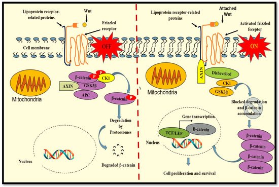
Figure 1) begins with the attachment of Wnt proteins to the seven-pass frizzled (Fz) transmembrane receptors and the co-receptor lipoprotein receptor-related proteins (LRP). When the Wnt ligand is not present (OFF), a protein complex consisting of axin, casein kinase (CK) 1, adenomatous polyposis coli (APC) and glycogen synthase kinase 3 beta (GSK3β) is formed. GSK3β causes the phosphorylation of β-catenin, tagging it for degradation by proteasomes. The attachment of Wnt to receptor Fz (ON) advances the stimulation of the dishevelled (Dvl) protein that is responsible for deactivating the axin protein complex. This results in the accumulation of cytoplasmic β-catenin, favouring its translocation to the nucleus and the formation of an active transcriptional complex with T cell-specific factor (TCF) and lymphoid enhancer-binding factor 1 (LEF1) for protein transcription [
5,
27]. Largely, Wnt3a is involved in activating the canonical Wnt/β-catenin pathway, and in vitro, synthetic Wnt3a activates the Wnt/β-catenin pathway for cell proliferation and differentiation [
28].
Figure 1. The Wnt/β-catenin pathway. Signal transduction begins with the attachment of Wnt proteins to the seven-pass frizzled (Fz) transmembrane receptors and the lipoprotein receptor-related proteins (LRP). When the Wnt ligand is not present (OFF), a protein complex consisting of axin, casein kinase (CK) 1, adenomatous polyposis coli (APC) and glycogen synthase kinase 3 beta (GSK3β) is formed. GSK3β phosphorylates β-catenin, tagging it for degradation. The attachment of Wnt to Fz (ON) causes the stimulation of the dishevelled (Dvl) protein, resulting in the deactivation of the axin protein complex and the accumulation of cytoplasmic β-catenin. β-catenin translocated into the nucleus forms an active transcriptional complex with T cell-specific factor (TCF)/lymphoid enhancer-binding factor 1 (LEF1) for protein transcription.
A large amount of active communication processes occurs in response to injury, eventually leading to wound restoration. An efficacious wound restoration process is largely governed by differentiation and proliferation of various cells including fibroblasts, epidermal stem cells (ESCs) and keratinocytes, achieved through different biological signalling pathways. Incorrect regulation of cellular signalling results in abnormal wound healing, including the development of chronic ulcers. Wnt signalling plays a significant role in controlling cell proliferation, movement and differentiation during tissue restoration [
5]. In fibroblasts, the Wnt/β-catenin pathway is inactive and is frequently activated due to injury [
29]. Wang et al. (2017) [
30] defined a feedback controlling loop joining basic fibroblast growth factor (bFGF) and Wnt signalling via β-catenin in fibroblasts. The bFGF Wnt-regulated pathway is implicated in cell proliferation, and inhibition of bFGF reduces Wnt-mediated influence on cell proliferation. Basic FGF proteins are influential mitogens in normal growth and wound healing [
31,
32].
3. Regulation of the Wnt/β-Catenin Pathway in Diabetic Wound Healing
A delay in wound restoration in DM is mainly due to mechanisms related to abnormal inflammation, irregular expression of matrix metalloproteinases (MMPs), reduced cell proliferation, disproportionate cell apoptosis and reduced expression of growth factors and their receptors [
3]. High protease levels significantly inhibit dermal reconstruction by reducing ECM components and fibroblast function. Fibroblasts from chronic diabetic wounds are exceedingly senescent, further contributing to reduced ECM deposition [
33]. In addition, the reduced healing process in diabetic wounds is worsened by reduced dermal cell neovascularisation, persistent infection and poor cell differentiation within the wound, largely affecting the treatment outcome [
28]. The Wnt/β-catenin signalling pathway directly participates in the alteration of various biological processes related to the manifestation and advancement of DM and its complications [
24].
During diabetic wound restoration, Wnt/β-catenin signalling stimulates skin thickness and pigmentation, and the literature reports that increased regulation of the Wnt/β-catenin pathway augments the action of high-glucose-suppressed cells [
6]. It is suggested that reduced activity of the Wnt/β-catenin pathway is due to decreased R-spondin (RSPO) instigated by DM and is one of the main reasons for the irregularity in diabetic wound healing [
10]. The RSPO protein family consist of RSPO 1 to 4 secreted proteins that are enhancers of the Wnt signalling pathway. RSPOs are responsible for the stabilisation of the Wnt receptors and their co-receptors via the inactivation of membrane-bound ubiquitin ligases ZNRF3 (zinc and ring finger 3) and RNF43 (ring finger 43) that antagonize the Wnt pathway by targeting the Wnt receptors for ubiquitylation-mediated disintegration [
34]. Adjustment or alteration of the Wnt/β-catenin pathway is known to enhance diabetic wound restoration, and it is suggested that transplanting Wnt signalling-activated cells promotes diabetic wound restoration [
28,
35]. In diabetic wounds, there is a significant decrease in the activity of GSK3β, caspase 3, NF-κB and β-catenin pathways [
36].
GSK3β, a serine/threonine kinase, is ubiquitously expressed as a strategic regulator of various signalling pathways for cellular proliferation and survival and plays a critical role in phosphorylating the Wnt receptors on LRP5/6, in that way causing stabilization of the Wnt/β-catenin pathway [
37]. Inhibition of GSK3β is critical in cell proliferation and differentiation during the wound restorative process, and modulation of GSK3β-mediated Wnt/β-catenin pathway advances diabetic wound healing [
38].
This entry is adapted from the peer-reviewed paper 10.3390/ijms23084210

