Image-Based Annotation of Chemogenomic Libraries for Phenotypic Screening: History
Please note this is an old version of this entry, which may differ significantly from the current revision.

Phenotypical screening is a widely used approach in drug discovery for the identification of small molecules with cellular activities. However, functional annotation of identified hits often poses a challenge. The development of small molecules with narrow or exclusive target selectivity such as chemical probes and chemogenomic (CG) libraries, greatly diminishes this challenge, but non-specific effects caused by compound toxicity or interference with basic cellular functions still pose a problem to associate phenotypic readouts with molecular targets. Hence, each compound should ideally be comprehensively characterized regarding its effects on general cell functions. Here, we report an optimized live-cell multiplexed assay that classifies cells based on nuclear morphology, presenting an excellent indicator for cellular responses such as early apoptosis and necrosis. This basic readout, in combination with the detection of other general cell damaging activities of small molecules such as changes in cytoskeletal morphology, cell cycle and mitochondrial health, provides a comprehensive time-dependent characterization of the effect of small molecules on cellular health in a single experiment. The developed high-content assay offers multi-dimensional comprehensive characterization that can be used to delineate generic effects regarding cell functions and cell viability, allowing an assessment of compound suitability for subsequent detailed phenotypic and mechanistic studies.

  • phenotypic screening
  • high content imaging
  • chemogenomics
  • machine learning
  • cell cycle

1. Introduction

Phenotypic screening has recently experienced a resurgence in drug discovery after many years of focus on target based approaches [1]. In particular, methods such as cell painting [2][3][4] or phenomics are gaining interest due to their ability to detect disease relevant morphological and expression signatures. These exciting new technologies provide insights into the biological effects of small molecules on cellular systems and the suitability of identified hits for translational studies. One of the main advantages of phenotypic screening lies in the potential of identifying functionally active chemical modulators without the need to know their precise mode of action (MoA). However, the lack of detailed mechanistic insight complicates the rational development of identified hit matter and validation studies [5]. One way to circumvent these complications is the use of better annotated chemical libraries, consisting of highly target-specific chemical probes [6][7][8] or chemogenomics libraries which contain well-characterized inhibitors with narrow but not exclusive target selectivity [9][10]. In particular, the latter have gained increasing interest as a new approach in drug discovery [11][12] as chemogenomic libraries may cover a large diversity of targets and a larger fraction of druggable proteins. Thus, chemogenomic compounds (CGCs) can supplement chemical probe collections that are not available for many targets due to their stringent quality criteria [13]. In cellular studies, the use of several CGCs directed towards one target but with diverse additional activities, will allow deconvolution of phenotypic readouts and identification of the target causing the cellular effect [14][15]. In addition, compounds from diverse chemical scaffolds may enable an easier identification of off-targets from different families. Further validation such as proteomic-based approaches or quantitative structure-activity relationships (QSAR) may be required [16]. The importance of chemogenomics for drug development has recently been demonstrated by a call of the Innovative Medicines Initiative (IMI), which resulted in the funding of the EUbOPEN project. One aim of this project is to assemble an open access chemogenomic library covering more than 1000 proteins by well annotated CGCs as well as chemical probes [17]. The expansion of this CGC collection to cover the entire druggable proteome will be the goal of the Target 2035 [18].
While target selectivity is an important parameter, there is a need for a comprehensive annotation of CGCs in terms of quality of the used chemical matter, such as structural identity, purity and solubility. In addition, the effects of CGCs on basic cellular functions such as cell viability, mitochondrial health, membrane integrity, cell cycle and interference with cytoskeletal functions which may be affected by non-specific binding of CGCs to tubulin should be considered [19]. Computational as well as screening approaches have been employed to predict the (unspecific) toxicity of libraries used for screening [20]. Although it is not always easy to distinguish between on-target and off-target effects in a cell viability assay, adding information on chemical and biological quality to CGC libraries will help to differentiate between target specific and unspecific effects [10]. New technology developments such as automated image analysis systems and machine learning algorithms enabled high-content techniques to become the method of choice for the essential annotation of chemogenomic libraries. Here, the researchers present a modular live-cell high-content cellular viability assay, which the researchers expanded to include assessment of CGC effects on the cell cycle, tubulin, mitochondrial health and membrane integrity. In contrast to the Cell Painting assay, which captures a multitude of morphological features of fixed cells at a given time point and requires extensive downstream analysis [3], the purpose of the researchers assay is to describe cell health in living cells, providing the opportunity for real-time measurement over a long time-period. The modular nature of the assay offers the opportunity for an expansion such as adding compound-safety assays and other cellular stress reporter systems without the need for additional informatics capacities [21].

2. Image-Based Annotation of Chemogenomic Libraries for Phenotypic Screening

Microscopy-based high-content screening, as a strategy for drug discovery, allows monitoring of multiple phenotypes in a fast and economical way [22]. Phenotypic screening has regained attention in drug discovery in recent years. In comparison to target-based drug discovery methods, phenotypic screening does not rely on the knowledge of a specific target per se and works as a tool to address complex relations of poorly understood diseases [5]. Extracting information from biological images collected during phenotypic screening and reducing them to a multidimensional profile, a process called image-based profiling, can be used to identify new disease-associated phenotypes, provide a better understanding about target effects and to predict compound activity, toxicity and mechanism of actions [23]. HighVia Extend is a live-cell, expandable, unbiased, image-based profiling assay, suitable for real-time measurements [24]. Similar to HighVia, HighVia Extend is modular in nature, inexpensive and flexible, providing the possibility to add additional fluorescent dyes for further readouts or adaptations for the use in different cell lines. Importantly, the assay is applicable for kinetic measurement for over 72 h and can therefore differentiate between primary target effects and secondary phenotypic results caused by the compound treatment. The lack of kinetic information is a frequent problem in phenotypic screens, which monitor endpoints [25]. Using a single readout, Hoechst33342, to assess cell nuclei, the researchers were able to identify healthy cells with high confidence, which enabled the use of additional stains to detect changes in tubulin appearance and mitochondrial content, respectively. Adding the FUCCI system, additional information regarding compounds affecting the cell cycle could be obtained. However, compared to Cell Painting, which uses mostly fixed cells and is based on the generation and evaluation of thousands of features [3] the researchers assay provides comprehensive information about cytotoxicity with considerably less features. Thus, the subsequent data processing is less demanding on bioinformatics capabilities while providing additional kinetic aspects. The modular nature of the assay allows for free combination with other dyes or a pre-screening of compounds with only Hoechst33342 and nuclear gating of the cells to reduce the costs of live-cell dyes. the researchers also successfully combined this experiment with other less complex cytotoxic screens as primary screens, such as proliferation experiments using a plate-reader based readout assessing the metabolic state of cells.
The presented assay offers a suitable annotation for (chemogenomic) libraries, providing information on the effect of these compounds on cellular health. It can be used in combination with assays assessing other aspects of cellular health, such as proteome stress involving protein misfolding and aggregation to better annotate a compound library [26]. the researchers assay thus helps to distinguish between false-positive or false-negative results of subsequent phenotypic assays [27][28]. False negative results can for example be caused by compounds with low solubility or precipitation of a compound as well as low permeability properties. Poorly soluble compounds can also cause false positive results, which may arise by causing unspecific cell death. Another potential source of false negative data might arise due to the missing expression of certain proteins in the tested cell line. The use of several cell lines in parallel as well as assessing the expression profiles using mRNA sequencing databases can, to a certain extent, offset this bias. Other compounds may cause false positive signals in cell assays due to reactivity of structural groups under applied conditions such as redox effects, complex formation, intrinsic fluorescence, degradation and others [28][29]. In the literature, already a large number of small molecules have been annotated as substances to frequently interfere with different assays [30]. Additional unspecific effects on cellular viability have been described for compounds binding to tubulin, e.g., Gul et al. showed that the preclinical used MTH1 inhibitor TH588 showed decreased tumor growth due to involvement in microtubule spindle regulation instead of the first investigated target effect [19][31]. The assessment of the tubulin modulating properties of compounds in a library can thus provide an alert with respect to the downstream effect on cell viability, which is particularly important for cancer cell biology.
For compounds without specific binding information to a protein as well as for target validation, the assay can provide a simple profile for each compound in a time dependent manner. By comparing the effect on cellular health for compounds targeting the same protein, unspecific effects can be easily detected using further analysis and clustering of results. Testing a well-annotated compound collection can thus be used to identify new biology mechanisms for known targets or even find new target correlations.

2.2. Multiplex Protocol

HEK293T (ATCC® CRL-1573™) and U2OS (ATCC®HTB-96™) were cultured in DMEM plus L-Glutamine (High glucose) supplemented by 10% FBS (Gibco) and Penicillin/Streptomycin (Gibco). MRC-9 fibroblasts (ATCC® CCL-2™) were cultured in EMEM plus L-Glutamine supplemented by 10% FBS (Gibco) and Penicillin/Streptomycin (Gibco). One day prior to compound exposure, cells were stained simultaneously to seeding with 60 nM Hoechst33342 (Thermo Scientific, MA, USA), 75 nM Mitotracker red (Invitrogen, MA, USA), 0.3 µL/well Annexin V Alexa Fluor 680 conjugate (Invitrogen, MA, USA) and 25 nL/well BioTracker™ 488 Green Microtubule Cytoskeleton Dye (EMD Millipore, MA, USA). Cells were seeded at a density of 2000 cells per well in a 384 well plates in culture medium (Cell culture microplate, PS, f-bottom, µClear®, 781091, Greiner, Frickenhausen, Germany), with a volume of 50 µL per well. All outer wells were filled with 100 µL PBS-buffer (Gibco).
Using the CQ1 high-content confocal microscope (Yokogawa), cellular shape and fluorescence was measured before and 12 h as well as 24 h after compound treatment. All compounds were diluted in DMSO to a concentration of 10 mM. Compounds were added directly to the cells in a 1:1000 dilution (50 nL/well) using an Echo 550 (LabCyte, San Josef, CA, USA).
For image acquisition, the following parameters were used: Ex 405 nm/Em 447/60 nm, 500 ms, 50%; Ex 561 nm/Em 617/73 nm, 100 ms, 40%; Ex 488/Em 525/50 nm, 50 ms, 40%; Ex 640 nm/Em 685/40, 50 ms, 20%; bright field, 300 ms, 100% transmission, one centered field per well, seven z-stacks per well with a total of 55 µm spacing. The rather large spacing distance was used to create a robust readout, compensating potential plate variations and enabling automated screening without the use of autofocus. The overlap of the fluorescence emission spectra of the dyes was neglectable for all but the MitoTracker Red and Annexin V Alexa Fluor 680. However, this overlap does not influence the analysis, since the excitation maxima of these two dyes are well separated and the gating algorithm analyses only the Mitotracker Red intensity in Annexin 5 negative cells.
All images were analyzed using the CellPathfinder software (Yokogawa). Segmentation of cells was performed as described earlier. First, the cells are classified in Hoechst High Intensity Objects or Normal Intensity Objects . All normal gated cells are further classified in healthy, fragmented or pyknosed nuclei . The pyknosed cells are gated in mitotic or apoptotic cells using seven features for the cell body and five features for the cell nuclei according to their Annexin V staining intensity . All cells that were classified as including a healthy nucleus are further gated into three phenotypic classes. They are gated in tubulin effect or no tubulin effect , mitochondrial mass increased or not increased and membrane permeability/membrane normal. Growth rate was calculated against non-treated cells and cells treated with DMSO 0.1% [32].

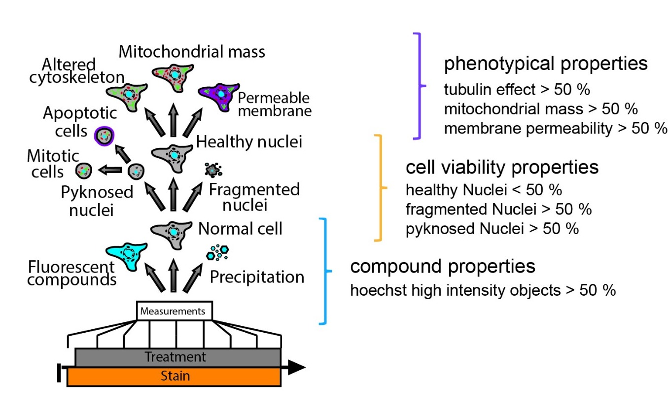
Figure 1.: General workflow of Multiplex High Via protocol analysis with property thresholds.

This entry is adapted from the peer-reviewed paper 10.3390/molecules27041439

References

  1. Haasen, D.; Schopfer, U.; Antczak, C.; Guy, C.; Fuchs, F.; Selzer, P. How Phenotypic Screening Influenced Drug Discovery: Lessons from Five Years of Practice. ASSAY Drug Dev. Technol. 2017, 15, 239–246.
  2. Rietdijk, J.; Tampere, M.; Pettke, A.; Georgiev, P.; Lapins, M.; Warpman-Berglund, U.; Spjuth, O.; Puumalainen, M.-R.; Carreras-Puigvert, J. A phenomics approach for antiviral drug discovery. BMC Biol. 2021, 19, 156.
  3. Bray, M.-A.; Singh, S.; Han, H.; Davis, C.T.; Borgeson, B.; Hartland, C.; Kost-Alimova, M.; Gustafsdottir, S.M.; Gibson, C.C.; Carpenter, A. Cell Painting, a high-content image-based assay for morphological profiling using multiplexed fluorescent dyes. Nat. Protoc. 2016, 11, 1757–1774.
  4. Schiff, L.; Migliori, B.; Chen, Y.; Carter, D. Deep learning and automated Cell Painting reveal Parkinson’s disease-specific signatures in primary patient fibroblasts. bioRxiv 2020.
  5. Moffat, J.G.; Vincent, F.; Lee, J.A.; Eder, J.; Prunotto, M. Opportunities and challenges in phenotypic drug discovery: An industry perspective. Nat. Rev. Drug Discov. 2017, 16, 531–543.
  6. Arrowsmith, C.; Audia, J.; Austin, C.; Baell, J.; Bennett, J.; Blagg, J.; Bountra, C.; Brennan, P.; Brown, P.; Bunnage, M.E.; et al. The promise and peril of chemical probes. Nat. Chem. Biol. 2015, 11, 536–541.
  7. Brown, P.J.; Müller, S. Open access chemical probes for epigenetic targets. Futur. Med. Chem. 2015, 7, 1901–1917.
  8. Drewes, G.; Knapp, S. Chemoproteomics and Chemical Probes for Target Discovery. Trends Biotechnol. 2018, 36, 1275–1286.
  9. Bunnage, M.E.; Chekler, E.L.P.; Jones, L. Target validation using chemical probes. Nat. Chem. Biol. 2013, 9, 195–199.
  10. Wells, C.I.; Al-Ali, H.; Andrews, D.M.; Asquith, C.R.M.; Axtman, A.D.; Dikic, I.; Ebner, D.; Ettmayer, P.; Fischer, C.; Frederiksen, M.; et al. The Kinase Chemogenomic Set (KCGS): An Open Science Resource for Kinase Vulnerability Identification. Int. J. Mol. Sci. 2021, 22, 566.
  11. Canham, S.M.; Wang, Y.; Cornett, A.; Auld, D.S.; Baeschlin, D.K.; Patoor, M.; Skaanderup, P.R.; Honda, A.; Llamas, L.; Wendel, G.; et al. Systematic Chemogenetic Library Assembly. Cell Chem. Biol. 2020, 27, 1124–1129.
  12. Dafniet, B.; Cerisier, N.; Boezio, B.; Clary, A.; Ducrot, P.; Dorval, T.; Gohier, A.; Brown, D.; Audouze, K.; Taboureau, O. Development of a chemogenomics library for phenotypic screening. J. Chemin. 2021, 13, 91.
  13. Müller, S.; Ackloo, S.; Arrowsmith, C.H.; Bauser, M.; Baryza, J.L.; Blagg, J.; Boettcher, J.; Bountra, C.; Brown, P.; Bunnage, M.; et al. Donated chemical probes for open science. eLife 2018, 7, 7.
  14. Bredel, M.; Jacoby, E. Chemogenomics: An emerging strategy for rapid target and drug discovery. Nat. Rev. Genet. 2004, 5, 262–275.
  15. Jones, L.; Bunnage, M.E. Applications of chemogenomic library screening in drug discovery. Nat. Rev. Drug Discov. 2017, 16, 285–296.
  16. Caron, P.R.; Mullican, M.D.; Mashal, R.D.; Wilson, K.P.; Su, M.S.; Murcko, M. Chemogenomic approaches to drug discovery. Curr. Opin. Chem. Biol. 2001, 5, 464–470.
  17. >EUbOPEN. Available online: https://www.eubopen.org/ (accessed on 5 January 2022).
  18. Carter, A.J.; Kraemer, O.; Zwick, M.; Mueller-Fahrnow, A.; Arrowsmith, C.H.; Edwards, A.M. Target 2035: Probing the human proteome. Drug Discov. Today 2019, 24, 2111–2115.
  19. Kawamura, T.; Kawatani, M.; Muroi, M.; Kondoh, Y.; Futamura, Y.; Aono, H.; Tanaka, M.; Honda, K.; Osada, H. Proteomic profiling of small-molecule inhibitors reveals dispensability of MTH1 for cancer cell survival. Sci. Rep. 2016, 6, 26521.
  20. Sun, H.; Wang, Y.; Cheff, D.M.; Hall, M.D.; Shen, M. Predictive models for estimating cytotoxicity on the basis of chemical structures. Bioorg. Med. Chem. 2020, 28, 115422.
  21. Tang, H.; Duggan, S.; Richardson, P.L.; Marin, V.; Warder, S.E.; McLoughlin, S.M. Target Identification of Compounds from a Cell Viability Phenotypic Screen Using a Bead/Lysate-Based Affinity Capture Platform. J. Biomol. Screen. 2015, 21, 201–211.
  22. Boutros, M.; Heigwer, F.; Laufer, C. Microscopy-Based High-Content Screening. Cell 2015, 163, 1314–1325.
  23. Chandrasekaran, S.N.; Ceulemans, H.; Boyd, J.D.; Carpenter, A.E. Image-based profiling for drug discovery: Due for a machine-learning upgrade? Nat. Rev. Drug Discov. 2021, 20, 145–159.
  24. Cole, R. Live-cell imaging. Cell Adhes. Migr. 2014, 8, 452–459.
  25. Neumann, B.; Held, M.; Liebel, U.; Erfle, H.; Rogers, P.; Pepperkok, R.; Ellenberg, J. High-throughput RNAi screening by time-lapse imaging of live human cells. Nat. Methods 2006, 3, 385–390.
  26. Liu, Y.; Fares, M.; Dunham, N.P.; Gao, Z.; Miao, K.; Jiang, X.; Bollinger, S.S.; Boal, A.K.; Zhang, X. AgHalo: A Facile Fluorogenic Sensor to Detect Drug-Induced Proteome Stress. Angew. Chem. Int. Ed. 2017, 56, 8672–8676.
  27. Baell, J.B.; Nissink, J.W.M. Seven Year Itch: Pan-Assay Interference Compounds (PAINS) in 2017—Utility and Limitations. ACS Chem. Biol. 2018, 13, 36–44.
  28. Chakravorty, S.J.; Chan, J.; Greenwood, M.N.; Popa-Burke, I.; Remlinger, K.S.; Pickett, S.D.; Green, D.V.S.; Fillmore, M.C.; Dean, T.W.; Luengo, J.I.; et al. Nuisance Compounds, PAINS Filters, and Dark Chemical Matter in the GSK HTS Collection. SLAS Discov. Adv. Sci. Drug Discov. 2018, 23, 532–545.
  29. Jasial, S.; Hu, Y.; Bajorath, J. How Frequently Are Pan-Assay Interference Compounds Active? Large-Scale Analysis of Screening Data Reveals Diverse Activity Profiles, Low Global Hit Frequency, and Many Consistently Inactive Compounds. J. Med. Chem. 2017, 60, 3879–3886.
  30. Baell, J.B.; Holloway, G.A. New Substructure Filters for Removal of Pan Assay Interference Compounds (PAINS) from Screening Libraries and for Their Exclusion in Bioassays. J. Med. Chem. 2010, 53, 2719–2740.
  31. Gul, N.; Karlsson, J.; Tängemo, C.; Linsefors, S.; Tuyizere, S.; Perkins, R.; Ala, C.; Zou, Z.; Larsson, E.; Bergö, M.O.; et al. The MTH1 inhibitor TH588 is a microtubule-modulating agent that eliminates cancer cells by activating the mitotic surveillance pathway. Sci. Rep. 2019, 9, 14667.
  32. Hafner, M.; Niepel, M.; Chung, M.; Sorger, P.K. Growth rate inhibition metrics correct for confounders in measuring sensitivity to cancer drugs. Nat. Methods 2016, 13, 521–527.
More
This entry is offline, you can click here to edit this entry!
Video Production Service