Exosomes and Diabetes: History
Please note this is an old version of this entry, which may differ significantly from the current revision.
Contributor:

Diabetes is part of a group of metabolic disorders characterized by long-term high blood glucose levels due to either inadequate production of insulin (type 1) or poor response of the recipient cell to insulin (type 2). Organ dysfunctions are the main causes of morbidity and mortality due to high glucose levels. Understanding the mechanisms of organ crosstalk may help us improve our basic knowledge and find novel strategies to better treat the disease. Exosomes are part of a newly emerged research area and have attracted a great deal of attention for their capacity to regulate communications between cells. In conditions of diabetes, exosomes play important roles in the pathological processes in both T1DM and T2DM, such as connecting the immune cell response to pancreatic tissue injury, as well as adipocyte stimulation to insulin resistance of skeletal muscle or liver. Furthermore, in recent years, nucleic acids containing exosomes—especially microRNAs (miRNAs) and long noncoding RNAs (lncRNAs)—have been shown to mainly regulate communications between organs in pathological processes of diabetes, including influencing metabolic signals and insulin signals in target tissues, affecting cell viability, and modulating inflammatory pancreatic cells. Moreover, exosome miRNAs show promise in their use as biomarkers or in treatments for diabetes and diabetic complications.  

  • exosomes
  • miRNA
  • diabetes

1. Extracellular Vesicles and Exosomes

Communication between organs is crucial for their normal functioning and pathological physiology [1,2,3]. In addition to the traditional known factors that affect organ communication such as cytokines, hormones, and chemokines, a new family involving extracellular vesicles (EVs) has emerged as a group of important modulators. The generic term “extracellular vesicles” was proposed to define all lipid bilayer-enclosed extracellular structures. EVs can be secreted by almost all cell types, including plant cells, bacteria, and fungi [4,5,6,7,8]. These small vesicles can be released in response to various stimulation, for example, changes in cell physical environments (PH, temperature, irradiation) or cell stress and activation induced by chemical agents [9,10,11,12,13]. Accruing literature reveals four major classes of EVs, which are sorted by size, subcellular origin, and content, named microvesicles, apoptotic bodies, virus-like particles, and exosomes [14,15,16,17,18]. The first three types of EVs are formed by outward budding of the plasma membrane. Exosomes, which are unlike other EVs, range from 30 to 100 nm in size and are formed by an intracellular endocytic trafficking pathway involving fusion of multivesicular late endocytic compartments [multivesicular bodies (MVBs)] with the plasma membrane. The generation of exosomes is initially formed as intraluminal vesicles (ILVs)  [19,20]. When an MVB fuses with the plasma membrane, the intraluminal vesicles (also called exosomes) are released to the extracellular space. The molecular mechanisms of MVB formation include two distinct pathways: the endosomal sorting complex required for transport (ESCRT) dependent pathway, and the ESCRT independent pathway. The ESCRT pathway requires formation of a complex by ESCRT, the sorting protein, Vps4, and the constitutive heat-shock protein, Hsp-70 [21,22,23]. The ESCRT is a family of proteins that associate in successive complexes (ESCRT-0, -I, -II, and -III) at the membranes of MVBs to regulate the formation of ILVs as well as their cargo [4]. The typical tumor susceptibility gene 101 protein (Tsg101) and Alix (encoded by PDCD6IP) for exosome identification belong to the ESCRT complex. The other pathway involves ESCRT-independent pathways. This pathway regulates the assembly of exosomes and requires Hsp70-phospholipid interactions and the activity of acid sphingomyelinase (nSMase), an enzyme that hydrolyzes sphingomyelin without the presence of ESCRTs [24,25].

1. Exosomes and Diabetes

1.1. Exosomes and Type 1 Diabetes

The pathology of type 1 diabetes (T1DM) is known to be related to autoimmune disorders. A complex interaction between pancreatic β-cells and an innate or an adaptive immune system leads to the irreversible destruction of insulin-producing cells in pancreatic islets [44,45,46,47]. With more in-depth research, a relationship between exosomes and type 1 diabetes has emerged in recent years [48]. On one hand, exosomes carry active immune molecules (normally proteins or nuclei) that can activate immune cells, such as T cells and B cells, and induce β-cell apoptosis, thereby contributing to T1DM development [49,50] (Figure 1). On the other hand, researchers found that rat and human pancreatic islets also release intracellular β-cell autoantigens in human T1DM, GAD65, IA-2, and proinsulin in exosomes, which are taken up by and activate dendritic cells [13]. Moreover, insulinoma-derived exosomes stimulate the innate immune response in the Myd88-dependent pathway, which is an inflammatory signaling downstream of members of the Toll-like receptor (TLR) and the interleukin-1 (IL-1) receptor families [51], and exosomes derived from islet mesenchymal stem cell (MSCs) directly activate the T cell response and stimulate the release of interferon gamma (IFN-γ) to induce inflammation, which plays a role in the initiation of autoimmune responses in T1D [52,53] (Figure 1). In addition, some exosomes are promising as effective and practical candidates for T1D therapy. For instance, adipocyte-derived stem cell exosomes exert ameliorative effects on T1DM by multiple pathways, including modulating the immune cell response [54], attenuating podocyte damage [55], and promoting proangiogenic properties [56]. Nonetheless, exosomes can be developed as an effective tool to improve islet transplant by modulating the immune response or as a biomarker of recurrent autoimmunity for islet transplant diagnosis [57,58].
Figure 1. Exosomes regulate diabetic pathological process. Schematic representation of exosomes participating in the pathologies of type 1 diabetes (T1D) and type 2 diabetes (T2D). (a) Immune cell-derived exosomes can activate immune cells and induce β-cell apoptosis and cell death. (b) In certain models, pancreatic islets also release intracellular β-cell autoantigens in exosomes in turn to favor the immune response. (c) Adipocyte-derived exosomes carrying sonic hedgehog (SHH), a protein known to modulate immunity, induce proinflammatory or M1 polarization of bone marrow-derived macrophages and induce insulin resistance in the main insulin-sensitive organs (skeletal muscle, liver, adipose).

1.2. Exosomes and Type 2 Diabetes

Type 2 is the main phenotype of diabetes, accounting for more than 90% of the diabetic population [59]. It is characterized by high plasma glucose due to insulin resistance. Excessive accumulation of fat due to obesity in adipose tissue has been considered a major driver in the pathophysiology of insulin resistance [60,61,62]. Recent studies have shown that adipocytes can produce and release exosomes containing bioactive cargo and acting as a mechanism of cell-to-cell communication, which contributes to the incidence of insulin resistance. For example, adipocyte-derived exosomes carrying sonic hedgehog (SHH), a protein known to modulate immunity, induce proinflammatory or M1 polarization of bone marrow-derived macrophages and RAW 264.7 macrophages via the Ptch/PI3K signaling pathway and contribute to insulin resistance [63] (Figure 2). Additionally, exosomes derived from insulin-resistant adipocytes promote plaque burden and plaque vulnerability partly by inducing vasa vasorum angiogenesis in human umbilical vein endothelial cells [64] (Figure 2). In addition to adipocytes, exosomes derived from other cells or organs also contribute to insulin resistance, including bone marrow mesenchymal cells, human placental cells, and cardiomyocytes [65,66,67,68]. Moreover, exosomes are promising as a therapeutic agent for type 2 diabetes. Recent studies have shown that exosomes derived from umbilical cord MSCs relieve β-cell destruction and alleviate type 2 diabetic nephropathy [69,70]. Another investigation also related exosomes to the mechanism underlying the beneficial effects of exercise or bariatric surgery on type 2 diabetes [71,72,73].

2. MiRNA in Exosomes and Their Roles in Diabetes

MicroRNAs (miRNAs) are small, noncoding RNA molecules containing approximately 22 nucleotides. miRNAs are found in plants, animals, and some viruses that function in RNA silencing and posttranscriptional regulation of gene expression [76]. Many microRNAs, such as let-7, miR-223, miR-29, miR-103, and miR-107, are known to regulate metabolic disorders (including diabetes) through various molecular pathways, such as modulation of lipid or glucose metabolism, liver gluconeogenesis, insulin secretion, and autophagy [77,78,79,80,81]. MiRNA-carrying exosomes have advantages that work at distant tissues more efficiently. Thus, some exosomal miRNAs are pathological factors in diabetes by targeting key proteins and acting as crucial modulators of insulin sensitivity. In cells, insulin combined with its substrate, insulin receptor substrate-1 (IRS-1), stimulates a cascade signal, including phosphorylation of protein kinase B (PKB/AKT), at serine 473, leading to phosphorylated AKT promoting the translocation of glucose transporter-4 (Glut4) from the cytosol to the membrane for glucose uptake [82]. Katayama and colleagues examined the miRNA expression profile of serum-derived exosome-enriched extracellular vesicles in healthy controls and type 2 diabetic patients. They found that exosome-derived extracellular miR-20b-5p is highly abundant in type 2 diabetic patients, and further in vitro studies showed that exosomal miR-20b-5p targeted AKT-interacting protein (AKTIP), which interacts directly with AKT and modulates AKT activity by enhancing the phosphorylation of AKT regulatory sites and reducing glycogen accumulation in primary human skeletal muscle, resulting in insulin resistance [
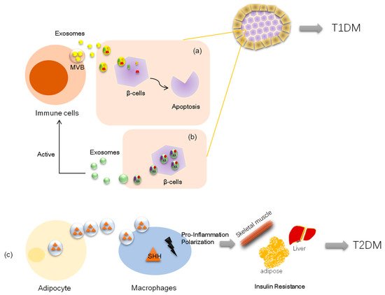
83]. Consistent with the above findings, another study demonstrated that pancreatic cancer-derived exosomes enter C2C12 myotubes and inhibit insulin and PI3K/Akt signaling, thereby preserving insulin-induced FoxO1 nuclear exclusion and impairing Glut4 trafficking. In this study, microarray methods revealed that certain exosomal microRNAs (miR-450b-3p and miR-151-3p) likely play key roles in this process (Figure 1) [84]. Further, exosome miR-155 derived from adipose tissue macrophages in obesity-induced diabetic mice causes glucose intolerance and insulin resistance by targeting PPARγ, a transcription factor and key regulator of lipid metabolism, when administered to lean mice [85]. Similarly, another elegant experiment on obese mice showed that exosomes from obese mice could induce glucose intolerance in lean mice; in this study, the authors transfected exosomes with the selected miRNA (miR-192, miR-122, miR-27a-3p, and miR-27b-3p; all were demonstrated to be increased in obese mice) and then injected the reconstituted exosomes into lean mice. Surprisingly, the lean mice developed glucose intolerance and insulin resistance as well. The data further showed that exosomal miRNAs induced diabetes in white adipose tissues in lean mice by targeting PPARα [86], which is also a transcription factor and a major regulator of lipid metabolism.
Inadequate pancreatic β-cell numbers is a pathological manifestation of type 1 diabetes. Researchers have found that lymphocyte-derived exosomes miR-142-3p and miR-142-5p from prediabetic mice transferred to pancreatic β-cells trigger the expression of genes involved in chemokine signaling, including Ccl2, Ccl7, and Cxcl10. The induction of these genes may promote the recruitment of immune cells and exacerbate β-cell death during an autoimmune attack, which also contributes to diabetes development [49]. Nonetheless, a recent study demonstrated that bone marrow mesenchymal stem cells derived exosome miR-29b-3p from aged mice mediated insulin resistance in young mice [65], which indicated that exosomal miRNAs could exert their role independently. Thus, exosome cargo with miRNAs is also considered a novel therapeutic agent. Exosome miR-222 was reduced in rabbits with type 1 diabetes; however, MSC-derived exosomes containing miR-222 injected subconjunctivally and intraocularly to these diabetic rabbits could attenuate retinal degeneration [87]. Similarly, intravenous administration of exosome miR-106b and miR-222 derived from bone narrow promoted post-injury β-cell proliferation through Cip/Kip family downregulation in insulin-deficient mice [88]. In addition, MSC-derived exosome miR-146a has anti-inflammatory effects on damaged astrocytes and prevents diabetes-induced cognitive impairment [89].
Consequently, many exosomal miRNAs have been found to be increased in the plasma or the urine of diabetic patients, making them promising circulating biomarkers associated with type 2 diabetes, such as exosomal miR-21-5p, miR-375-3p, miR-133b, miR-342, and miR-30, which are all upregulated in the serum of diabetic subjects [90,91,92], and miR-451-5p, let-7c-5p, miR-362-3p, miR-877-3p, miR-150-5p, and miR-15a-5p, which are upregulated in the urine of diabetic subjects [93,94,95]. It should be noted that, for certain miRNAs, total circulating miRNA levels are distinct from circulating extracellular vesicle miRNA content. For example, a clinical study including 19 children with type 1 diabetes along with 16 healthy controls showed that miR-21-5p derived from serum extracellular vesicles was increased threefold compared with that in nondiabetic individuals, while total serum miR-21-5p was reduced in diabetic participants [92], suggesting that exosomal RNA may be a distinct biomarker from total serum RNA levels.

This entry is adapted from the peer-reviewed paper 10.3390/cells8080853

This entry is offline, you can click here to edit this entry!
Video Production Service