Epigenetic Modifications and Carcinogenesis: History
Please note this is an old version of this entry, which may differ significantly from the current revision.
Subjects: Cell Biology
Contributor:

Epigenetics encompasses a group of dynamic, reversible, and heritable modifications that occur within cells that are independent of gene mutations. These alterations are highly influenced by the environment, from the environment that surrounds the human being to the internal microenvironments located within tissues and cells. The ways that pigenetic modifications promote the initiation of the tumorigenic process have been widely demonstrated. Similarly, it is well known that carcinogenesis is supported and prompted by a strong proinflammatory environment. At the same time, cancer cells can alter their epigenetic profile to generate a positive loop in the promotion of the inflammatory process. Therefore, an in-depth understanding of the epigenetic networks between the tumor microenvironment and cancer cells might highlight new targetable mechanisms that could prevent tumor progression. 

  • epigenetics
  • carcinogenesis
  • inflammation
  • tumor microenvironment

1. Introduction: The Complexity of Carcinogenesis

Carcinogenesis is a complex multistep process that leads to the onset of tumors in normal tissue in vivo. Indeed, normal cells can become transformed by accumulating several gene mutations. This carcinogenic process can be divided into three different parts: initiation, promotion, and progression. Carcinogenesis is usually initiated by the progressive accumulation of sporadic mutations that normally accumulate during the lifespan of a cell. Most of these mutations are supposed to have no relevant role in tumor illness and can also be found in normal tissues. However, a limited number of mutations, called “driver” mutations, confer a growth advantage to the cell, which in turn, through a modest but significant increase in the replication rate, can further prompt the appearance of other driver mutations, leading to cancer promotion. This process can require years, or decades, to originate a primitive tumor. Generally, mutations in the metastatic cancer are not particularly different from those of the primitive tumor, raising the problem of which genetic alteration can support the metastatic phenotype. In addition, somatic driver mutations affect a very limited number of genes and intracellular pathways [1]. These findings, together with the increasing consciousness that DNA expression and cellular phenotype are regulated by other factors in addition to gene sequence, prompted the exploration of other mechanisms that could induce cancer onset, such as epigenetic regulation. In contrast to gene mutation, most epigenetic regulation modifies gene expression without permanent changes in the genomic sequence. More importantly, epigenetic modulation is reversible and faster regulated than the establishment of gene mutation, namely genomic evolution [2]. Substantially, epigenetics controls the interactions among DNA, RNA, and the nucleosome with modifying proteins and without inducing gene mutations. One of the possible epigenetic controls is the modulation of transcription factors by limiting their accessibility to the DNA filament and determining which genes will be expressed. The other main epigenetic events are DNA and RNA methylation–demethylation and chromatin remodeling by histone acetylation–deacetylation. Because epigenetic modulation is centered on reversible interactions with different structures, it is also modifiable by environmental influences, aging, and drugs. This aspect is of particular interest because, although DNA alterations are not amenable to pharmacotherapy, there is a growing number of small molecules that can be used as epigenomic drugs for anticancer purposes and that are less toxic than traditional chemotherapy [3][4]. In fact, promising results have been recently reported on the combined use of DNA methylation inhibitors and histone deacetylase inhibitors in clinical trials [5]. The influence of the surrounding environment in determining the epigenome of a cancer cell once more highlights the importance of the tumor microenvironment (TME) in defining the initiation and progression of tumor growth. The TME consists of a supporting structure and cells that surround the tumor foci, creating a niche that can either foster or suppress tumor growth. In the TME, inflammatory cells play a pivotal role by releasing different stromal factors, such as chemokines and cytokines, which modulate carcinogenesis. Therefore, it is not surprising that the crosstalk between the TME and cancer cells is strictly mediated by epigenetic modifications [6]. In this research, the focus is describing how epigenetic modifications regulating inflammation are able to influence cancer onset and progression.

2. Overview of Epigenetic Modifications

The mechanisms determining epigenetic modifications can be divided into four groups: DNA methylation, RNA methylation, histone posttranslational modifications, and the broad family of epigenetic regulators constituted by noncoding RNAs (ncRNAs) (Figure 1).
/media/item_content/202203/6231402bb6708cancers-14-01221-g001.png
Figure 1. Overview of epigenetic modifications. Schematic representation of the four major epigenetic modifications, DNA methylation, histone modifications, RNA methylation, and miRNAs, as part of the broad family of noncoding RNAs (ncRNAs). Me: methylation; Ac: acetylation, Lct: lactylation. Created with BioRender.com.

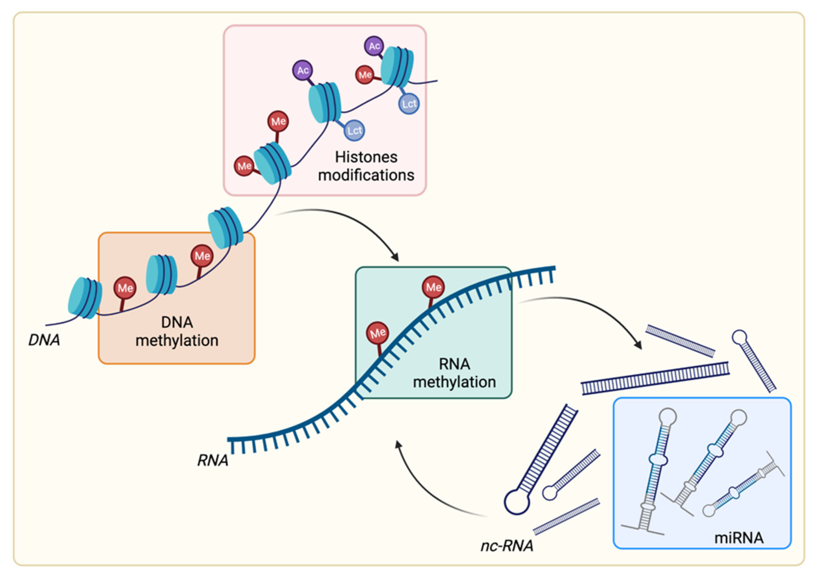
2.1. DNA Methylation

Epigenetic regulation can act at different levels in the flux of information from DNA to the cellular periphery. At the first level of transcriptional control, DNA methylation is found. DNA methylation occurs directly at the DNA level and allows a reduction in DNA transcription without affecting its gene sequence [7]. DNA methylation implies the covalent modification of a nucleotide and, in mammals, typically but not exclusively involves the methylation of cytosine at position 5 of the pyrimidine ring (5mC) [8]. Interestingly, 5mC is preferentially located in CpG dinucleotides, and is normally concentrated in large clusters named CpG islands, of which there are reported to be approximately 29 million in the human genome, 70–80% of which are methylated in somatic cells [9]. Non-CpG methylation occurs more frequently in human embryonic or induced pluripotent stem cells [10]. As a general rule, DNA methylation behaves as a negative regulator, reducing promoter accessibility and, consequently, the transcription of downstream genes [11]. DNA hypomethylation shows a clear, positive correlation with higher transcriptional activity. While the methylation of a promoter correlates with transcriptional repression, it has been shown that methylation on the coding gene body is related to an increase in gene expression [12].
The proteins involved in DNA methylation can be grouped into three main categories, which cover and ensure all the aspects of this regulatory process. These groups involve (i) DNA methyl transferases (DNMTs), often called the “writers”, which ensure de novo methylation (such as DNMT3A and DNMT3B) or its maintenance over time and during DNA replication (such as DNMT1); (ii) a broad set of proteins involved in 5mC recognition termed the “readers” (such as methyl-CpG binding protein 2 (MECP 2), methyl-CpG binding domain protein (MBD) 1–6, the Kaiso family, and ubiquitin-like proteins (UHRF1 and UHRF2)), which can recognize the methylation mark and can be recruited at the chromatin level mediating the silencing of the target gene; and (iii) several DNA demethylases (such as ten-eleven translocation (TET) enzymes) that are involved in the process of the removal of the methyl group, called the “erasers” [13]. As far as tumorigenesis is concerned, there are two types of changes in DNA methylation that can occur: first, the demethylation of oncogene promoters, and second, the de novo methylation of selected CpG islands [14]. Accordingly, several types of cancer present wide demethylation zones, while others, such as gliomas, cholangiocarcinomas and lymphomas, are characterized by mutations in the isocitrate dehydrogenase (IDH) genes, which induce hypermethylation [15]. Interestingly, de novo methylation of CpG islands is a widespread programmed process mediated by polycomb-targeting complexes, a group of epigenetic repressors that operates by recruiting the de novo methylases DNMT3A and DNMT3B in tumors [16][17][18]. Notably, the DNA methylation pattern is continuously reformed throughout human life based on the crosstalk between DNMT and TET. This indicates that DNA methylation is a dynamic activity and is therefore susceptible to environmental influences, such as nutrient availability, physical effort, illnesses, and interactions with drugs. Moreover, during aging, this fine-tuned mechanism becomes progressively unbalanced, suggesting that aging cells could represent a favorable environment, which, if hit by protumoral mutations, can more easily move toward carcinogenesis [19]. For example, abnormal hypermethylation of the MutL homologus 1 (MLH1) promoter, a gene involved in hereditary nonpolyposis colorectal cancer has been related to gene silencing and microsatellite instability, which in turn could initiate genetic instability in colorectal and endometrial cancers [20][21][22][23]. Interestingly, most large bowel adenocarcinomas with MLH1 methylation display widespread methylation of the promoter of several genes, the so-called CIMP phenotype, and the BRAF-V600E mutation. The CIMP phenotype has been described in several tumor types; however, its molecular bases are poorly defined [24].

2.2. RNA Methylation

In addition to DNA, RNA also represents an effective target for epigenetic regulation via both methylation and ncRNA interference. RNA methylation occurs at N6-methyladenosine (m6A) and has been reported to affect the complexity of cancer progression by regulating RNA processing, nuclear export, and RNA translation. Moreover, m6A modification also occurs on ncRNAs, indicating that it controls RNA functions both directly and indirectly [25]. For DNA methylation, m6A modifications are also reversible and dynamic. Correspondingly, the major players can be categorized into (i) writers, such as different methyltransferase-like proteins (METTL3, METTL 14, METTL16), RNA binding motif protein (RMB) 15/15B, and others; (ii) readers, such as YT521-B homology (YTH) domain-containing proteins, eukaryotic initiation factor 3 (eIF3), and heterogenous nuclear ribonucleoprotein (HNRNP) protein family members; and (iii) erasers, such as alkylation repair homolog 5 (ALKBH5) and fat mass and obesity-associated protein (FTO) [6][26][27][28]. An increasing number of studies have shown that aberrant m6A modifications are closely associated with different types of cancer, such as glioblastoma, cervical and endometrial cancer, hepatocellular carcinoma, acute myeloid leukemia, breast cancer, pancreatic cancer, and prostate cancer (fully reviewed in [29]).

2.3. Histone Posttranslational Modifications

As previously described, DNA methylation acts directly on DNA, while histone modification implies the chemical variation of the proteins that form the nucleosome, determining whether and when specific genes will be transcribed or silenced.
Histone proteins are the targets of a broad set of chemical modifications, including acetylation, methylation, phosphorylation, ubiquitination, ADP-ribosylation, citrullination, SUMOylation, and others. All these modifications are reversible and, together with the presence of different histone isoforms, permit the existence of a broad and only partially clarified panel of different spatial chromatin rearrangements [30]. As a whole, these histone modifications constitute a truly complex chromatin-based signaling system, often referred to as histone code. This is based on changes in the charge, density, and hydrophobicity of the strands, which induce a conformational shift in the protein structure and permit the docking of specific regulatory proteins [31].
Among these modifications, the most studied is histone acetylation, which acts on the charge of the lysine residues. On the one hand, histones typically have a cumulative positive charge, which allows a strong interaction with the negatively charged DNA strand. On the other hand, acetylation occurs at the N-terminal extremity of the proteins, neutralizes the lysine charge, and reduces the histone-DNA interaction. Acetylation represents a steady state between the action of histone acetyltransferases (HATs), which transfer an acetyl group from acetyl-CoA onto lysine to form ε-N-acetyl lysine and histone deacetylases (HDACs), which remove this acetyl group. HATs are represented by many proteins, often presenting a bromodomain, which display their activity directly on histones packaged into the nucleosomes or previously in the cytoplasm on unassembled histones. Primarily, HATs target histones H3 and H4; however, H2A and B can also be acetylated on several lysine residues [32]. The classic HAT p300 has been found to be mutated in several cancers [33], and in a similar way, PCAF, a p300/CREB binding protein (CBP)-associated factor, is negatively associated with lung or gastric cancer [34][35]. Consistently, general hypoacetylation occurs during carcinogenesis, together with altered acetylation patterns, such as for histone H4K16 or other residues [36][37][38]. Consequently, many HDAC inhibitors (HDACis), also defined as epidrugs, have been used in cancer therapy, as reviewed elsewhere [39].
Another important histone modification is methylation, which can occur on all histone proteins on the nitrogen atoms of different lysine (K) or arginine (R) residues. In addition, each residue, be it methylated, trimethylated, acetylated, or modified in a different way, admits the docking of several reader proteins involved in the packaging or unpackaging of chromatin. These processes can involve different portions of the DNA filament, influencing the accessibility of the gene sequence and of gene promoters or enhancers [40]. This activity, together with proper DNA methylation, contributes to determining cell identity and tissue enrollment [41] and their maintenance over time. Moreover, the DNA sequence and its methylation status act as factors regulating nucleosome occupancy. In fact, repetitive DNA regions enriched in methylated CpG islands have been reported to strongly affect interaction with the octamer, while unmethylated CpG islands or unmethylated transcription binding sites show the lowest occupancy levels [42]. Inactivated regions appear to be characterized by polycomb protein attachment [43][44]. In particular, enhancer of Zeste homolog 2 (EZH2) is a histone methyltransferase member of polycomb repressive complex 2 (PRC2), which normally methylates lysine 27 of histone H3 (H3K27) [45]. For this reason, its activity keeps the chromatin in a repressed state throughout the cell cycle and, together with the methylation of lysine 9 of histone 3 (H3K9) and of other laminin-associated factors, helps to transmit cell identity and tissue commitment to daughter cells. Therefore, it is not surprising that mutations in this enzyme are frequent in cancer [45]. Consistently, EZH2 is frequently mutated in B cell lymphoma and melanoma [46]. EZH2 somatic mutations induce hypermethylation activity on H3K27, followed by the depletion of other critical genes, leading B cells to remain in a permanent proliferative state [47].
Last, the ATP-dependent remodeling of nucleosome position is led by switch–sucrose non-fermentable (SWI–SNF) complexes, which are a large protein family of ATP-dependent chromatin remodeling complexes. This family has been regarded as having tumor suppressor activity and was found to be frequently mutated in several malignancies, including chronic and acute leukemia, lymphomas, rhabdoid tumors, and ovarian cancers [48]. In particular, AT-rich interactive domain-containing protein 1A (ARID1A), a member of the SWI-SNF family, was found to be mutated in gastric and pancreatic cancers and is related to breast cancer metastasis and indicative of trastuzumab resistance [49][50].
Histone lactylation is a recently studied posttranslational modification [51]. Zhang et al. found that lactic acid, already known to promote gene expression and histone acetylation [52], can directly tag lysine residues on H3, H4, H2A, and H2B histones. Histone lactylation also occurs in lung tumors and melanoma cells. Moreover, exogenous lactate decreases the HDAC content in the nucleus, HDAC activity, and chromatin methylation [52][53]. HDAC inhibition occurs at IC50 values which are not only lower than those of other pharmacological inhibitors, but also higher than reported intracellular physiological lactate concentrations. Collectively, these data suggest that lactate can potentially transduce the modifications induced by hypoxia and glucose fermentative metabolism that normally occur in the tumoral environment at the chromatin level. Further studies are needed to precisely define the exact role of lactate as an epigenetic factor in cancer onset and progression.
Altogether, the acquisition of a permissive chromatin arrangement not only predisposes cells to carcinogenesis, but also transmits them after replication to the succeeding generations, creating a cell clone with higher replication potential. Thus, inside the tumor mass, epigenetic plasticity can contribute to creating intratumor heterogeneity, which represents a valuable tool to address the variability of environmental conditions due to tumor spreading among different tissues in distant organs or during the selective conditions imposed by medical treatments.

2.4. Noncoding RNA: Focus on microRNA

Another level of epigenetic regulation acts directly on transcriptional activity and involves a different category of molecules, i.e., the broad class of ncRNAs. It has been estimated that only 1–2% of RNA is messenger RNA and can codify for proteins, while the remaining part consists of ncRNA. The class of ncRNA gathers several different types of RNA, such as housekeeping RNA with structural and well-characterized functions. For example, ribosomal or transfer RNA, and many different ncRNAs, present to a minor extent with a not completely clarified regulatory role. This latter group could be further divided according to size into small ncRNAs (sncRNAs, <200 nt) and long ncRNAs (lncRNAs, >200 nt) [54]. The best characterized sncRNA is miRNA, a highly conserved single-stranded RNA with ~20 nucleotides.
miRNAs play a relevant role in cancer pathogenesis through two outstanding aspects of their activity: on the one hand, the ability to control the synthesis of almost all cellular proteins, and on the other hand, the deep interdependence with the other epigenetic control mechanisms. These aspects, together with their numerosity, create a complicated network of reciprocal influences, where all the components regulate each other, and the perturbation of one or more elements often leads to some strongly dysregulated patterns. For instance, miR-15/16 are interesting examples of these interrelationships. In fact, they are listed among tumor suppressive miRNAs and are downregulated in several tumors, including chronic lymphocytic leukemia (CLL); multiple myeloma; prostate, colon, lung, and ovarian cancers; and other tumors. In CLL, miR-15/16 downregulation is due to miRNA deletion at 13q14, which occurs with high incidence in this cancer [55]. miR-15/16 deletion leads to the overexpression of several target genes depending on the specific tissue, including the anti-apoptotic factor B cell lymphoma 2 (BCL2) in CLL [56]; the cyclooxygenase-2 (COX-2) gene in colon cancer [57]; cyclin D1, the proto-oncogene protein WNT3, in prostate cancer [58]; VEGFa in multiple myeloma; and genes, such as C-MYC and ALK in other tumors [59][60]. The activity of miR-15/16 can mediate the action of P53, which in turn can both transcriptionally and posttranscriptionally control miRNAs, such as miR-34 and miR-200 [61]. Accordingly, P53 mutations, which are highly represented in a wide variety of cancers, lead to the downregulation of several miRNA families followed by an increase in the expression and activity of their target oncogenes. In particular, miR-34 controls BCL2, NOTCH, and the high mobility group AT-Hook 2 (HMGA2) in gastric cancer and MYC and MET in ovarian cancer [62][63], while miR-200 controls zinc finger E-box binding homeobox (ZEB)1, BM1, CNNB1, FN1, LEPR, and NTRK2, and inhibits cellular growth and metastasis in several cancers, including nasopharyngeal, pancreatic, and breast cancer [64][65].
Similar to mutated P53, several other oncogenes exert part of their action, dysregulating the balance of the miRNA network, by downregulating tumor suppressive miRNA. As an example, MYC is a well-known transcription factor with oncogenic activity that regulates the transcription of a broad number of miRNAs, downregulating miR-15a/16-1, miR-26a, miR-34, and let-7 family members, and consequently reducing their proapoptotic and antiproliferative effects [66]. Interestingly, the miRNA let-7 family is responsible for the ablation of MYC in Burkitt lymphoma, thus inhibiting cancer cell progression, while it targets interleukin (IL)-6 in breast cancer, the transcription factor E2F2 in prostate cancer, and the anti-apoptotic BCL, namely, BCL-XL in the liver [67]. Similarly, in Kirsten rat sarcoma (RAS) mutant pancreatic cancers, the RAS oncogene binds, through its RAS-responsive element-binding (RREB1), the promoter of the miR-143/145 family, which normally act as repressors of the same RAS and RREB1 transcription factors. Therefore, by inhibiting its inhibitors, mutated RAS strongly potentiates its own oncogenic activities [68]. The same mechanism of reciprocal inhibition occurs between the ZEB1 and miR-200 family; thus, by blocking miRNA translation, the ZEB1 and ZEB2 proteins can upregulate their expression in several cancers [69].
In addition to mutated transcription factors, several epigenetic mechanisms can alter the normal balance of miRNA in cancer. First, variation in DNA methylation strongly affects the miRNA network. For example, the loci of the miR-34, miR-124, and miR200 families are hypermethylated and epigenetically silenced in a vast number of different tumors [70][71][72]. According to the positive feedback model already seen in other dysregulated scenarios, many miRNAs include among their targets the mRNA for methylases or demethylases. This scenario can change the methylation status of numerous different gene loci, leading to the repression of tumor suppressors, the enhancement of oncogenes, and disequilibria in miRNA production, with a strong multiplicative effect on the dysregulation of cell activities.

This entry is adapted from the peer-reviewed paper 10.3390/cancers14051221

References

  1. Vogelstein, B.; Papadopoulos, N.; Velculescu, V.E.; Zhou, S.; Diaz, L.A.; Kinzler, K.W. Cancer genome landscapes. Science 2013, 339, 1546–1558.
  2. Easwaran, H.; Tsai, H.-C.; Baylin, S.B. Cancer epigenetics: Tumor heterogeneity, plasticity of stem-like states, and drug resistance. Mol. Cell 2014, 54, 716–727.
  3. Miranda Furtado, C.L.; Dos Santos Luciano, M.C.; da Silva Santos, R.; Furtado, G.P.; Moraes, M.O.; Pessoa, C. Epidrugs: Targeting epigenetic marks in cancer treatment. Epigenetics 2019, 14, 1164–1176.
  4. Topper, M.J.; Vaz, M.; Marrone, K.A.; Brahmer, J.R.; Baylin, S.B. The emerging role of epigenetic therapeutics in immuno-oncology. Nat. Rev. Clin. Oncol. 2020, 17, 75–90.
  5. Chen, Q.W.; Zhu, X.Y.; Li, Y.Y.; Meng, Z.Q. Epigenetic regulation and cancer (review). Oncol. Rep. 2014, 31, 523–532.
  6. Lu, Y.; Chan, Y.-T.; Tan, H.-Y.; Li, S.; Wang, N.; Feng, Y. Epigenetic regulation in human cancer: The potential role of epi-drug in cancer therapy. Mol. Cancer 2020, 19, 79.
  7. Cedar, H.; Bergman, Y. Programming of DNA methylation patterns. Annu. Rev. Biochem. 2012, 81, 97–117.
  8. Lister, R.; Pelizzola, M.; Dowen, R.H.; Hawkins, R.D.; Hon, G.; Tonti-Filippini, J.; Nery, J.R.; Lee, L.; Ye, Z.; Ngo, Q.-M.; et al. Human DNA methylomes at base resolution show widespread epigenomic differences. Nature 2009, 462, 315–322.
  9. Ziller, M.J.; Müller, F.; Liao, J.; Zhang, Y.; Gu, H.; Bock, C.; Boyle, P.; Epstein, C.B.; Bernstein, B.E.; Lengauer, T.; et al. Genomic distribution and inter-sample variation of non-CpG methylation across human cell types. PLoS Genet. 2011, 7, e1002389.
  10. Ramsahoye, B.H.; Biniszkiewicz, D.; Lyko, F.; Clark, V.; Bird, A.P.; Jaenisch, R. Non-CpG methylation is prevalent in embryonic stem cells and may be mediated by DNA methyltransferase 3a. Proc. Natl. Acad. Sci. USA 2000, 97, 5237–5242.
  11. Jones, P.A. Functions of DNA methylation: Islands, start sites, gene bodies and beyond. Nat. Rev. Genet. 2012, 13, 484–492.
  12. Yang, X.; Han, H.; De Carvalho, D.D.; Lay, F.D.; Jones, P.A.; Liang, G. Gene body methylation can alter gene expression and is a therapeutic target in cancer. Cancer Cell 2014, 26, 577–590.
  13. Panneerdoss, S.; Eedunuri, V.K.; Yadav, P.; Timilsina, S.; Rajamanickam, S.; Viswanadhapalli, S.; Abdelfattah, N.; Onyeagucha, B.C.; Cui, X.; Lai, Z.; et al. Cross-talk among writers, readers, and erasers of m6A regulates cancer growth and progression. Sci. Adv. 2018, 4, eaar8263.
  14. Klutstein, M.; Nejman, D.; Greenfield, R.; Cedar, H. DNA Methylation in Cancer and Aging. Cancer Res. 2016, 76, 3446–3450.
  15. Welner, S.A.; Koty, Z.C.; Boksa, P. Chromaffin cell grafts to rat cerebral cortex reverse lesion-induced memory deficits. Brain Res. 1990, 527, 163–166.
  16. Schlesinger, Y.; Straussman, R.; Keshet, I.; Farkash, S.; Hecht, M.; Zimmerman, J.; Eden, E.; Yakhini, Z.; Ben-Shushan, E.; Reubinoff, B.E.; et al. Polycomb-mediated methylation on Lys27 of histone H3 pre-marks genes for de novo methylation in cancer. Nat. Genet. 2007, 39, 232–236.
  17. Viré, E.; Brenner, C.; Deplus, R.; Blanchon, L.; Fraga, M.; Didelot, C.; Morey, L.; Van Eynde, A.; Bernard, D.; Vanderwinden, J.-M.; et al. The Polycomb group protein EZH2 directly controls DNA methylation. Nature 2006, 439, 871–874.
  18. Chan, H.L.; Morey, L. Emerging Roles for Polycomb-Group Proteins in Stem Cells and Cancer. Trends Biochem. Sci. 2019, 44, 688–700.
  19. Issa, J.-P. Aging and epigenetic drift: A vicious cycle. J. Clin. Investig. 2014, 124, 24–29.
  20. Ma, Y.; Chen, Y.; Petersen, I. Expression and promoter DNA methylation of MLH1 in colorectal cancer and lung cancer. Pathol. Res. Pract. 2017, 213, 333–338.
  21. Botezatu, A.; Iancu, I.V.; Plesa, A.; Manda, D.; Popa, O.; Bostan, M.; Mihaila, M.; Albulescu, A.; Fudulu, A.; Vladoiu, S.V.; et al. Methylation of tumour suppressor genes associated with thyroid cancer. Cancer Biomark. 2019, 25, 53–65.
  22. Yokoyama, T.; Takehara, K.; Sugimoto, N.; Kaneko, K.; Fujimoto, E.; Okazawa-Sakai, M.; Okame, S.; Shiroyama, Y.; Yokoyama, T.; Teramoto, N.; et al. Lynch syndrome-associated endometrial carcinoma with MLH1 germline mutation and MLH1 promoter hypermethylation: A case report and literature review. BMC Cancer 2018, 18, 576.
  23. Bonora, M.; Missiroli, S.; Perrone, M.; Fiorica, F.; Pinton, P.; Giorgi, C. Mitochondrial Control of Genomic Instability in Cancer. Cancers 2021, 13, 1914.
  24. Hughes, L.A.E.; Melotte, V.; de Schrijver, J.; de Maat, M.; Smit, V.T.H.B.M.; Bovée, J.V.M.G.; French, P.J.; van den Brandt, P.A.; Schouten, L.J.; de Meyer, T.; et al. The CpG island methylator phenotype: What’s in a name? Cancer Res. 2013, 73, 5858–5868.
  25. Ma, S.; Chen, C.; Ji, X.; Liu, J.; Zhou, Q.; Wang, G.; Yuan, W.; Kan, Q.; Sun, Z. The interplay between m6A RNA methylation and noncoding RNA in cancer. J. Hematol. Oncol. 2019, 12, 121.
  26. Liu, J.; Yue, Y.; Han, D.; Wang, X.; Fu, Y.; Zhang, L.; Jia, G.; Yu, M.; Lu, Z.; Deng, X.; et al. A METTL3-METTL14 complex mediates mammalian nuclear RNA N6-adenosine methylation. Nat. Chem. Biol. 2014, 10, 93–95.
  27. Zheng, G.; Dahl, J.A.; Niu, Y.; Fedorcsak, P.; Huang, C.-M.; Li, C.J.; Vågbø, C.B.; Shi, Y.; Wang, W.-L.; Song, S.-H.; et al. ALKBH5 is a mammalian RNA demethylase that impacts RNA metabolism and mouse fertility. Mol. Cell 2013, 49, 18–29.
  28. Zaccara, S.; Ries, R.J.; Jaffrey, S.R. Reading, writing and erasing mRNA methylation. Nat. Rev. Mol. Cell Biol. 2019, 20, 608–624.
  29. Sun, T.; Wu, R.; Ming, L. The role of m6A RNA methylation in cancer. Biomed. Pharmacother. 2019, 112, 108613.
  30. Sadakierska-Chudy, A.; Filip, M. A comprehensive view of the epigenetic landscape. Part II: Histone post-translational modification, nucleosome level, and chromatin regulation by ncRNAs. Neurotox. Res. 2015, 27, 172–197.
  31. Jenuwein, T.; Allis, C.D. Translating the histone code. Science 2001, 293, 1074–1080.
  32. Voss, A.K.; Thomas, T. Histone Lysine and Genomic Targets of Histone Acetyltransferases in Mammals. Bioessays 2018, 40, e1800078.
  33. Gayther, S.A.; Batley, S.J.; Linger, L.; Bannister, A.; Thorpe, K.; Chin, S.F.; Daigo, Y.; Russell, P.; Wilson, A.; Sowter, H.M.; et al. Mutations truncating the EP300 acetylase in human cancers. Nat. Genet. 2000, 24, 300–303.
  34. Fei, H.-J.; Zu, L.-D.; Wu, J.; Jiang, X.-S.; Wang, J.-L.; Chin, Y.E.; Fu, G.-H. PCAF acts as a gastric cancer suppressor through a novel PCAF-p16-CDK4 axis. Am. J. Cancer Res. 2016, 6, 2772–2786.
  35. Wan, J.; Xu, W.; Zhan, J.; Ma, J.; Li, X.; Xie, Y.; Wang, J.; Zhu, W.-G.; Luo, J.; Zhang, H. PCAF-mediated acetylation of transcriptional factor HOXB9 suppresses lung adenocarcinoma progression by targeting oncogenic protein JMJD6. Nucleic Acids Res. 2016, 44, 10662–10675.
  36. Fraga, M.F.; Ballestar, E.; Villar-Garea, A.; Boix-Chornet, M.; Espada, J.; Schotta, G.; Bonaldi, T.; Haydon, C.; Ropero, S.; Petrie, K.; et al. Loss of acetylation at Lys16 and trimethylation at Lys20 of histone H4 is a common hallmark of human cancer. Nat. Genet. 2005, 37, 391–400.
  37. Seligson, D.B.; Horvath, S.; McBrian, M.A.; Mah, V.; Yu, H.; Tze, S.; Wang, Q.; Chia, D.; Goodglick, L.; Kurdistani, S.K. Global levels of histone modifications predict prognosis in different cancers. Am. J. Pathol. 2009, 174, 1619–1628.
  38. Elsheikh, S.E.; Green, A.R.; Rakha, E.A.; Powe, D.G.; Ahmed, R.A.; Collins, H.M.; Soria, D.; Garibaldi, J.M.; Paish, C.E.; Ammar, A.A.; et al. Global histone modifications in breast cancer correlate with tumor phenotypes, prognostic factors, and patient outcome. Cancer Res. 2009, 69, 3802–3809.
  39. Sarkar, R.; Banerjee, S.; Amin, S.A.; Adhikari, N.; Jha, T. Histone deacetylase 3 (HDAC3) inhibitors as anticancer agents: A review. Eur. J. Med. Chem. 2020, 192, 112171.
  40. Zee, B.M.; Levin, R.S.; Xu, B.; LeRoy, G.; Wingreen, N.S.; Garcia, B.A. In vivo residue-specific histone methylation dynamics. J. Biol. Chem. 2010, 285, 3341–3350.
  41. Bai, L.; Charvin, G.; Siggia, E.D.; Cross, F.R. Nucleosome-depleted regions in cell-cycle-regulated promoters ensure reliable gene expression in every cell cycle. Dev. Cell 2010, 18, 544–555.
  42. Collings, C.K.; Anderson, J.N. Links between DNA methylation and nucleosome occupancy in the human genome. Epigenet. Chromatin 2017, 10, 18.
  43. Soufi, A.; Donahue, G.; Zaret, K.S. Facilitators and impediments of the pluripotency reprogramming factors’ initial engagement with the genome. Cell 2012, 151, 994–1004.
  44. Onder, T.T.; Kara, N.; Cherry, A.; Sinha, A.U.; Zhu, N.; Bernt, K.M.; Cahan, P.; Marcarci, B.O.; Unternaehrer, J.; Gupta, P.B.; et al. Chromatin-modifying enzymes as modulators of reprogramming. Nature 2012, 483, 598–602.
  45. Comet, I.; Riising, E.M.; Leblanc, B.; Helin, K. Maintaining cell identity: PRC2-mediated regulation of transcription and cancer. Nat. Rev. Cancer 2016, 16, 803–810.
  46. Kim, K.H.; Roberts, C.W.M. Targeting EZH2 in cancer. Nat. Med. 2016, 22, 128–134.
  47. Béguelin, W.; Popovic, R.; Teater, M.; Jiang, Y.; Bunting, K.L.; Rosen, M.; Shen, H.; Yang, S.N.; Wang, L.; Ezponda, T.; et al. EZH2 is required for germinal center formation and somatic EZH2 mutations promote lymphoid transformation. Cancer Cell 2013, 23, 677–692.
  48. Hodges, C.; Kirkland, J.G.; Crabtree, G.R. The Many Roles of BAF (mSWI/SNF) and PBAF Complexes in Cancer. Cold Spring Harb. Perspect. Med. 2016, 6, a026930.
  49. Berns, K.; Sonnenblick, A.; Gennissen, A.; Brohée, S.; Hijmans, E.M.; Evers, B.; Fumagalli, D.; Desmedt, C.; Loibl, S.; Denkert, C.; et al. Loss of ARID1A Activates ANXA1, which Serves as a Predictive Biomarker for Trastuzumab Resistance. Clin. Cancer Res. 2016, 22, 5238–5248.
  50. Yates, L.R.; Knappskog, S.; Wedge, D.; Farmery, J.H.R.; Gonzalez, S.; Martincorena, I.; Alexandrov, L.B.; Van Loo, P.; Haugland, H.K.; Lilleng, P.K.; et al. Genomic Evolution of Breast Cancer Metastasis and Relapse. Cancer Cell 2017, 32, 169–184.e7.
  51. Zhang, D.; Tang, Z.; Huang, H.; Zhou, G.; Cui, C.; Weng, Y.; Liu, W.; Kim, S.; Lee, S.; Perez-Neut, M.; et al. Metabolic regulation of gene expression by histone lactylation. Nature 2019, 574, 575–580.
  52. Latham, T.; Mackay, L.; Sproul, D.; Karim, M.; Culley, J.; Harrison, D.J.; Hayward, L.; Langridge-Smith, P.; Gilbert, N.; Ramsahoye, B.H. Lactate, a product of glycolytic metabolism, inhibits histone deacetylase activity and promotes changes in gene expression. Nucleic Acids Res. 2012, 40, 4794–4803.
  53. Genders, A.J.; Martin, S.D.; McGee, S.L.; Bishop, D.J. A physiological drop in pH decreases mitochondrial respiration, and HDAC and Akt signaling, in L6 myocytes. Am. J. Physiol. Cell Physiol. 2019, 316, C404–C414.
  54. Djebali, S.; Davis, C.A.; Merkel, A.; Dobin, A.; Lassmann, T.; Mortazavi, A.; Tanzer, A.; Lagarde, J.; Lin, W.; Schlesinger, F.; et al. Landscape of transcription in human cells. Nature 2012, 489, 101–108.
  55. Calin, G.A.; Dumitru, C.D.; Shimizu, M.; Bichi, R.; Zupo, S.; Noch, E.; Aldler, H.; Rattan, S.; Keating, M.; Rai, K.; et al. Frequent deletions and down-regulation of micro- RNA genes miR15 and miR16 at 13q14 in chronic lymphocytic leukemia. Proc. Natl. Acad. Sci. USA 2002, 99, 15524–15529.
  56. Lovat, F.; Fassan, M.; Sacchi, D.; Ranganathan, P.; Palamarchuk, A.; Bill, M.; Karunasiri, M.; Gasparini, P.; Nigita, G.; Distefano, R.; et al. Knockout of both miR-15/16 loci induces acute myeloid leukemia. Proc. Natl. Acad. Sci. USA 2018, 115, 13069–13074.
  57. Young, L.E.; Moore, A.E.; Sokol, L.; Meisner-Kober, N.; Dixon, D.A. The mRNA stability factor HuR inhibits microRNA-16 targeting of COX-2. Mol. Cancer Res. 2012, 10, 167–180.
  58. Bonci, D.; Coppola, V.; Musumeci, M.; Addario, A.; Giuffrida, R.; Memeo, L.; D’Urso, L.; Pagliuca, A.; Biffoni, M.; Labbaye, C.; et al. The miR-15a-miR-16-1 cluster controls prostate cancer by targeting multiple oncogenic activities. Nat. Med. 2008, 14, 1271–1277.
  59. Sun, C.-Y.; She, X.-M.; Qin, Y.; Chu, Z.-B.; Chen, L.; Ai, L.-S.; Zhang, L.; Hu, Y. miR-15a and miR-16 affect the angiogenesis of multiple myeloma by targeting VEGF. Carcinogenesis 2013, 34, 426–435.
  60. Huang, E.; Liu, R.; Chu, Y. miRNA-15a/16: As tumor suppressors and more. Future Oncol. 2015, 11, 2351–2363.
  61. Hünten, S.; Siemens, H.; Kaller, M.; Hermeking, H. The p53/microRNA network in cancer: Experimental and bioinformatics approaches. Adv. Exp. Med. Biol. 2013, 774, 77–101.
  62. Ji, Q.; Hao, X.; Meng, Y.; Zhang, M.; Desano, J.; Fan, D.; Xu, L. Restoration of tumor suppressor miR-34 inhibits human p53-mutant gastric cancer tumorspheres. BMC Cancer 2008, 8, 266.
  63. Warne, M.S.; Connell, D.W.; Hawker, D.W. Comparison of the critical concentration and critical volume hypotheses to model non-specific toxicity of individual compounds. Toxicology 1991, 66, 187–195.
  64. Xia, H.; Ng, S.S.; Jiang, S.; Cheung, W.K.C.; Sze, J.; Bian, X.-W.; Kung, H.-F.; Lin, M.C. miR-200a-mediated downregulation of ZEB2 and CTNNB1 differentially inhibits nasopharyngeal carcinoma cell growth, migration and invasion. Biochem. Biophys. Res. Commun. 2010, 391, 535–541.
  65. Pecot, C.V.; Rupaimoole, R.; Yang, D.; Akbani, R.; Ivan, C.; Lu, C.; Wu, S.; Han, H.-D.; Shah, M.Y.; Rodriguez-Aguayo, C.; et al. Tumour angiogenesis regulation by the miR-200 family. Nat. Commun. 2013, 4, 2427.
  66. Bui, T.V.; Mendell, J.T. Myc: Maestro of MicroRNAs. Genes Cancer 2010, 1, 568–575.
  67. Di Leva, G.; Garofalo, M.; Croce, C.M. MicroRNAs in cancer. Annu. Rev. Pathol. 2014, 9, 287–314.
  68. Kent, O.A.; Chivukula, R.R.; Mullendore, M.; Wentzel, E.A.; Feldmann, G.; Lee, K.H.; Liu, S.; Leach, S.D.; Maitra, A.; Mendell, J.T. Repression of the miR-143/145 cluster by oncogenic Ras initiates a tumor-promoting feed-forward pathway. Genes Dev. 2010, 24, 2754–2759.
  69. Burk, U.; Schubert, J.; Wellner, U.; Schmalhofer, O.; Vincan, E.; Spaderna, S.; Brabletz, T. A reciprocal repression between ZEB1 and members of the miR-200 family promotes EMT and invasion in cancer cells. EMBO Rep. 2008, 9, 582–589.
  70. Lujambio, A.; Calin, G.A.; Villanueva, A.; Ropero, S.; Sánchez-Céspedes, M.; Blanco, D.; Montuenga, L.M.; Rossi, S.; Nicoloso, M.S.; Faller, W.J.; et al. A microRNA DNA methylation signature for human cancer metastasis. Proc. Natl. Acad. Sci. USA 2008, 105, 13556–13561.
  71. Lujambio, A.; Ropero, S.; Ballestar, E.; Fraga, M.F.; Cerrato, C.; Setién, F.; Casado, S.; Suarez-Gauthier, A.; Sanchez-Cespedes, M.; Git, A.; et al. Genetic unmasking of an epigenetically silenced microRNA in human cancer cells. Cancer Res. 2007, 67, 1424–1429.
  72. Neves, R.; Scheel, C.; Weinhold, S.; Honisch, E.; Iwaniuk, K.M.; Trompeter, H.-I.; Niederacher, D.; Wernet, P.; Santourlidis, S.; Uhrberg, M. Role of DNA methylation in miR-200c/141 cluster silencing in invasive breast cancer cells. BMC Res. Notes 2010, 3, 219.
More
This entry is offline, you can click here to edit this entry!
Video Production Service