Plant Cytogenetics in the Micronuclei Investigation: History
Please note this is an old version of this entry, which may differ significantly from the current revision.
Subjects: Toxicology
Contributor:

Cytogenetics is the branch of genetics, cytology, and cell biology that analyses the nuclear genomes at the chromosome level. Cytogenetics makes the chromosome a substantial target in elementary plant cell biology and other fields such as mutagenesis and genotoxicity studies. Standard cytogenetic methods were, and are still, commonly used. Modern cytogenetic technologies involving advanced microscopy and imaging methods, that progress in the analyses on epigenetic DNA and histone modifications as well as DNA damage by using fluorescent antibodies benefit plant genome structure, dynamics, and evolution. They have also served the comprehensive evaluation of the effects of various mutagens on the plant genome that are observed as chromosome aberrations, including micronuclei (MN).

  • cytogenetics
  • micronuclei
  • plant

1. The Importance of the Micronucleus Assay in Plants

Micronuclei (MN) are structural chromosome aberrations that are detected in non-dividing cells during interphase. Among numerous genotoxicity assays, the micronucleus (MN) test is especially recommended to evaluate the genotoxic effects of chemical and physical agents, as well as mixtures of substances. Since 1959 when the MN assay was first applied in kidney beans, followed the treatment with gamma-ray [1], it served as a well-established, fast, and reliable routine system for measuring the genome damage that is caused by genotoxic agents in mitotic and meiotic plant cells [2][3]. Currently, the MN test is still successfully used in testing many agents, including pesticides, nitroaromatic compounds, polyaromatic hydrocarbons, nitrosamines, heavy metals, ionizing radiation, and industrial chemicals, as well as other environmental samples [4][5][6]. Nowadays, the interactions of nanoparticles with plants have become a new field in micronuclei assays [7][8][9]. Currently, the testing is mainly performed in Allium, Nicotiana, and Vicia [10][11][12][13] and other model plants [7][14][15][16][17].
Compared to the chromosomal aberrations (CA) assays that are applied to mitotically divided cells, the MN test is less time-consuming and easier to perform. Most mutagens decrease mitotic activity, thus making the chromosome aberrations analyses in dividing cells, especially in metaphases, challenging and often impossible.
Although the knowledge on different aspects of the origin, structure, genetic activity, and micronuclei in plants has been explored in recent years, there is still much less that is known than in humans and animals. There are many reviews of MN in humans and animals, also from the last few years [18][19][20][21][22][23][24][25][26][27]. Micronuclei have become a potential linkage biomarker to cancer and aging-related diseases [28]. The MN test is now quite widely described in plants [29], although there are still no reviews that summarize all the data on MN, with particular reference to the latest methodological developments in the field of molecular cytogenetics.

2. Micronuclei—The Formation and Fate

Micronuclei (MN) are small, extranuclear bodies that are located next to the parental nucleus in the cytoplasm. Micronuclei are detected in the meristematic interphase cells of the shoots or roots, in the next cell cycle, followed by treatment with the mutagen.
MN can originate in two ways. They can arise from acentric fragments resulting from double-strand breaks (DSBs) which are not repaired or repaired improperly. The micronuclei could also occur from the entire chromosome(s) that does not attach to the mitotic spindle at metaphase. Likewise, micronuclei that have arisen from entire chromosomes could result from kinetochore damage, failure of the cell cycle control system, or centromeric DNA hypomethylation. Thus, the knowledge on the origin of micronuclei allows for assessing the mutagen’s mechanism of action as clastogenic or aneugenic. Changes in the structure of the chromosomes, such as chromosome fragments and delayed chromosomes, can also be detected during mitosis; however, their detection is not as straightforward as during interphase. Some micronuclei might also be derived from the breakage of anaphase bridges that are formed from dicentric chromosomes, concatenated ring chromosomes, the union of sister chromatids, unresolved sister chromatid connections, or chromosomes that have merged by telomere fusion.
The number of micronuclei in a single cell is most often one, but sometimes cells with a higher MN number are observed, depending on the number of chromosome fragments or delayed chromosomes. It still needs to be emphasized that, taking into account the mechanism of micronucleus formation, the frequency of dividing cells after the mutagenic treatment influences the frequency of micronuclei. Cells need to divide so that chromosome fragments can be removed outside the newly formed daughter nuclei and create an MN.
From the data that are available for animals and humans, micronuclei can be lost from the cells and incorporated into the nucleus [30]. There are no specific data on the fate of micronuclei in plants cells.

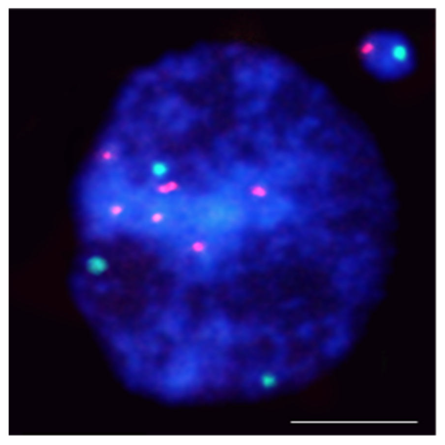
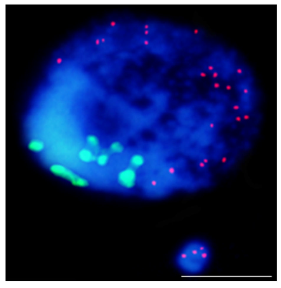
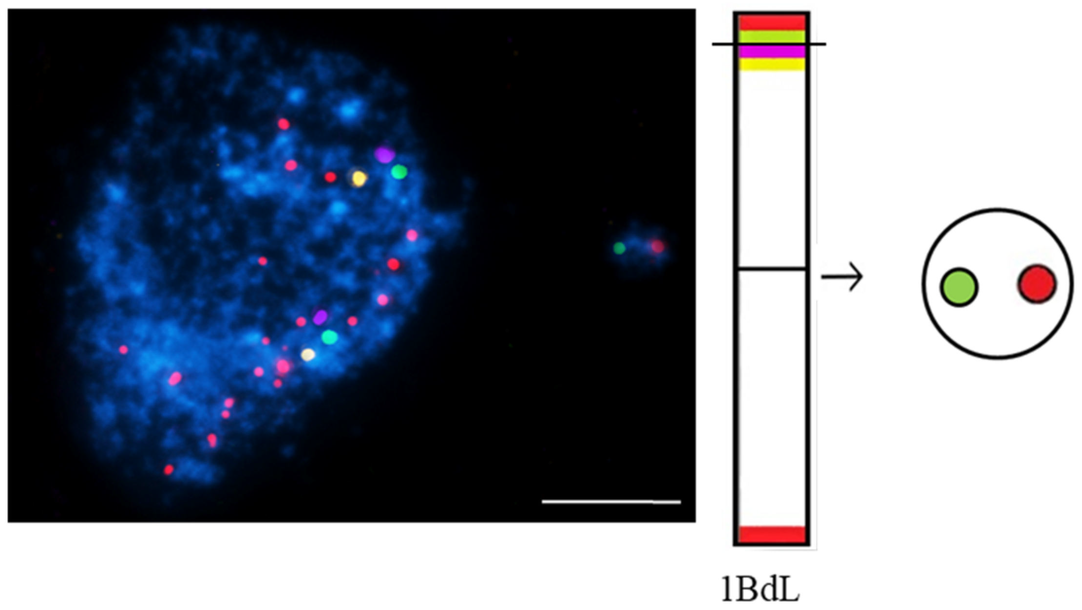
3. Fluorescence In Situ Hybridization Serves to Understand the Origin of Micronuclei

A breakthrough in the analyses of the localization of DNA damage at the chromosomal level in plants came with applying fluorescence in situ hybridization (FISH). It provides information on the possible ‘hot spots’ in plant genomes for DNA damage after the action of mutagens. Also, it gives information on the mechanisms of the biological effect of the individual agents that induce DNA damage. This knowledge is particularly crucial in plant mutagenesis, as the use of chemical and physical mutagens is the most common way to obtain mutants. This technique could detect even extremely small aberrations in dividing and non-dividing cells.
There is only one morphological type of micronuclei that may differ in size. The size of the micronucleus does not provide any information on whether it originated from chromosome fragments or entire chromosome(s), as the size may be related to the different degrees of the chromatin condensation. A more detailed analysis of the involvement of a specific chromosome or chromosome fragments in micronuclei formation is possible using fluorescence in situ hybridization (FISH). So far, FISH has not found such a wide application in the study of chromosome aberrations, including MN, in plants, as it has in humans [31][32][33]. Different types of DNA probes for FISH are applied in plants, e.g., repetitive DNA sequences, single-locus chromosome-specific BAC clones, partial (e.g., arm), and whole chromosome paints. The limitations of the chromosome-specific DNA sequences in plants make the comprehensive identification of chromosome fragments in micronuclei using FISH still limited to a few species. Among the repetitive DNA sequences, centromere, Arabidopsis thaliana (Arabidopsis)-type (TTTAGGG)n telomeric sequences, and ribosomal DNA (rDNA), which give strong and easily observed FISH signals, have found application in the detailed characterization of MN. These DNA sequences’ advantages are evolutionary conservation and location at a specific chromosome region. Repetitive dispersed DNA sequences are not a good source for probe pool for fluorescence in situ hybridization to study the origin of micronuclei.
FISH using 45S rDNA as the probe was first applied to localize the chromatin aberrations, such as translocations [34] and anaphase bridges [35], in Arabidopsis thaliana. Applying the rDNA as probes showed rules regarding gamma-ray–induced MN formation in barley (Hordeum vulgare) (Figure 1).
Figure 1. Hordeum vulgare interphase nuclei with the micronucleus induced by X-radiation. The nucleus was subjected to mcFISH with 5S rDNA (red) and 25S rDNA (green) probes. The micronucleus has one 5S rDNA and one 25S rDNA. Chromatin is stained with DAPI (blue). The bar represents = 10 µm.
5S rDNA-bearing chromosomes are shown to be more often involved in MN formation than NOR chromosomes in barley [36][37]. Similar rules regarding radiation-induced MN formation have been found in Brachypodium distachyon [38]. The hot spots for chromosome breakage in Lolium multiflorum were not correlated with rDNA sites [39].
The use of the centromere and telomere-specific DNA sequences for FISH also provided some rules regarding the origin of MN. It confirmed that the gamma ray-induced MN may originate from acentric fragments or whole lagging chromosomes. Thus, this approach allows the distinguishing of the micronuclei being a clastogenic and aneugenic effect of mutagens. However, most MN had only telomeric DNA signals, indicating that terminal deletion is the primary type of chromosome aberration leading to their formation (Figure 2).
Figure 2. Results of mcFISH with telomeric (red) and centromeric (green) probes. Brachypodium distachyon interphase nuclei with micronucleus that were induced by X-radiation; micronucleus shows only telomeric DNA signals. The bar represents 5 µm. Micrograph by A. Kus.
Comparing the contribution of particular chromosome fragments in MN that are induced by different chemical clastogens, the maleic acid hydrazide (MH) and nitroso-N-methyl-urea (MNU) have shown the difference in the size of the chromosome fragments that are involved in the MN. Most MH-induced MN originated from large acentric fragments, whereas MNU-induced MN is from small terminal chromosome fragments [36][37].
FISH provides much more information about MN formation with DNA probes that are dedicated to different chromosomes or particular chromosomes. Standard A- and B-chromosome-specific probes were successfully used in the rye gamma-irradiated cells (Secale cereale L.) [40] for the detection of the translocations between the A- and B-chromosomes.
One of the FISH approaches that is used to detect and characterize micronuclei in plants is multicolor FISH (mcFISH). It is based on the two consecutive FISH experiments that use a pair or pairs of probe sets that are removed after each experiment and include the reprobing step. Combining more than two differently labeled DNA probes on the same nuclei slide makes this technique more informative [41]. For the first time, mcFISH has been applied in human carcinogenicity studies [42], then it has found application in mammalian cells [43]. mcFISH is a common technique that is widely used in plants; however, it has narrow application in plant mutagenesis and genotoxicity. For the first time, this approach was applied in the analysis of the involvement of four different DNA sequences: 5S rDNA, 25S rDNA, the Arabidopsis-type (TTTAGGG)n telomeric sequence, and the Brachypodium-originated centromeric BAC clone CB33J12 in the micronuclei formation in Brachypodium distachyon root-tip cells that were subjected to a chemical mutagen [38].
The most advanced FISH-based approach in plants is chromosome painting (CP), which permits the selective visualization of entire chromosomes or their specific segments during mitosis as well the interphase [44][45][46][47][48][49]. The wide use of this technique for humans and mammals to determine the involvement of specific chromosomes in the formation of micronuclei showed that they preferentially comprise particular chromosomes that are related to the chromatin organization [50]. The large amounts of repetitive DNA on all chromosomes are obstacles to CP on plants. CP is limited to a few plant species: Arabidopsis [51], Brachypodium [52], and few other species that are characterized by a small genome. mcFISH and CP with low repeat (small and large pools of bacterial artificial chromosomes (BAC)) clones that are specific for selected chromosomes, were applied to improve the ‘standard’ MN test in Brachypodium distachyon (Figure 3).
Figure 3. Brachypodium distachyon interphase nuclei with micronuclei that were induced by MH-treatment that were subjected to mcFISH with the following probes: telomeric sequence (red), I BAC pool (green), II BAC pool (violet), and III BAC pool (yellow). Chromatin is stained with DAPI (blue). The diagram next to the photomicrographs shows the putative origins of the micronuclei. Transverse dashed lines indicate chromosome breakpoint. The scale bar = 5 µm. Micrographs by A. Kus.
BAC-FISH-based chromosome painting provides new information on the composition, origin, and mechanisms of micronuclei formation that is induced by MH-treatment and X-radiation in Brachypodium by showing the ‘fragile spots’ of DNA breaks [53]. Site-specific DNA breaks in chromosomes Bd4 and Bd5 were shown [54].
To summarize, FISH provides new insights into the localization of DNA breaks on plant chromosomes, proving the non-random distributions of chromosome aberrations. The reasons for this non-random distribution may be the spatial organization of the nucleus at the interphase, the diverse transcriptional activity of specific chromosome regions, and chromosome size. Single BAC-FISH-based chromosome barcoding and ‘chromosome painting’ approaches have proven to be effective in analyzing the mechanism of micronuclei formation in plants after mutagenic treatment. The advantages of the FISH technique in terms of accuracy and quality of quantitative analyses make the technique one that is likely to become more widespread in DNA damage studies in plants.

This entry is adapted from the peer-reviewed paper 10.3390/ijms23031306

References

  1. Evans, H.; Neary, G.; Williamson, F. The Relative Biological Efficiency of Single Doses of Fast Neutrons and Gamma-rays on Vicia Faba Roots and the Effect of Oxygen. Int. J. Radiat. Biol. Relat. Stud. Phys. Chem. Med. 1959, 1, 216–229.
  2. Araldi, R.P.; de Melo, T.C.; Mendes, T.B.; de Sá Júnior, P.L.; Nozima, B.H.N.; Ito, E.T.; de Carvalho, R.F.; de Souza, E.B.; de Cassia Stocco, R. Using the comet and micronucleus assays for genotoxicity studies: A review. Biomed. Pharmacother. 2015, 72, 74–82.
  3. Mišík, A.; Nersesyan, A.; Mišíková, K.; Knasmueller, S. Micronucleus Assays with Meiotic Pollen Tetrad Cells of Tradescantia and with Mitotic Root Tip Cells of Allium cepa and Vicia faba Issues in Toxicology. In The Micronucleus Assay in Toxicology; Knasmüller, S., Fenech, M., Eds.; The Royal Society of Chemistry: London, UK, 2019.
  4. Chen, L.; Yuan, S.; Liu, X.; Zhou, X.; Zhou, Y.; Song, Y. Genotoxicity response of Vicia faba seedlings to cadmium in soils as characterized by direct soil exposure and micronucleus test. Ecotoxicology 2019, 29, 65–74.
  5. Oubane, M.; Khadra, A.; Ezzariai, A.; El Fels, L.; Kouisni, L.; Hafidi, M. Micronucleus assay based on Vicia faba roots as a tool to assess the performances of wastewater treatment systems. Environ. Technol. Innov. 2020, 19, 100903.
  6. Klein, P.; Chauvey, L.; Kallerhoff, J.; Pinelli, E.; Morard, M.; Silvestre, J. A Tool Derived from the Vicia faba Micronucleus Assay, to Assess Genotoxicity, Cytotoxicity or Biostimulation of Novel Compounds Used in Agriculture. Agronomy 2021, 11, 321.
  7. Ghosh, M.; Jana, A.; Sinha, S.; Jothiramajayam, M.; Nag, A.; Chakraborty, A.; Mukherjee, A.; Mukherjee, A. Effects of ZnO nanoparticles in plants: Cytotoxicity, genotoxicity, deregulation of antioxidant defenses, and cell-cycle arrest. Mutat. Res. Toxicol. Environ. Mutagen. 2016, 807, 25–32.
  8. Scherer, M.D.; Sposito, J.C.V.; Falco, W.F.; Grisolia, A.B.; Andrade, L.H.C.; Lima, S.M.; Machado, G.; Nascimento, V.A.; Gonçalves, D.A.; Wender, H.; et al. Cytotoxic and genotoxic effects of silver nanoparticles on meristematic cells of Allium cepa roots: A close analysis of particle size dependence. Sci. Total Environ. 2019, 660, 459–467.
  9. Gosh, M.; Ghosh, I.; Godderis, L.; Hoet, P.; Mukherjee, A. Genotoxicity of engineered nanoparticles in higher plants. Mutat. Res. Genet. Toxicol. Environ. Mutagen. 2019, 842, 132–145.
  10. Prabhu, S.; Kumar, V.; Bharath, J.; Thaha, M.; Rajeswari, D. A Review on Vicia faba as a plant test system in toxicity evaluation of various metals: Vicia-micronucleus test (Vicia-MCN). Int. J. Chem. Tech. Res. 2017, 10, 961–967.
  11. Jiang, X.; Chen, H.; Liao, Y.; Ye, Z.; Li, M.; Klobu, G. Ecotoxicity and genotoxicity of polystyrene microplastics on higher plant Vicia faba. Environ. Pollut. 2019, 251, 831–838.
  12. Kaur, M.; Sharma, A.; Soodan, R.K.; Chahal, V.; Kumar, V.; Katnoria, J.K.; Nagpal, A.K. Allium cepa Root Chromosomal Aberration Assay: A Tool to Assess Genotoxicity of Environmental Contaminants. In Environmental Contaminants and Natural Products: A Human Health Perspective; Sharma, A., Kumar, M., Kaur, S., Nagpal, A.K., Eds.; Bentham Science: Sharjah, United Arab Emirates, 2019; pp. 65–93.
  13. Zgórska, A.; Borgulat, A. Genotoxicity of wastewater samples from the textile industry detected by broad bean (Vicia faba) micronucleus test assay. Appl. Ecol. Environ. Res. 2020, 18, 5315–5323.
  14. Abdelsalam, N.R.; Abdel-Megeed, A.; Alic, H.M.; Saleme, M.Z.M.; Al-Hayalif, M.F.A.; Elshikh, M.S. Genotoxicity effects of silver nanoparticles on wheat (Triticum aestivum L.) root tip cells. Ecotoxicol. Environ. Saf. 2018, 155, 76–85.
  15. Alvarenga, I.F.S.; dos Santos, F.E.; Silveira, G.L.; Andrade-Vieira, L.F.; Martin, G.C.; Guilherme, L.R.G. Investigating arsenic toxicity in tropical soils: A cell cycle and DNA fragmentation approach. Sci. Total Environ. 2020, 698, 134272.
  16. Korshikov, I.I.; Belonozhko, Y.A.; Lapteva, E.V. The Use of a Micronucleus Test in Pinus pallasiana D. Don and Picea abies (L.) Karst. for the Assessment of Technogenic Pollution’s Influence. Cytol. Genet. 2019, 53, 106–112.
  17. Mishra, K. Assessment of Cytotoxic and Genotoxic Potential of Heavy Metals in Plants: A Review. Int. J. Plant Environ. 2020, 6, 152–155.
  18. Heddle, J.A.; Fenech, M.; Hayashi, M.; MacGregor, J.T. Reflections on the development of micronucleus assays. Mutagenesis 2010, 26, 3–10.
  19. Luzhna, L.; Kathiria, P.; Kovalchuk, O. Micronuclei in genotoxicity assessment: From genetics to epigenetics and beyond. Front. Genet. 2013, 4, 131.
  20. Kwon, M.; Leibowitz, M.L.; Lee, J.H. Small but mighty: The causes and consequences of micronucleus rupture. Exp. Mol. Med. 2020, 52, 1777–1786.
  21. Nersesyan, A.; Fenech, M.; Bolognesi, C.; Misik, M.; Setayesh, T.; Wultsch, G.; Bonass, S.; Thomas, P.; Knasmuller, S. Use of the lymphocyte cytokinesis-block micronucleus assay in occupational biomonitoring of genome damage caused by in vivo exposure to chemical genotoxins: Past, present and future. Mutat. Res. 2016, 770, 1–11.
  22. McClelland, S.E. Role of chromosomal instability in cancer progression. Endocr.-Relat. Cancer 2017, 24, T23–T31.
  23. Saks, M.; Upreti, S.; Rajendra, S.V.; Dang, R. Genotoxicity: Mechanisms, testing guidelines and methods. Glob. J. Pharm. Pharm. Sci. 2017, 1, 133–138.
  24. Kirsch-Volders, M.; Fenech, M.; Bolognesi, C. Validity of the lymphocyte cytokinesis-block micronucleus assay (L-CBMN) as biomarker for human exposure to chemicals with different modes of action: A synthesis of systematic reviews. Mutat. Res. Genet. Toxicol. Environ. Mutagen. 2018, 836, 47–52.
  25. Ye, C.J.; Sharpe, Z.; Alemara, S.; MacKenzie, S.; Liu, G.; Abdallah, B.; Horne, S.; Regan, S.; Heng, H.H. Micronuclei and Genome Chaos: Changing the System Inheritance. Genes 2019, 10, 366.
  26. Norppa, H.; Falck, G.C.M. What do human micronuclei contain? Mutagenesis 2003, 18, 221–233.
  27. Sommer, S.; Buraczewska, I.; Kruszewski, M. Micronucleus Assay: The State of Art, and Future Directions. Int. J. Mol. Sci. 2020, 21, 1534.
  28. Fenech, M. Cytokinesis-block micronucleus cytome assay evolution into a more comprehensive method to measure chromosomal instability. Genes 2020, 11, 1203.
  29. Hayashi, M. The micronucleus test—Most widely used in vivo genotoxicity test. Genes Environ. 2016, 38, 18.
  30. Hintzsche, H.; Hemmann, U.; Poth, A.; Utesch, D.; Lott, J.; Stopper, H. Fate of micronuclei and micronucleated cells. Mutat. Res. 2017, 771, 85–98.
  31. Liehr, T. Fluorescence In Situ Hybridization (FISH)—Application Guide, 2nd ed.; Springer: Berlin/Heidelberg, Germany, 2017.
  32. Liehr, T. Is molecular cytogenetic diagnostic of are diseases in Europe close to extinction. J. Genet. Genom. 2020, 4, 2.
  33. Florian, R.T.; Kraft, F.; Leitão, E.; Kaya, S.; Klebe, S.; Magnin, E.; van Rootselaar, A.-F.; Buratti, J.; Kühnel, T.; Schröder, C.; et al. Unstable TTTA/TTTCA expansions in MARCH6 are associated with familial adult myoclonic epilepsy type 3. Nat. Commun. 2019, 10, 4919.
  34. Weiss, H.; Maluszynska, J. Chromosomal rearrangement in autotetraploid plants of Arabidopsis thaliana. Hereditas 2000, 133, 255–261.
  35. Siroky, J.; Zluvova, J.; Riha, K.; Shippen, R.E.; Vyskot, B. Rearrangements of ribosomal DNA clusters in late generation telomerase-deficient Arabidopsis. Chromosoma 2003, 112, 116–123.
  36. Juchimiuk, J.; Hering, B.; Maluszynska, J. Multicolour FISH in an analysis of chromosome aberrations induced by N-nitroso-N-methylurea and maleic hydrazide in barley cells. J. Appl. Genet. 2007, 48, 99–106.
  37. Juchimiuk-Kwasniewska, J.; Brodziak, L.; Maluszynska, J. FISH in analysis of gamma ray-induced micronuclei formation in barley. J. Appl. Genet. 2010, 52, 23–29.
  38. Kus, A.; Kwasniewska, J.; Hasterok, R. Brachypodium distachyon—A Useful Model in the Qualification of Mutagen-Induced Micronuclei Using Multicolor FISH. PLoS ONE 2017, 12, e0170618.
  39. Rocha, L.C.; Mittelmann, A.; Houben, A.; Techio, V.H. Fragile sites of 45S rDNA of Lolium multiflorum are not hotspots for chromosomal breakages induced by X-ray. Mol. Biol. Rep. 2016, 43, 659–665.
  40. Hasterok, R.; Jenkins, G.; Langdon, T.; Jones, R.N. The nature and destiny of translocated B-chromosome-specific satellite DNA of rye. Chromosome Res. 2002, 10, 83–86.
  41. Hasterok, R.; Langdon, T.; Taylor, S.; Jenkins, G. Combinatorial labeling of DNA probes enables multicolor fluorescence in situ hybridization in plants. Folia Histochem. Cytobiol. 2002, 40, 319–323.
  42. Nederlof, P.M.; Robinson, D.; Abuknesha, R.; Wiegant, J.; Hopman, A.H.N.; Tanke, H.J.; Raap, A.K. Three-color fluorescence in situ hybridization for the simultaneous detection of multiple nucleic acid sequences. Cytom. J. Int. Soc. Anal. Cytol. 1989, 10, 20–27.
  43. Natarajan, A.; Boei, J. Formation of chromosome aberrations: Insights from FISH. Mutat. Res. Mol. Mech. Mutagen. 2003, 544, 299–304.
  44. Lusinska, J.; Majk, J.; Betekhtin, A.; Susek, K.; Wolny, E.; Hasterok, R. Chromosome identification and reconstruction of evolutionary rearrangements in Brachypodium distachyon, B. stacei and B. hybridum. Ann. Bot. 2018, 122, 445–459.
  45. He, L.; Braz, G.T.; Torres, G.; Jiang, J. Chromosome painting in meiosis reveals pairing of specific chromosomes in polyploid Solanum species. Chromosoma 2018, 127, 505–513.
  46. Hou, L.; Xu, M.; Zhang, T.; Xu, Z.; Wang, W.; Zhang, J.; Yu, M.; Ji, W.; Zhu, C.; Gong, Z.; et al. Chromosome painting and its applications in cultivated and wild rice. BMC Plant Biol. 2018, 18, 110.
  47. Chen, F.; Song, Y.; Li, X.; Chen, J.; Mo, L.; Zhang, X.; Lin, Z.; Zhang, L. Genome sequences of horticultural plants: Past, present, and future. Hortic. Res. 2019, 6, 112.
  48. Albert, P.S.; Zhang, T.; Semrau, K.; Rouillard, J.-M.; Kao, Y.-H.; Wang, C.-J.R.; Danilova, T.V.; Jiang, J.; Birchler, J.A. Whole-chromosome paints in maize reveal rearrangements, nuclear domains, and chromosomal relationships. Proc. Natl. Acad. Sci. USA 2019, 116, 1679–1685.
  49. Meng, Z.; Wang, Q.; Khurshid, H.; Raza, G.; Han, J.; Wang, B.; Wang, K. Chromosome Painting Provides Insights Into the Genome Structure and Evolution of Sugarcane. Front. Plant Sci. 2021, 12, 731664.
  50. Hovhannisyan, G.; Aroutiounian, R.; Liehr, T. Chromosomal Composition of Micronuclei in Human Leukocytes Exposed to Mitomycin C. J. Histochem. Cytochem. 2012, 60, 316–322.
  51. Lysak, M.A.; Berr, A.; Pecinka, A.; Schmidt, R.; McBreen, K.; Schubert, I. Mechanisms of chromosome number reduction in Arabidopsis thaliana and related Brassicaceae species. Proc. Natl. Acad. Sci. USA 2006, 103, 5224–5229.
  52. Idziak, D.; Hazuka, I.; Poliwczak, B.; Wiszynska, A.; Wolny, E.; Hasterok, R. Insight into the Karyotype Evolution of Brachypodium Species Using Comparative Chromosome Barcoding. PLoS ONE 2014, 9, e93503.
  53. Kus, A.; Kwasniewska, J.; Szymanowska-Pulka, J.; Hasterok, R. Dissecting the chromosomal composition of mutagen-induced micronuclei in Brachypodium distachyon using multicolour FISH. Ann. Bot. 2018, 122, 1161–1171.
  54. Kus, A.; Szymanowska-Pułka, J.; Kwasniewska, J.; Hasterok, R. Detecting Brachypodium distachyon Chromosomes Bd4 and Bd5 in MH- and X-Ray-Induced Micronuclei Using mcFISH. Int. J. Mol. Sci. 2019, 20, 2848.
More
This entry is offline, you can click here to edit this entry!
Video Production Service