Dysregulation of the Gut Barrier Function during ALD: History
Please note this is an old version of this entry, which may differ significantly from the current revision.
Contributor:

Chronic alcohol consumption and alcohol-associated liver disease (ALD) represent a major public health problem worldwide. Only a minority of patients with an alcohol-use disorder (AUD) develop severe forms of liver disease (e.g., steatohepatitis and fibrosis) and finally progress to the more advanced stages of ALD, such as severe alcohol-associated hepatitis and decompensated cirrhosis. Emerging evidence suggests that gut barrier dysfunction is multifactorial, implicating microbiota changes, alterations in the intestinal epithelium, and immune dysfunction. This failing gut barrier ultimately allows microbial antigens, microbes, and metabolites to translocate to the liver and into systemic circulation. Subsequent activation of immune and inflammatory responses contributes to liver disease progression.

  • intestinal barrier
  • chronic alcohol consumption
  • gut-liver axis
  • intestinal immunity
  • inflammation

1. Introduction

Chronic alcohol consumption is one of the leading causes of chronic liver disease and liver-related deaths worldwide. Although more than 90–95% of people with excessive alcohol drinking develop fatty liver, only a minority (10–20%) of subjects with an alcohol-use disorder (AUD) ultimately progress to more advanced stages of alcohol-associated liver disease (ALD) (e.g., severe alcohol-associated hepatitis and decompensated cirrhosis) and its related complications [1]. Some factors that are correlated with disease progression have been identified, including sex, drinking patterns, genetics, obesity, and dietary factors. Those factors either increase susceptibility to liver damage or aggravate disease by acting synergistically with alcohol [2][3]. The interaction between obesity and alcohol, in particular for mild or moderate consumption, is controversial in humans [4]. The incidence of obesity and the associated metabolic syndrome is increasing worldwide. A recent cohort study showed that patients with excessive alcohol consumption have increased prevalence of obesity and metabolic syndrome [5], which might increase the risk for liver disease progression. It has been reported that moderate alcohol consumption increases the risk of development of hepatic steatosis in association with obesity. In contrast, moderate alcohol consumption reduces mortality in normal body mass index (BMI) and overweight individuals. This beneficial effect was, however, lost in obese subjects [6]. In rodents, the synergism between alcohol and obesity might be linked to alterations in immune responses, metabolism, and gut microbiota composition [3]. Recent animal data where alcohol binges have been added on top of a non-alcoholic steatohepatitis model support a synergistic effect for liver damage (for a detailed review see [7]). Even if some advances have been made in researchers' understanding of the factors involved in the development of ALD, there are still no FDA-approved therapies for treating patients with ALD and abstinence is considered the most effective treatment option [2]. Drugs aiming at reducing the immune responses have been studied for patients with severe forms of liver disease, especially in severe alcohol-associated hepatitis [2]. However, none of these approaches have proven to be safe and efficient [2].
The pathophysiology of gut barrier dysfunction associated with alcohol abuse is likely multifactorial. Changes in the intestinal microbiome, including bacteria, fungi, and viruses, have been associated with ALD [8][9][10][11]. This so-called intestinal dysbiosis together with increased intestinal permeability is thought to lead to microbial translocation. Microbes and/or pathogen-associated molecular patterns (PAMPs) reach the liver and serve as a trigger for the initiation of an inflammatory response. Consequently, studies targeted the gut microbes as part of the intestinal barrier for the potential treatment of ALD with some success (for a more detailed review see [12]). Other reports have described how targeting intestinal permeability might prove beneficial also as a potential treatment for liver diseases [13].
This general concept primarily derived from animal studies is, however, challenged by observations made in humans indicating that only approximately 40% of subjects with alcohol abuse have measurable intestinal dysbiosis [14]. Moreover, increased intestinal permeability in humans does not seem to be a prerequisite for microbial translocation to occur [15]. These observations imply that additional factors beyond microbiota changes and intestinal permeability are likely implicated in the pathophysiology of ALD. They also highlight the difficulties with extrapolating animal data to human pathology for several reasons: (1) animals have a natural aversion to alcohol, requiring ways of administration that do not mimic human alcohol-seeking behavior [16][17]; (2) rodents have a 5-times faster ethanol metabolism, making it difficult to reach stable high alcohol levels [18]; (3) compared to humans, rodents are characterized by profound differences in their immune system [19] and their microbiota [20]; and (4) animals only develop mild forms of ALD upon chronic alcohol feeding and even the most “sophisticated models” do not resume the liver-damage pattern observed in humans [21].

2. The Gut Barrier

2.1. Gut Microbiome

The gut is the natural habitat for 100 trillion microorganisms (bacteria, archaea, fungi, and viruses) [22]. This community of microbe’s genome—the microbiome—encodes 100 times more genes than the human genome [23]. These microbes exert a remarkable influence on the host during homeostasis and disease. Indeed, the gut microbiota can modulate multiple physiological functions of the host, such as strengthening gut integrity and shaping the intestinal epithelium, harvesting energy [24], protecting against pathogens [25], and regulating immune response [26].
The gut microbiome represents our first line of defense against potential pathogens [27]. Intestinal microbes compete with pathogens for nutrients and produce antimicrobial molecules called bacteriocins [28], which shape the composition of the gut microbiota. Commensal microbes further contribute to the development of the mucosal immune system via direct interactions with host epithelial and immune cells [29]. In addition, the gut microbiome is responsible for a number of vital metabolic and signaling functions for the host, such as biotransformation of primary bile acids [30], production of short-chain fatty acids [24], synthesis of all B vitamins and vitamin K [31], as well as synthesis of essential amino acids [32].

2.2. Intestinal Epithelium

The principal functions of the intestinal barrier are nutrient absorption and defense against invading pathogens and potentially harmful substances. Mucosal cell types dynamically contribute to a balance between protective immunity and prevention of excessive immune responses against nutrients and commensal microbes. A first single-layer of cells, called the intestinal epithelium, facilitate absorption of nutrients while preventing microbial attachment and their translocation into the blood circulation [33].
The small intestinal epithelium is organized in villi and crypts of Lieberkühn. Adult stem cells reside at the base of the crypts and give rise to all absorptive and secretory cells making up the epithelial layer [34]. Gut barrier functions are insured by different epithelial cell types. Secretory goblet cells produce mucus—a viscous fluid enriched in mucin glycoproteins that form a first physical barrier against luminal microbes. Commensal bacteria also degrade glycans to extract the energy content, which then they share with the host in a mutualistic relationship [35]. Moreover, goblet cells produce antimicrobial peptides, such as trefoil factor [36] and resistin-like molecule beta [37], to dampen the mucosal attachment of microbes. In addition, they are also able to present bacterial antigens to dendritic cells via the so-called “goblet cells antigen-associated passages” (GAPs), thus educating the intestinal immune system [38]. Paneth cells, which reside at the base of the crypts, contribute to gut barrier function by producing and releasing into the crypt lumen antimicrobial molecules such as antimicrobial peptides, the regenerating islet-derived 3 (Reg3) family of proteins, lysozyme, and secretory phospholipase A2 [39]. Microfold cells, also known as M cells, are located in the follicle-associated epithelium. They uptake antigens and microorganisms from the lumen and deliver to organized lymphoid tissues within the mucosa [40]. Absorptive enterocytes also contribute to defense mechanisms by regulating paracellular permeability with tight junction proteins and directly recognizing a variety of PAMPs with different pattern recognition receptors, including Toll-like receptors (TLRs) and nucleotide-binding oligomerization domain-containing proteins (NODs) [41]. Furthermore, a dense glycocalyx consisting of extended and heavily glycosylated membrane mucins covers the surface of enterocytes, reinforcing the protective properties of the intestinal mucus [42].

2.3. Intestinal Immune System

The mucosal immune system plays an important role in orchestrating intestinal defense mechanisms against potential pathogenic microorganisms by coordinating innate and adaptive responses. Immune cells strategically localize in different compartments of the gut mucosa according to the expression of different receptors and the capacity to sense directly or indirectly microbes or their products.
Macrophages are in part found under the epithelium in the lamina propria where they engulf and kill invading microorganisms and subsequently expose their antigens to adaptive immune cells. Unlike phagocytes in other parts of the body, gut macrophages produce large amounts of regulatory cytokines, such as IL-10, and contribute to maintain a tolerant status in the intestine while avoiding excessive inflammatory reactions [43]. The intestinal macrophages are continuously replaced by circulating monocytes. However, a recent report sheds light on the existence of long-lived tissue-resident gut macrophages that are not replaced by infiltrating monocytes [44].
Dendritic cells (DCs) orchestrate mucosal immunity by acquiring antigens in the epithelium through goblet or microfold epithelial cells or by directly capturing and sampling luminal antigens. They then processed antigens to T cells and B cells in the mucosa or in organized secondary lymphoid structures known as Peyer’s Patches, as well as in mesenteric lymph nodes [45]. Tolerogenic dendritic cells promote differentiation of regulatory T cells (Treg) [46], which in turn are able to induce tolerogenic DCs via IL-10, TGF-β, and other surface molecules [47]. DCs in gut-associated lymphoid tissue (GALT) and mesenteric lymph nodes further promote differentiation of B cells into immunoglobulin A (IgA)-producing plasma cells and memory B cells. Dimers of immunoglobulin A are transported through intestinal epithelial cells and secreted into the lumen where it binds commensal and pathogenic microbes, without initiating a pro-inflammatory response [48], and therefore contributes to mucosal defense.
Recent investigations have revealed the existence of CD8+ and CD4+ T resident memory cells (TRM) and their importance in the maintenance of gut microbial immune surveillance. These lymphocytes are localized in the intestinal epithelium and lamina propria and allow the fast initiation of targeted immune responses against potential invaders [49] (Figure 1A).
Figure 1. (A) The small intestinal barrier in healthy conditions. The intestinal barrier is composed of three main layers: the mucus, the epithelium, and the lamina propria. The epithelium, which is overlaid by a thin and discontinuous mucus layer, together with enterocytes and specialized cells constitute a first line of defense. Immune cells in the mucosa further fine-tune the defense mechanisms against pathogens. Macrophages underlying the epithelium regulate immune response by phagocytosis of microbes and production of large amounts of anti-inflammatory cytokines, thus preventing excessive immune responses. Dendritic cells capture, process, and present microbial antigens to different adaptive immune cells. T lymphocytes rapidly act against pathogens by killing infected cells, producing cytokines, and coordinating immune responses. (B) The small intestinal barrier in patients with an alcohol-use disorder. Chronic alcohol consumption is associated with alterations in the composition of the gut microbiota, increased attachment of microbes to the intestinal mucosa, and their translocation into the portal and systemic circulation. This process is enhanced by impairment of various epithelial and immune defense mechanisms. A loose, thickened mucus layer as well as reduced production of antimicrobial molecules and a diminution of macrophages and T lymphocytes all contribute to the failing gut barrier in AUD patients. Figures were created with Biorender.com (14 November 2021).

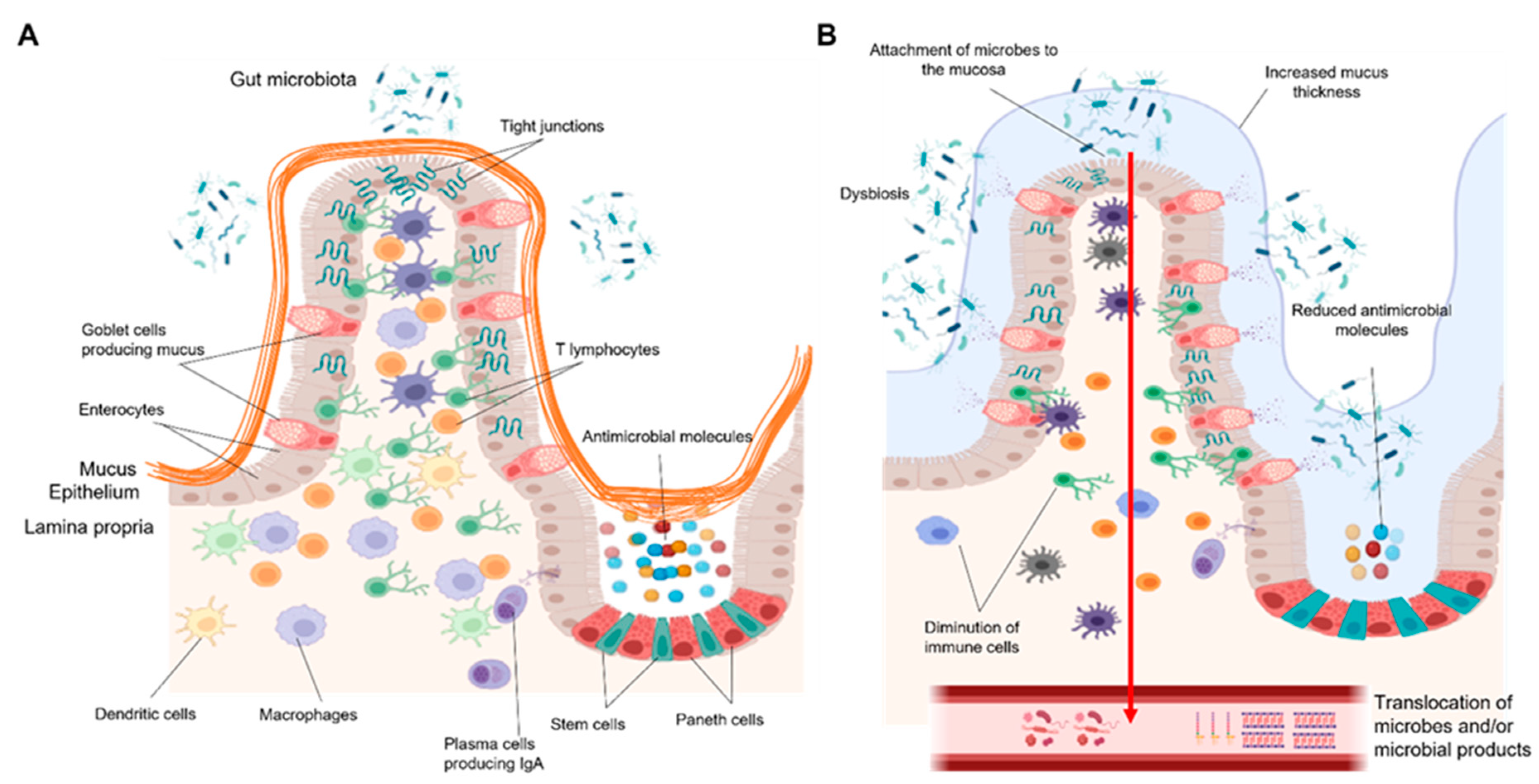
3. How Can Gut Barrier Dysfunction Contribute to ALD Progression?

The intestine and liver communicate via bidirectional links through the biliary tract, portal vein, and systemic circulation. The liver synthesizes and transports primary bile acids and antimicrobial molecules to the intestinal lumen through the bile duct. In the gut, the host and microbes metabolize endogenous (bile acids and amino acids) and exogenous substrates (dietary and environmental factors) into products that are then transported to the liver through the portal vein. Furthermore, the systemic circulation connects the gut-liver axis by transporting liver metabolites into the intestine [50]. Many of those products of metabolites (e.g., bile acids, fatty acids, ethanol metabolites) might act as signaling molecules at the local and distant sites where they activate various signaling processes (e.g., cell growth and differentiation, host metabolism, and immune responses) involved in regulation of the intestinal barrier [51] (Figure 2). Changes in the levels and in the structure of these signaling molecules, for example, a different proportion of secondary and primary bile acids, can negatively affect the processes (e.g., intestinal permeability, epithelial differentiation, absorption of nutrients and immunity) essential for defense mechanisms in the gut. As a result, breakdown of gut barrier function allows microbes and/or their products to get into the liver and systemic circulation, where they are recognized by different pattern recognition receptors in various cell types with subsequent activation of immune responses. According to the stage of ALD, the cell types, and signaling pathways involved, activation of a pro-inflammatory response against microbial antigens might be beneficial or detrimental for liver disease progression [52]. Researchers here discuss some recent findings that support the link between gut barrier dysfunction, systemic inflammation, and impaired hepatic immunity in the pathogenesis of early and advanced stages of ALD.
Figure 2. Bidirectional communication between the intestine and the liver. The liver produces primary bile acids, antimicrobial molecules, and IgA that are released in the intestine through the bile duct. In the intestine, these molecules contribute to shape the microbiota. In addition, primary bile acids are converted into secondary bile acids by the gut microbiota. Bile acids, which are reabsorbed in the terminal ileum, microbial products and metabolites, as well as viable microbes are transported to the liver through the portal vein. Once in the liver, they are implicated in triggering immune and inflammatory responses that might lead to liver disease. Moreover, the gut–liver axis is connected via the systemic circulation where ethanol, ethanol-derived metabolites, as well as other inflammatory mediators (cytokines, metabolites, etc.) can reach the two organs, thus influencing their functions. Figures were created with Biorender.com (14 November 2021).

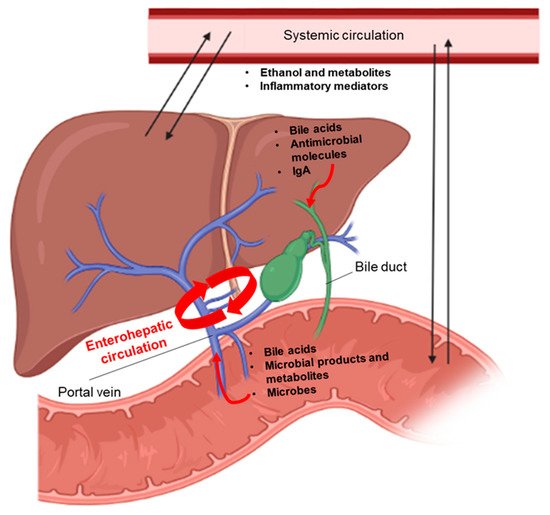
3.1. Systemic Inflammatory Responses

Increased systemic translocation of bacterial products, such as lipopolysaccharides (LPS) and peptidoglycans (PGN), have been shown to be associated with elevated plasma levels of inflammatory cytokines (IL1β, IL8, IL18, TNF-α, and IL6) in AUD patients [53]. Mechanistic analyses have shown that in AUD patients, LPS and to a greater extent PGN can contribute to the activation of peripheral blood mononuclear cells (PBMCs). PBMCs are likely contributing to PGN-triggered release of IL1β, IL8, and IL18 into the blood [53]. In contrast, TNF-α and IL6 were not elevated in PBMCs, suggesting that their increased plasma levels might originate from other sources [54]. Inflammatory chemokine monocyte chemoattractant protein 1 (MCP-1) levels in the plasma are increased in actively drinking subjects compared to the controls [55]. This may promote monocyte recruitment and infiltration into the liver where they transform into inflammatory monocyte-derived macrophages. This process has been observed in mice chronically fed ethanol [56] and it is believed to play an important role in the early pathogenesis of ALD [57]. Future studies are needed to clarify the pathways linked to this process. In contrast to the early stages of alcohol-associated liver disease, severe alcohol-associated hepatitis is characterized by massive hepatic infiltration of neutrophils coming from the circulation [57]. However, a recent investigation found that alcohol-associated hepatitis patients with more neutrophils had a better outcome than those with less neutrophil infiltration [58], probably due to defective bacterial clearance and impaired liver regeneration.

3.2. Impairment of Immune Responses Related to Microbes in the Liver

Increasing evidence supports the role of unbalanced immunity in the different stages of ALD. When the gut barrier is disrupted, higher amounts of microbes and/or their products translocate to the liver [59], leading to the activation of the hepatic immune system through pattern recognition receptors like Toll-like receptors (TLRs) [60]. TLRs are expressed widely in immune and non-immune cells [61] in the liver and their activation by different PAMPs coming from the intestine contributes to ALD progression [62].
It has been proposed that activation of the TLR4 signaling pathway by LPS plays a pivotal role in disease progression. Mice deficient in TLR4, CD14, and LPS-binding protein (LBP) show resistance to alcohol-induced liver injury [63]. Other studies in mice show the pathophysiological importance of TLR4 in resident macrophages (e.g., Kupffer cells), monocyte-derived macrophages, and hepatic stellate cells [64]. The concept of TLR4 activation with subsequent TNF-α release has conducted to translational approaches such as the anti-TNF-α therapies for patients with severe alcoholic hepatitis. The disappointing outcome of these clinical trials [65] highlight two main facts in the field of alcohol-associated liver disease: firstly, data obtained in animal models of ALD cannot be necessarily extrapolated to humans [16]; secondly, but not in order of importance, inflammatory responses can be beneficial or detrimental depending on the cell type involved and stage of liver disease. In this context, researchers have recently revealed that intracellular TLRs, in particular TLR7, increased at early stages of ALD in humans, whereas TLR4 did not, and was correlated with liver disease severity [55]. TLR7 upregulation occurred concomitantly with activation of interferon signaling predominantly in hepatocytes [55]. In contrast, specific TLR7 activation did not exacerbate liver disease in a chronic ethanol-feeding model [66].
Kupffer cells and monocyte-derived macrophages also exhibit different functions in different stages of ALD. Animal models show an important role of TLR4 activation with release of pro-inflammatory cytokines in alcohol-induced liver injury [67]. Pro-inflammatory cytokines produced by hepatic macrophages also seem to be important in the early pathogenesis of ALD [68]. However, defective macrophages in more advanced stages of liver disease may not allow optimal clearance of microbes by phagocytosis, leading to severe infections, a feature often encountered in patients with severe alcohol-associated hepatitis [69] and decompensated cirrhosis [70].

This entry is adapted from the peer-reviewed paper 10.3390/ijms222312687

References

  1. World Health Organization. Global Status Report on Alcohol and Health 2018; World Health Organization: Geneva, Switzerland, 2018.
  2. O’Shea, R.S.; Dasarathy, S.; McCullough, A.J.; Practice Guideline Committee of the American Association for the Study of Liver Diseases; Practice Parameters Committee of the American College of Gastroenterology. Alcoholic liver disease. Hepatology 2010, 51, 307–328.
  3. Åberg, F.M.F.; Färkkilä, M. Drinking and Obesity: Alcoholic Liver Disease/Nonalcoholic Fatty Liver Disease Interactions. Semin. Liver Dis. 2020, 4, 154–162.
  4. Traversy, G.; Chaput, J.-P. Alcohol Consumption and Obesity: An Update. Curr. Obes. Rep. 2015, 4, 122–130.
  5. Singh, A.; Amin, H.; Garg, R.; Gupta, M.; Lopez, R.; Alkhouri, N.; McCullough, A. Increased Prevalence of Obesity and Metabolic Syndrome in Patients with Alcoholic Fatty Liver Disease. Dig. Dis. Sci. 2020, 65, 3341–3349.
  6. Peeraphatdit, T.B.; Ahn, J.C.; Choi, D.H.; Allen, A.M.; Simonetto, D.A.; Kamath, P.S.; Shah, V.H. A Cohort Study Examining the Interaction of Alcohol Consumption and Obesity in Hepatic Steatosis and Mortality. In Mayo Clinic Proceedings; Elsevier: Amsterdam, The Netherlands, 2020; Volume 95, pp. 2612–2620.
  7. Hwang, S.; Ren, T.; Gao, B. Obesity and binge alcohol intake are deadly combination to induce steatohepatitis: A model of high-fat diet and binge ethanol intake. Clin. Mol. Hepatol. 2020, 26, 586–594.
  8. Lang, S.; Schnabl, B. Microbiota and Fatty Liver Disease—The Known, the Unknown, and the Future. Cell Host Microbe 2020, 28, 233–244.
  9. Jiang, L.; Stärkel, P.; Fan, J.-G.; Fouts, D.E.; Bacher, P.; Schnabl, B. The gut mycobiome: A novel player in chronic liver diseases. J. Gastroenterol. 2021, 56, 1–11.
  10. Yang, A.-M.; Inamine, T.; Hochrath, K.; Chen, P.; Wang, L.; Llorente, C.; Bluemel, S.; Hartmann, P.; Xu, J.; Koyama, Y.; et al. Intestinal fungi contribute to development of alcoholic liver disease. J. Clin. Investig. 2017, 127, 2829–2841.
  11. Jiang, L.; Lang, S.; Duan, Y.; Zhang, X.; Gao, B.; Chopyk, J.; Schwanemann, L.K.; Ventura-Cots, M.; Bataller, R.; Bosques-Padilla, F.; et al. Intestinal Virome in Patients With Alcoholic Hepatitis. Hepatology 2020, 72, 2182–2196.
  12. Zhou, Z.; Zhong, W. Targeting the gut barrier for the treatment of alcoholic liver disease. Liver Res. 2017, 1, 197–207.
  13. Khoshbin, K.; Camilleri, M. Effects of dietary components on intestinal permeability in health and disease. Am. J. Physiol. Liver Physiol. 2020, 319, G589–G608.
  14. Leclercq, S.; Matamoros, S.; Cani, P.D.; Neyrinck, A.; Jamar, F.; Stärkel, P.; Windey, K.; Tremaroli, V.; Bäckhed, F.; Verbeke, K.; et al. Intestinal permeability, gut-bacterial dysbiosis, and behavioral markers of alcohol-dependence severity. Proc. Natl. Acad. Sci. USA 2014, 111, E4485–E4493.
  15. Maccioni, L.; Gao, B.; Leclercq, S.; Pirlot, B.; Horsmans, Y.; De Timary, P.; Leclercq, I.; Fouts, D.; Schnabl, B.; Stärkel, P. Intestinal permeability, microbial translocation, changes in duodenal and fecal microbiota, and their associations with alcoholic liver disease progression in humans. Gut Microbes 2020, 12, 1–23.
  16. Wilkin, R.J.; Lalor, P.F.; Parker, R.; Newsome, P.N. Murine Models of Acute Alcoholic Hepatitis and Their Relevance to Human Disease. Am. J. Pathol. 2016, 186, 748–760.
  17. Tabakoff, B.; Hoffman, P.L. Animal Models in Alcohol Research. Alcohol Res. Health J. Natl. Inst. Alcohol Abus. Alcohol. 2000, 24, 77–84.
  18. Cederbaum, I.A. Metabolism Alcohol. Clin. Liver Dis. 2013, 16, 667–685.
  19. Mestas, J.; Hughes, C.C.W. Of mice and not men: Differences between mouse and human immunology. J. Immunol. 2004, 172, 2731–2738.
  20. Nguyen, T.L.A.; Vieira-Silva, S.; Liston, A.; Raes, J. How informative is the mouse for human gut microbiota research? Dis. Model. Mech. 2015, 8, 1–16.
  21. Gao, B.; Xu, M.-J.; Bertola, A.; Wang, H.; Zhou, Z.; Liangpunsakul, S. Animal Models of Alcoholic Liver Disease: Pathogenesis and Clinical Relevance. Gene Expr. 2017, 17, 173–186.
  22. Clemente, J.C.; Ursell, L.K.; Parfrey, L.W.; Knight, R. The Impact of the Gut Microbiota on Human Health: An Integrative View. Cell 2012, 148, 1258–1270.
  23. Sender, R.; Fuchs, S.; Milo, R. Are We Really Vastly Outnumbered? Revisiting the Ratio of Bacterial to Host Cells in Humans. Cell 2016, 164, 337–340.
  24. Canfora, E.E.; Jocken, J.W.; Blaak, E.E. Short-chain fatty acids in control of body weight and insulin sensitivity. Nat. Rev. Endocrinol. 2015, 11, 577–591.
  25. Kamada, N.; Chen, G.Y.; Inohara, N.; Núñez, G. Control of pathogens and pathobionts by the gut microbiota. Nat. Immunol. 2013, 14, 685–690.
  26. Fulde, M.; Hornef, M.W. Maturation of the enteric mucosal innate immune system during the postnatal period. Immunol. Rev. 2014, 260, 21–34.
  27. Ubeda, C.; Djukovic, A.; Isaac, S. Roles of the intestinal microbiota in pathogen protection. Clin. Transl. Immunol. 2017, 6, e128.
  28. Cotter, P.D.; Ross, R.; Hill, C. Bacteriocins—A viable alternative to antibiotics? Nat. Rev. Genet. 2013, 11, 95–105.
  29. Takiishi, T.; Fenero, C.M.; Câmara, N.O.S. Intestinal barrier and gut microbiota: Shaping our immune responses throughout life. Tissue Barriers 2017, 5, e1373208.
  30. Staley, C.; Weingarden, A.R.; Khoruts, A.; Sadowsky, M.J. Interaction of gut microbiota with bile acid metabolism and its influence on disease states. Appl. Microbiol. Biotechnol. 2017, 101, 47–64.
  31. LeBlanc, J.G.; Milani, C.; de Giori, G.S.; Sesma, F.; van Sinderen, D.; Ventura, M. Bacteria as vitamin suppliers to their host: A gut microbiota perspective. Curr. Opin. Biotechnol. 2013, 24, 160–168.
  32. Lin, R.; Liu, W.; Piao, M.; Zhu, H. A review of the relationship between the gut microbiota and amino acid metabolism. Amino Acids 2017, 49, 2083–2090.
  33. Allaire, J.; Crowley, S.M.; Law, H.T.; Chang, S.-Y.; Ko, H.-J.; Vallance, B.A. The Intestinal Epithelium: Central Coordinator of Mucosal Immunity. Trends Immunol. 2018, 39, 677–696.
  34. Gehart, H.; Clevers, H. Tales from the crypt: New insights into intestinal stem cells. Nat. Rev. Gastroenterol. Hepatol. 2019, 16, 19–34.
  35. Hansson, G.C. Mucins and the Microbiome. Annu. Rev. Biochem. 2020, 89, 769–793.
  36. Belle, N.M.; Ji, Y.; Herbine, K.; Wei, Y.; Park, J.; Zullo, K.; Hung, L.-Y.; Srivatsa, S.; Young, T.; Oniskey, T.; et al. TFF3 interacts with LINGO2 to regulate EGFR activation for protection against colitis and gastrointestinal helminths. Nat. Commun. 2019, 10, 1–13.
  37. Herbert, D.R.; Yang, J.-Q.; Hogan, S.P.; Groschwitz, K.; Khodoun, M.; Munitz, A.; Orekov, T.; Perkins, C.; Wang, Q.; Brombacher, F.; et al. Intestinal epithelial cell secretion of RELM-β protects against gastrointestinal worm infection. J. Exp. Med. 2009, 206, 2947–2957.
  38. McDole, J.R.; Wheeler, L.W.; McDonald, K.G.; Wang, B.; Konjufca, V.; Knoop, K.; Newberry, R.D.; Miller, M.J. Goblet cells deliver luminal antigen to CD103+ dendritic cells in the small intestine. Nat. Cell Biol. 2012, 483, 345–349.
  39. Lu, R.; Zhang, Y.-G.; Xia, Y.; Zhang, J.; Kaser, A.; Blumberg, R.; Sun, J. Paneth Cell Alertness to Pathogens Maintained by Vitamin D Receptors. Gastroenterology 2020, 160, 1269–1283.
  40. George, J.J.; Oittinen, M.; Martin-Diaz, L.; Zapilko, V.; Iqbal, S.; Rintakangas, T.; Martins, F.T.A.; Niskanen, H.; Katajisto, P.; Kaikkonen, M.U.; et al. Polycomb Repressive Complex 2 Regulates Genes Necessary for Intestinal Microfold Cell (M Cell) Development. Cell. Mol. Gastroenterol. Hepatol. 2021, 12, 873–889.
  41. Wells, J.M.; Rossi, O.; Meijerink, M.; van Baarlen, P. Epithelial crosstalk at the microbiota-mucosal interface. Proc. Natl. Acad. Sci. USA 2011, 108, 4607–4614.
  42. Pelaseyed, T.; Hansson, G.C. Membrane mucins of the intestine at a glance. J. Cell Sci. 2020, 133, jcs240929.
  43. Bain, C.C.; Schridde, A. Origin, Differentiation, and Function of Intestinal Macrophages. Front. Immunol. 2018, 9, 2733.
  44. De Schepper, S.; Verheijden, S.; Aguilera-Lizarraga, J.; Viola, M.F.; Boesmans, W.; Stakenborg, N.; Voytyuk, I.; Schmidt, I.; Boeckx, B.; de Casterlé, I.D.; et al. Self-Maintaining Gut Macrophages Are Essential for Intestinal Homeostasis. Cell 2018, 175, 400–415.e13.
  45. Bekiaris, V.; Persson, E.K.; Agace, W.W. Intestinal dendritic cells in the regulation of mucosal immunity. Immunol. Rev. 2014, 260, 86–101.
  46. Hilpert, C.; Sitte, S.; Arnold, H.; Lehmann, C.H.K.; Dudziak, D.; Mattner, J.; Voehringer, D. Dendritic Cells Control Regulatory T Cell Function Required for Maintenance of Intestinal Tissue Homeostasis. J. Immunol. 2019, 203, 3068–3077.
  47. Aliberti, J. Immunity and Tolerance Induced by Intestinal Mucosal Dendritic Cells. Mediat. Inflamm. 2016, 2016, 1–8.
  48. Tezuka, H.; Ohteki, T. Regulation of IgA Production by Intestinal Dendritic Cells and Related Cells. Front. Immunol. 2019, 10, 1891.
  49. Paap, E.M.; Müller, T.M.; Sommer, K.; Neurath, M.F.; Zundler, S. Total Recall: Intestinal TRM Cells in Health and Disease. Front. Immunol. 2021, 11, 1–8.
  50. Stärkel, P.; Schnabl, B. Bidirectional Communication between Liver and Gut during Alcoholic Liver Disease. Semin. Liver Dis. 2016, 36, 331–339.
  51. Tripathi, A.; Debelius, J.; Brenner, D.A.; Karin, M.; Loomba, R.; Schnabl, B.; Knight, R. The gut–liver axis and the intersection with the microbiome. Nat. Rev. Gastroenterol. Hepatol. 2018, 15, 397–411.
  52. Gao, B.; Tsukamoto, H. Inflammation in Alcoholic and Nonalcoholic Fatty Liver Disease: Friend or Foe? Gastroenterology 2016, 150, 1704–1709.
  53. Leclercq, S.; de Saeger, C.; Delzenne, N.; de Timary, P.; Stärkel, P. Role of inflammatory pathways, blood mononuclear cells, and gut-derived bacterial products in alcohol dependenc. Biol. Psychiatry 2014, 76, 725–733.
  54. Leclercq, S.; De Timary, P.; Delzenne, N.; Stärkel, P. The link between inflammation, bugs, the intestine and the brain in alcohol dependence. Transl. Psychiatry 2017, 7, e1048.
  55. Stärkel, P.; Schnabl, B.; Leclercq, S.; Komuta, M.; Bataller, R.; Argemi, J.; Palma, E.; Chokshi, S.; Hellerbrand, C.; Maccioni, L.; et al. Deficient IL-6/Stat3 Signaling, High TLR7, and Type I Interferons in Early Human Alcoholic Liver Disease: A Triad for Liver Damage and Fibrosis. Hepatol. Commun. 2019, 3, 867–882.
  56. Wang, M.; You, Q.; Lor, K.; Chen, F.; Gao, B.; Ju, C. Chronic alcohol ingestion modulates hepatic macrophage populations and functions in mice. J. Leukoc. Biol. 2014, 96, 657–665.
  57. Gao, B.; Ahmad, M.F.; Nagy, L.E.; Tsukamoto, H. Inflammatory pathways in alcoholic steatohepatitis. J. Hepatol. 2019, 70, 249–259.
  58. Altamirano, J.; Miquel, R.; Katoonizadeh, A.; Gonzalez-Abraldes, J.; Duarte–Rojo, A.; Louvet, A.; Augustin, S.; Mookerjee, R.; Michelena, J.; Smyrk, T.C.; et al. A Histologic Scoring System for Prognosis of Patients With Alcoholic Hepatitis. Gastroenterology 2014, 146, 1231–1239.e6.
  59. Stärkel, P.; Leclercq, S.; de Timary, P.; Schnabl, B. Intestinal dysbiosis and permeability: The yin and yang in alcohol dependence and alcoholic liver disease. Clin. Sci. 2018, 132, 199–212.
  60. Yang, L.; Eseki, E. Toll-Like Receptors in Liver Fibrosis: Cellular Crosstalk and Mechanisms. Front. Physiol. 2012, 3, 138.
  61. Takeda, K.; Akira, S. Toll-Like receptors. Curr. Protoc. Immunol. 2015, 2015, 14.12.1–14.12.10.
  62. Szabo, G. Gut–Liver Axis in Alcoholic Liver Disease. Gastroenterology 2015, 148, 30–36.
  63. Hritz, I.; Mandrekar, P.; Velayudham, A.; Catalano, D.; Dolganiuc, A.; Kodys, K.; Kurt-Jones, E.; Szabo, G. The critical role of toll-like receptor (TLR) 4 in alcoholic liver disease is independent of the common TLR adapter MyD88. Hepatology 2008, 48, 1224–1231.
  64. Inokuchi, S.; Tsukamoto, H.; Park, E.; Liu, Z.-X.; Brenner, D.A.; Seki, E. Toll-Like Receptor 4 Mediates Alcohol-Induced Steatohepatitis Through Bone Marrow-Derived and Endogenous Liver Cells in Mice. Alcohol. Clin. Exp. Res. 2011, 35, 1509–1518.
  65. Boetticher, N.C.; Peine, C.J.; Kwo, P.; Abrams, G.A.; Patel, T.; Aqel, B.; Boardman, L.; Gores, G.J.; Harmsen, W.S.; McClain, C.J.; et al. A Randomized, Double-Blinded, Placebo-Controlled Multicenter Trial of Etanercept in the Treatment of Alcoholic Hepatitis. Gastroenterology 2008, 135, 1953–1960.
  66. Gustot, T.; Lemmers, A.; Moreno, C.; Nagy, N.; Quertinmont, E.; Nicaise, C.; Franchimont, D.; Louis, H.; Devière, J.; Le Moine, O. Differential liver sensitization to Toll-like receptor pathways in mice with alcoholic fatty liver. Hepatology 2006, 43, 989–1000.
  67. Tacke, F. Targeting hepatic macrophages to treat liver diseases. J. Hepatol. 2017, 66, 1300–1312.
  68. Ju, C.; Mandrekar, P. Macrophages and Alcohol-Related Liver Inflammation. Alcohol Res. Curr. Rev. 2015, 37, 251–262.
  69. Michelena, J.; Altamirano, J.; Abraldes, J.G.; Affò, S.; Morales-Ibanez, O.; Sancho-Bru, P.; Dominguez, M.; Garcia-Pagan, J.C.; Fernández, J.; Arroyo, V.; et al. Systemic inflammatory response and serum lipopolysaccharide levels predict multiple organ failure and death in alcoholic hepatitis. Hepatology 2015, 62, 762–772.
  70. Piano, S.; Singh, V.; Caraceni, P.; Maiwall, R.; Alessandria, C.; Fernandez, J.; Soares, E.C.; Kim, D.J.; Kim, S.E.; Marino, M.; et al. Epidemiology and Effects of Bacterial Infections in Patients With Cirrhosis Worldwide. Gastroenterology 2019, 156, 1368–1380.e10.
More
This entry is offline, you can click here to edit this entry!
Video Production Service