Polymer-Based Constructs for Flexor Tendon Repair: History
Please note this is an old version of this entry, which may differ significantly from the current revision.
Contributor:

A flexor tendon injury is acquired fast and is common for athletes, construction workers, and military personnel among others, treated in the emergency department. However, the healing of injured flexor tendons is stretched over a long period of up to 12 weeks, therefore, remaining a significant clinical problem. Postoperative complications, arising after traditional tendon repair strategies, include adhesion and tendon scar tissue formation, insufficient mechanical strength for early active mobilization, and infections. Various researchers have tried to develop innovative strategies for developing a polymer-based construct that minimalizes these postoperative complications, yet none are routinely used in clinical practice. Understanding the role such constructs play in tendon repair should enable a more targeted approach. 

  • flexor tendon repair
  • anti-inflammatory
  • antimicrobial
  • polymer-based constructs

1. Introduction

The flexor digitorum superficialis, or in short, the flexor tendon, is an irreplaceable part of the human body. Connecting muscle to bone provides strength and stability, the ability to withstand tension, transmit forces, and release stored energy. Since tendons are subjected to repeated motions and degeneration over time, they are vulnerable to acute and chronic injuries [1][2][3]. Hand tendon traumas comprise approximately 10% of all emergency department visits and up to 20% of all injuries treated [4][5]. Athletes, construction workers, military personnel, and others who make repetitive movements have a greater risk of injuring the flexor tendon by tearing or rupturing. A trauma impact directly to the hand could also lead to such an injury. Injured flexor tendons will exhibit a biological attempt to heal the inflicted damage. However, the speed at which this happens is greatly outpaced by the own capacity of accumulating further damage. Therefore, it should be noted that flexor tendons cannot undergo spontaneous healing and operational procedures are almost always required [6][7]. In addition, flexor tendons have an extended healing period of up to 12 weeks due to their limited blood flow and hypocellularity [8][9].
So far, multiple therapeutic reconstruction techniques such as suturing, auto-, allo-, and xenograft or replacement with a synthetic prosthesis have been used [10][11]. Unfortunately, none of these traditional techniques accomplish a long-term adequate solution for postoperative complications such as infection, wear, tendon scar tissue formation, mechanical failure, and excessive adhesion formation [12]. The success and effectiveness of these traditional repair techniques are mostly linked to the degree of undesired postoperative adhesion formation between surrounding tissue and the healing site [7]. It is important to note that the original mechanical properties are never fully restored after tendon repair due to scar tissue formation around the healing site. The scar tissue is inferior in mechanical properties due to the predominant presence of type III collagen, whereas healthy tendon tissue mainly consists of type I collagen. An excessive amount of type III collagen results in loosely organized fibrils [13]. These complications can be avoided by inducing a healing response that is faster than the rate of adhesion and scar tissue formation [14].
New treatment strategies have emerged to overcome these clinical challenges. Tissue engineering for flexor tendon repair by combining cells and growth factors on interactive scaffolds formed the next promising repair technique [15]. Tissue engineering has gained popularity in the field of regenerative medicine due to its bioactivity and biocompatibility. The scaffold is used as a biomaterial that enables critical functions such as cell adhesion, proliferation, cell-biomaterial responses, and cell differentiation in the body. Scaffold vascularization is often a problem occurring in tissue engineering. The supply of oxygen through the scaffold to the surrounding tissue is essential for maintaining cellular respiration. Additionally, cellular functions including proliferation and differentiation are only possible when essential nutrient exchange and removal of toxins and waste products from the scaffold is ensured [16]. The scaffold must be able to achieve these functions without inducing an immune reaction [17]. However, compared to the traditional solutions, similar problems, as described above, appear such as the lack of mechanical strength in vivo which is needed for flexor tendon repair since they support large mechanical stresses [18]. Therefore, an alternative route of current experimental research is more focused on producing a material-based mechanical construct that is placed around the damaged tendon area. The construct acts as a mechanical and physical barrier to minimize the formed adhesion, without compromising the diffusion of nutrients and by-products produced by the biodegradation of the construct [19]. The ideal construct should provide sufficient mechanical support as well as provide the tendon with a controlled environment to regrow and if needed reattach.

2. New Strategies for the Repair of Flexor Tendon Injuries

The traditional strategies for flexor tendon repair can lead to serious complications, as mentioned above, despite the positive outcomes in the short term. The need of solving the shortcomings of traditional strategies for flexor tendon repair techniques has prompted the research of alternatives such as construct designs from polymeric materials that wrap around the injured tendon, not to be confused with synthetic grafts which replace the injured tendon.

2.1. Biochemical Solutions for Postoperative Complications

Research has shown that several factors affect flexor tendon healing and cell adhesion formation due to the invasion of external fibroblasts. The formation of post-surgical scar tissue and cell adhesion between surrounding tissue and tendon constricts tendon gliding and motion, causing a loss in functionality. In some situations, although they are rare, infections occur after flexor tendon repair [20]. These postoperative complications are still major clinical challenges to overcome.

2.1.1. Peritendinous Adhesion Formation

Adhesion formation can be minimized or even prevented by optimizing the intrinsic healing mechanism. Past researchers believed that flexor tendon healing was strongly dependent on extrinsic cellular ingrowth, which relies on adhesion formation at the site of injury. However, it was documented that flexor tendons should have the ability to heal by intrinsic healing mechanisms alone [21]. Intrinsic healing can be optimized by using biochemical factors to achieve scarless healing. Current intrinsic healing optimization methods include physical and mechanical barriers to prevent adhesion formation as well as chemical and molecular compound addition against scar tissue formation [22]. Ideally, physical barriers are combined with chemical/biological modulation to produce a superior biomaterial construct to prevent peritendinous adhesion.
Physical barriers form the first method for the prevention of peritendinous adhesion formation. Placing an anti-adhesive material, acting as a barrier, between the healing site and the surrounding tissue limits the contact between the tendon injury site and its sheath, diminishing the amount of surface available for adhesion formation. Hereby, the tendon is restricted to intrinsic healing, healing only to itself and not to surrounding tissue or the tendon sheath [14].

2.1.2. Infections

Infection is most commonly caused by a significant degree of contamination during the initial injury [20]. Infection rates increase when the trauma was caused by for example maritime or agricultural activities. In addition, infections depend on the type of injury with increased infection rates for bite wounds, crush injuries, replantation, and injuries with accompanied fractures [23][24][25]. In 2018, a review paper [26] reported the most found microbial populations causing flexor tendon infections such as i.e., Streptococcus pyogenes, Mycobacterium tuberculosis, Mycobacterium tuberculosis, Staphylococcus, and many more with the latter being the most frequently isolated bacterium. Flexor tendon sheath infections can have a devastating effect leading to morbidity and even the loss of a finger. Therefore, it is important to address the presence of such contamination or early infection prior to the surgical procedure. Good results have been obtained using closed tendon sheath irrigation, antibiotics, and debridement [27]. However, patients with severe pyogenic flexor tenosynovitis (PFT) are still at risk of morbidity. Leaving PFT untreated will result in rapid deterioration of the gliding mechanism and will cause adhesion formation. Fast observation and treatment of PFT are essential to prevent the disruption of finger and hand functionality. A physical examination is needed in order to identify Kanavel’s four cardinal signs of infections as follows: (i) a finger held in slight flexion, (ii) fusiform swelling of the affected digit, (iii) tenderness along the flexor tendon sheath, and (iv) pain with passive extension of the digit [28][29]. Recent development indicates that it is possible to integrate an antimicrobial compound, such as silver nanoparticles (Ag NPs) [30][31][32][33][34], for example, into material-based constructs used for tendon repair in order to mitigate the infection risk.

2.2. Requirements of Polymeric Materials for Flexor Tendon Repair

Several polymeric (bio)materials have been explored as alternatives to the traditional repair strategies. Neighboring fields of cartilage and bone tissue engineering formed an inspiration for possible polymer-based constructs. In addition, some have been specifically developed to mimic the extracellular matrix (ECM) and biomechanical structure of the flexor tendon. The ideal polymeric material should cover several requirements such as: (i) biodegradability; (ii) biocompatibility; (iii) processability and suitable structure architecture, and (iv) sufficient mechanical properties [35].
(i) Biodegradability
Polymeric constructs used for flexor tendon repair are not intended to permanently remain in the human body. Therefore, the construct must be preferably biodegradable. Biodegradation of such polymeric constructs releases by-products that should be non-toxic and able to be absorbed by, and ultimately exit, the body through metabolic pathways. These by-products also may not interfere with other organs in the body [12][35][36]. The construct cannot lose its mechanical properties during the biodegradation prior to the complete healing of the tendon. Generally, flexor tendon injuries can take 9 to even up to 12 weeks to heal (maturation takes even longer up to 12 months) [20], which must be matched with the biodegradation rate. The biodegradation process is greatly influenced by the external environment where it takes place. Polymer degradation is initiated when it comes into contact with surrounding fluids inside the body. This degradation process then leads to the formation of lower molecular weight polymers, oligomers, and eventually monomers by chain scission [37].
Chemical degradation via hydrolysis (which can be enzyme-catalyzed) is possible on every degradable polymer due to the presence of hydrolyzable bonds [38][39]. The molar mass and the degree of crystallinity are proven to have a significant effect on the degradation rate of polymers and are therefore important parameters to consider when designing a certain construct. An increase in molecular weight results in an increase of scissions needed to degrade the material [40]. Similar results can be observed for the degree of crystallinity where an increase resulted in a decrease in the degradation rate. This can be attributed to the amorphous sections of the polymer which will degrade first [41][42].
Research has observed that mechanical loads have a significant accelerating effect on the material biodegradation rate. Observations were made that the biodegradation rate of poly(D, L-lactic acid) (PDLLA) increased while applying a continuous tensile load in comparison to no applied tensile load. In addition, the combination of both tensile and compressive loads had an even further increased effect on the biodegradation rate [43][44]. The mechanical loading should always be considered when the biodegradation rate of a biodegradable polymer is regulated.
(ii) Biocompatibility
Biocompatibility is essential when polymeric materials are used for any biomedical application. This means that the construct should have an appropriate response to the host for flexor tendon repair [45][46]. Other definitions describe biocompatibility as non-immunogenic, non-toxic, non-thrombogenic, and non-carcinogenic. In most cases, a cytotoxicity test is conducted to determine the biocompatibility of a material. Hereby, the effect of toxic agents derived from the polymeric material on cell viability and cells can be determined [47]. A polymeric material is believed to be cytotoxic when cell viability is <70%, measured during in vitro cell seeding tests [48].
(iii) Processability and Structure Architecture
Processability is another important requirement to get a construct commercially and clinically viable. The processing technique used to create the construct should be easy to scale up and more importantly, be cost-efficient. Polymers can be processed into films/sheets or tubular, nanofibrous membranes by electrospinning or directly injected in vivo [49]. It is important that the construct enables nutrient, growth factor, and cytokine permeation during tendon healing, which is possible due to the porosity of the membrane. In addition, porosity will allow permeation of the degradation by-products through the membrane so they can be metabolized by the body, avoiding high concentrations of by-products (acidic when polyesters are used) leading to cytotoxicity [19].
In addition, sufficient porosity must be achieved to be able to promote the formation of blood vessels. Finally, the construct should also resemble the native ECM as much as possible to avoid rejection of the body, which is an advantage of using nanofibrous membranes.
(iv) Mechanical Properties
When developing a construct, utilized for flexor tendon repair and healing, having good mechanical properties is an essential factor to take into consideration. Identification of the mechanical strength can be achieved by measuring the impact resistance of the final construct to maintain its integrity during implantation [50]. Tensile tests include the most common mechanical tests to evaluate a construct. During tissue remodeling, each application requires a different working range for mechanical properties as it is desirable that the construction resembles the mechanical properties of the native organs or tissue. For example, the flexor tendon has ultimate tensile strength (UTS) values between 2.98–3.98 MPa, in combination with an elongation at a break between 10–12% [51], which is needed for immediate active mobilization after repair, during the healing process [52][53]. Several research projects, focusing on polymer-based constructs for flexor tendon repair, have tried to achieve these optimum mechanical properties but often did not succeed, Chen et al. [54] for example achieved a UTS of 1.43 ± 0.13 MPa. However, last year (2021) Pien et al. [55] managed to achieve the required mechanical strength due to the innovative solution of working with a multi-layer construct, in which the middle layer of polyethylene acts as a Chinese finger trap, increasing the mechanical strength. The biomechanical stability also depends on other factors such as degradation rate, absorption at the interface, and elasticity, which should be considered.

2.3. Materials for Flexor Tendon Scaffold and Construct Designs

Polymeric materials used for flexor tendon repair can either be biological, synthetic (Figure 1), or semi-synthetic. Biological polymers have gained interest because of the therapeutic properties of the construct itself. Chitosan for example has both an anti-inflammatory as well as an antimicrobial response [19]. Problems such as toxicity and chronic immunological reactions, sometimes occurring with synthetic polymers, are frequently avoided using these biological materials [18][56]. However, synthetic polymers are widely understood in the field of biomedical applications, mainly for their mechanical properties and ease of processing into porous structures [57], compared to biological materials. Biological polymers do not have this wide variety of possible processing techniques, but their ability to mimic the native ECM and often better biocompatibility compared to synthetic polymers makes them very interesting [58]. Despite these advantages, biological polymers often have inferior mechanical properties [15] and (too) short biodegradation times compared to synthetic polymers. Finally, other major drawbacks of biological polymers are their high polydispersity, limited purity, and batch-to-batch molar mass variations [59] causing varying characteristics which is unacceptable from a biomedical point of view. Both polymer types have the ability to be functionalized by chemical and biological compounds, controlling their chemical, biological and physical properties [60].
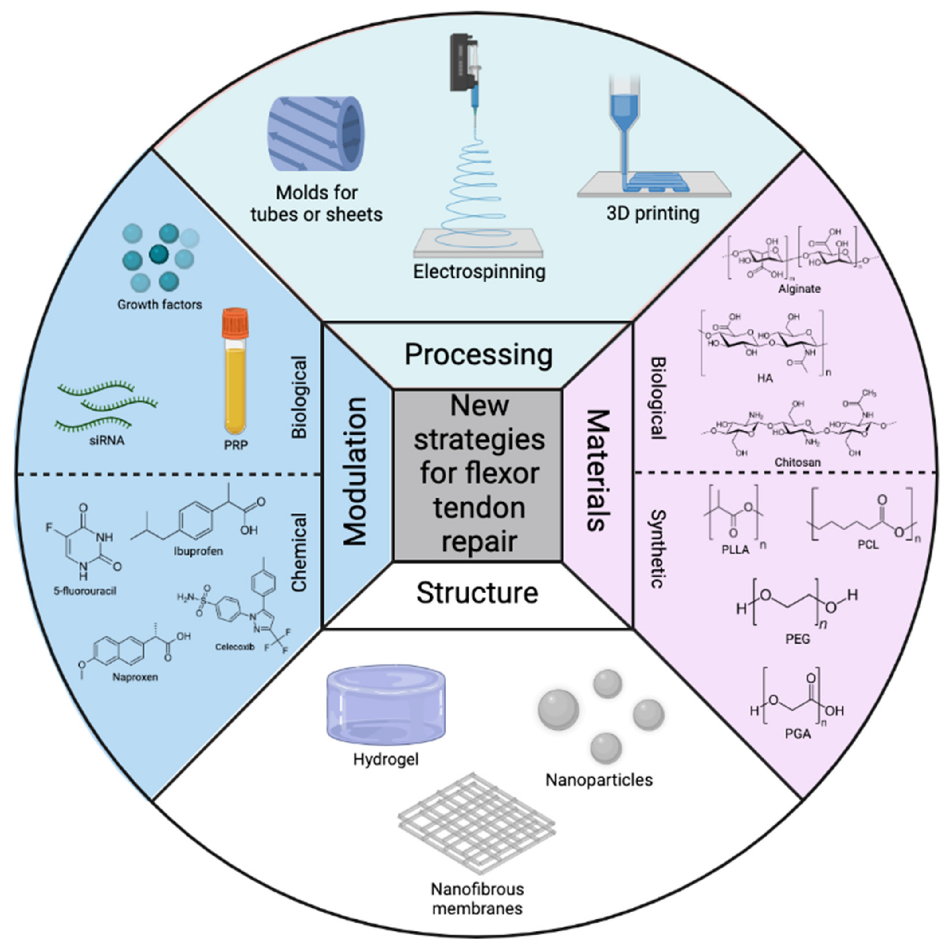
Figure 1. Overview of the most commonly used processing techniques, polymeric materials, structures, and modulations for flexor tendon repair here.

3. Conclusions and Perspectives

Flexor tendon injuries are common and mostly treated operationally. Hereby, several postoperative complications may occur such as infection, wear, tendon scar tissue formation, mechanical failure, and excessive adhesion formation. Multiple therapeutic reconstruction techniques, such as grafting or suturing, have been used in the past, but they don’t offer an adequate long-term solution. Current strategies for preventing the formation of peritendinous adhesion rely mainly on the inhibition of inflammation, using physical/mechanical barriers for averting wound contact with surrounding tissue, downregulating the ERK1/2 and SMAD2/3 phosphorylation, and inhibiting the excessive type I/III collagen and proliferation of fibroblasts. The use of polymer-based constructs can play a variety of important roles in preventing the formation of adhesion, not only by forming a physical barrier. Most current researchers focusing on flexor tendon repair develop a construct that is equipped with both anti-inflammatory as well as antimicrobial agents, solving the most severe postoperative complications occurring during tendon healing. The use of only synthetic or biological polymers is not sufficient for creating a multi-functional construct, therefore functional materials and combinations of several polymer types (semi-synthetic) have been engineered in combination with drugs, which are as a result highly effective in preventing these postoperative complications. Besides the material type, several techniques, structures, and even the incorporation of chemical and biological drugs have attracted more attention recently and were included. These new approaches in the development of new constructs are likely to result in an enhanced healing treatment for flexor tendon injuries. At present, a lot of these constructs already show great in vivo results in animal models, mostly mice or rabbits, although only limited studies have been performed on humans. The fast pace of development in the field will undoubtedly lead to the use of smart materials and multi-functional constructs, used in clinical practice. Although excellent results were obtained from past research, precise engineered constructs and the development of new drugs are not yet at their peak performance and the field of flexor tendon repair still requires new approaches and techniques.
Constructs can contain multiple drugs or biological compounds and fulfill combination treatment where the active payload release could be controlled in the future depending on the healing stage of the tendon. Hence, structures can heal the damaged or lacerated tendon at the first stage and later help in the maturation period of tendon healing. Active control of the drug release could also avoid commonly observed burst releases and avoid the accompanying possible side effects. Nano-based drug delivery has already proven to be successful in other medical fields to eradicate the problem of a burst release [61]. Future work may include the incorporation of nanoparticles into the nanofibrous constructs, i.e., electrospinning. The controlled active drug release can be accomplished in the future by engineering a smart polymeric construct that could have a trigger based on temperature, pH, electrical signals, magnetic field, and many more. Although such smart triggers have already been incorporated into other biomedical applications such as tumor immunotherapy [62], temperature-dependent drug and gene delivery [63], neural tissue engineering [64], skin wound healing [65], and many more, it is not yet developed for flexor tendon repair constructs.

This entry is adapted from the peer-reviewed paper 10.3390/polym14050867

References

  1. Kirkendall, D.T.; Garrett, W.E. Function and biomechanics of tendons. Scand. J. Med. Sci. Sport. 1997, 7, 62–66.
  2. Snedeker, J.G.; Foolen, J. Tendon injury and repair—A perspective on the basic mechanisms of tendon disease and future clinical therapy. Acta Biomater. 2017, 63, 18–36.
  3. Banik, B.L.; Lewis, G.S.; Brown, J.L. Multiscale Poly-(ϵ-caprolactone) Scaffold Mimicking Non-linearity in Tendon Tissue Mechanics. Regen. Eng. Transl. Med. 2016, 2, 1–9.
  4. Ghiya, M.N.; Murty, S.; Shetty, N.; D’Cunha, R. A descriptive study of hand injuries presenting to the adult emergency department of a tertiary care center in urban India. J. Emerg. Trauma. Shock 2017, 10, 19–25.
  5. Clark, D.P.; Scott, R.N.; Anderson, I.W. Hand problems in an accident and emergency department. J. Hand Surg. Br. 1985, 10, 297–299.
  6. Citeroni, M.R.; Ciardulli, M.C.; Russo, V.; Della Porta, G.; Mauro, A.; El Khatib, M.; Di Mattia, M.; Galesso, D.; Barbera, C.; Forsyth, N.R.; et al. Review in vitro innovation of tendon tissue engineering strategies. Int. J. Mol. Sci. 2020, 21, 1–78.
  7. Titan, A.L.; Foster, D.S.; Chang, J.; Longaker, M.T. Flexor Tendon: Development, Healing, Adhesion Formation, and Contributing Growth Factors. Plast. Reconstr. Surg. 2019, 144, 639e–647e.
  8. Legrand, A.; Kaufman, Y.; Long, C.; Fox, P.M. Molecular Biology of Flexor Tendon Healing in Relation to Reduction of Tendon Adhesions. J. Hand Surg. Am. 2017, 42, 722–726.
  9. Woo, S.L.Y.; Gelberman, R.H.; Cobb, N.G.; Amiel, D.; Lothringer, K.; Akeson, W.H. the importance of controlled passive mobilization on flexor tendon healing: A biomechanical study. Acta Orthop. 1981, 52, 615–622.
  10. Buschmann, J.; Meier Bürgisser, G. Autograft, allograft, and xenograft scaffolds for tendon and ligament repair. In Biomechanics of Tendons and Ligaments; Woodhead Publishing: Sawston, UK, 2017; ISBN 9780081004890.
  11. Rawson, S.; Cartmell, S.; Wong, J. Suture techniques for tendon repair; a comparative review. Muscles Ligaments Tendons J. 2013, 3, 220–228.
  12. Vasiliadis, A.V.; Katakalos, K. The role of scaffolds in tendon tissue engineering. J. Funct. Biomater. 2020, 11, 78.
  13. Sigal, I.R.; Grande, D.A.; Dines, D.M.; Dines, J.; Drakos, M. Biologic and Tissue Engineering Strategies for Tendon Repair. Regen. Eng. Transl. Med. 2016, 2, 107–125.
  14. Zhou, H.; Lu, H. Advances in the Development of Anti-Adhesive Biomaterials for Tendon Repair Treatment. Tissue Eng. Regen. Med. 2021, 18, 1–14.
  15. Beldjilali-Labro, M.; Garcia, A.G.; Farhat, F.; Bedoui, F.; Grosset, J.F.; Dufresne, M.; Legallais, C. Biomaterials in tendon and skeletal muscle tissue engineering: Current trends and challenges. Materials 2018, 11, 1116.
  16. Rademakers, T.; Horvath, J.M.; van Blitterswijk, C.A.; LaPointe, V.L.S. Oxygen and nutrient delivery in tissue engineering: Approaches to graft vascularization. J. Tissue Eng. Regen. Med. 2019, 13, 1815–1829.
  17. Cravedi, P.; Farouk, S.; Angeletti, A.; Edgar, L.; Tamburrini, R.; Duisit, J.; Perin, L.; Orlando, G. Regenerative immunology: The immunological reaction to biomaterials. Transpl. Int. 2017, 30, 1199–1208.
  18. No, Y.J.; Castilho, M.; Ramaswamy, Y.; Zreiqat, H. Role of Biomaterials and Controlled Architecture on Tendon/Ligament Repair and Regeneration. Adv. Mater. 2020, 32, 1904511.
  19. Chen, S.H.; Chen, C.H.; Fong, Y.T.; Chen, J.P. Prevention of peritendinous adhesions with electrospun chitosan-grafted polycaprolactone nanofibrous membranes. Acta Biomater. 2014, 10, 4971–4982.
  20. Pearce, O.; Brown, M.T.; Fraser, K.; Lancerotto, L. Flexor tendon injuries: Repair & Rehabilitation. Injury 2021, 52, 2053–2067.
  21. Beredjiklian, P.K. Biologic aspects of flexor tendon laceration and repair. J. Bone Jt. Surg. Ser. A 2003, 85, 539–550.
  22. McLaughlin, R.M. Complications After Treatment of Flexor Tendon Injuries. Small Anim. Surg. Secrets Second Ed. 2004, 14, 327–330.
  23. Kheiran, A.; Palial, V.; Rollett, R.; Wildin, C.J.; Chatterji, U.; Singh, H.P. Cat bite: An injury not to underestimate. J. Plast. Surg. Hand Surg. 2019, 53, 341–346.
  24. Malizos, K.N.; Papadopoulou, Z.K.; Ziogkou, A.N.; Rigopoulos, N.; Athanaselis, E.D.; Varitimidis, S.E.; Dailiana, Z.C. Infections of Deep Hand and Wrist Compartments. Microorganisms 2020, 8, 838.
  25. Pickrell, B.B.; Eberlin, K.R. Secondary Surgery Following Replantation and Revascularization. Hand Clin. 2019, 35, 231–240.
  26. Mamane, W.; Lippmann, S.; Israel, D.; Ramdhian-Wihlm, R.; Temam, M.; Mas, V.; Pierrart, J.; Masmejean, E.H. Infectious flexor hand tenosynovitis: State of knowledge. A study of 120 cases. J. Orthop. 2018, 15, 701–706.
  27. Stone, J.F.; Davidson, J.S. The role of antibiotics and timing of repair in flexor tendon injuries of the hand. Ann. Plast. Surg. 1998, 40, 7–13.
  28. Chapman, T.; Ilyas, A.M. Pyogenic Flexor Tenosynovitis: Evaluation and Treatment Strategies. J. Hand Microsurg. 2019, 11, 121–126.
  29. Chan, E.; Robertson, B.F.; Johnson, S.M. Kanavel signs of flexor sheath infection: A cautionary tale. Br. J. Gen. Pract. J. R. Coll. Gen. Pract. 2019, 69, 315–316.
  30. Trail, I.A.; Powell, E.S.; Noble, J. An evaluation of suture materials used in tendon surgery. J. Hand Surg. Br. 1989, 14, 422–427.
  31. Chen, C.H.; Chen, S.H.; Shalumon, K.T.; Chen, J.P. Dual functional core-sheath electrospun hyaluronic acid/polycaprolactone nanofibrous membranes embedded with silver nanoparticles for prevention of peritendinous adhesion. Acta Biomater. 2015, 26, 225–235.
  32. Shalumon, K.T.; Sheu, C.; Chen, C.H.; Chen, S.H.; Jose, G.; Kuo, C.Y.; Chen, J.P. Multi-functional electrospun antibacterial core-shell nanofibrous membranes for prolonged prevention of post-surgical tendon adhesion and inflammation. Acta Biomater. 2018, 72, 121–136.
  33. Bilal, M.; Rasheed, T.; Iqbal, H.M.N.; Li, C.; Hu, H.; Zhang, X. Development of silver nanoparticles loaded chitosan-alginate constructs with biomedical potentialities. Int. J. Biol. Macromol. 2017, 105, 393–400.
  34. Humayun, A.; Luo, Y.; Elumalai, A.; Mills, D.K. 3D printed antimicrobial PLA constructs functionalised with zinc- coated halloysite nanotubes-Ag-chitosan oligosaccharide lactate. Mater. Technol. 2022, 37, 28–35.
  35. Yang, G.; Rothrauff, B.B.; Tuan, R.S. Tendon and ligament regeneration and repair: Clinical relevance and developmental paradigm. Birth Defects Res. Part C Embryo Today Rev. 2013, 99, 203–222.
  36. Khan, F.; Tanaka, M. Designing smart biomaterials for tissue engineering. Int. J. Mol. Sci. 2018, 19, 17.
  37. Silva, M.; Ferreira, F.N.; Alves, N.M.; Paiva, M.C. Biodegradable polymer nanocomposites for ligament/tendon tissue engineering. J. Nanobiotechnol. 2020, 18, 23.
  38. Engineer, C.; Parikh, J.K.; Raval, A. Hydrolytic Degradation Behavior of Biodegradable Polymers from Controlled Drug Delivery System. Trends Biomater. Artif. Organs 2011, 25, 79–85.
  39. Vroman, I.; Tighzert, L. Biodegradable polymers. Materials 2009, 2, 307–344.
  40. Migliaresi, C.; Fambri, L.; Cohn, D. A study on the in vitro degradation of poly(lactic acid). J. Biomater. Sci. Polym. Ed. 1994, 5, 591–606.
  41. Duek, E.A.R.; Zavaglia, C.A.C.; Belangero, W.D. In vitro study of poly(lactic acid) pin degradation. Polymer 1999, 40, 6465–6473.
  42. Jenkins, M.J.; Harrison, K.L. The effect of crystalline morphology on the degradation of polycaprolactone in a solution of phosphate buffer and lipase. Polym. Adv. Technol. 2008, 19, 1901–1906.
  43. Guo, M.; Chu, Z.; Yao, J.; Feng, W.; Wang, Y.; Wang, L.; Fan, Y. The effects of tensile stress on degradation of biodegradable PLGA membranes: A quantitative study. Polym. Degrad. Stab. 2016, 124, 95–100.
  44. Fan, Y.B.; Li, P.; Zeng, L.; Huang, X.J. Effects of mechanical load on the degradation of poly(d,l-lactic acid) foam. Polym. Degrad. Stab. 2008, 93, 677–683.
  45. Cooper, J.A.; Lu, H.H.; Ko, F.K.; Freeman, J.W.; Laurencin, C.T. Fiber-based tissue-engineered scaffold for ligament replacement: Design considerations and in vitro evaluation. Biomaterials 2005, 26, 1523–1532.
  46. Goh, J.C.; Sahoo, S. Scaffolds for tendon and ligament tissue engineering. In Regenerative Medicine and Biomaterials for the Repair of Connective Tissues; Woodhead Publishing: Sawston, UK, 2010.
  47. Groth, T.; Falck, P.; Miethke, R.-R. Cytotoxicity of Biomaterials—Basic Mechanisms and In Vitro Test Methods: A Review. Altern. Lab. Anim. 1995, 23, 790–799.
  48. Cannella, V.; Altomare, R.; Leonardi, V.; Russotto, L.; Di Bella, S.; Mira, F.; Guercio, A. In Vitro Biocompatibility Evaluation of Nine Dermal Fillers on L929 Cell Line. Biomed Res. Int. 2020, 2020, 8676343.
  49. Yang, S.; Leong, K.F.; Du, Z.; Chua, K. The Design of Scaffolds for Use in Tissue Engineering. Part I. Traditional Factors. Tissue Eng. 2002, 7, 679–689.
  50. Song, A.; Rane, A.A.; Christman, K.L. Antibacterial and cell-adhesive polypeptide and poly(ethylene glycol) hydrogel as a potential scaffold for wound healing. Acta Biomater. 2012, 8, 41–50.
  51. Pring, D.J.; Amis, A.A.; Coombs, R.R.H. The mechanical properties of human flexor tendons in relation to artificial tendons. J. Hand Surg. Br. Eur. Vol. 1985, 10, 331–336.
  52. Verdan, C.; Potenza, A.D. Tendon Surgery of the Hand. Plast. Reconstr. Surg. 1980, 66, 1493–1503.
  53. Tang, J.B.; Gu, Y.T.; Rice, K.; Chen, F.; Pan, C.Z. Evaluation of four methods of flexor tendon repair for postoperative active mobilization. Plast. Reconstr. Surg. 2001, 107, 742–749.
  54. Chen, C.T.; Chen, C.H.; Sheu, C.; Chen, J.P. Ibuprofen-loaded hyaluronic acid nanofibrous membranes for prevention of postoperative tendon adhesion through reduction of inflammation. Int. J. Mol. Sci. 2019, 20, 5038.
  55. Pien, N.; Peeters, I.; Deconinck, L.; Van Damme, L.; De Wilde, L.; Martens, A.; Van Vlierberghe, S.; Dubruel, P.; Mignon, A. Design and development of a reinforced tubular electrospun construct for the repair of ruptures of deep flexor tendons. Mater. Sci. Eng. C 2021, 119, 111504.
  56. Mano, J.F.; Silva, G.A.; Azevedo, H.S.; Malafaya, P.B.; Sousa, R.A.; Silva, S.S.; Boesel, L.F.; Oliveira, J.M.; Santos, T.C.; Marques, A.P.; et al. Natural origin biodegradable systems in tissue engineering and regenerative medicine: Present status and some moving trends. J. R. Soc. Interface 2007, 4, 999–1030.
  57. Sensini, A.; Cristofolini, L. Biofabrication of electrospun scaffolds for the regeneration of tendons and ligaments. Materials 2018, 11, 1963.
  58. Narayanan, N.; Kuang, L.; Del Ponte, M.; Chain, C.; Deng, M. 1—Design and fabrication of nanocomposites for musculoskeletal tissue regeneration. In Nanocomposites for Musculoskeletal Tissue Regeneration; Liu, H., Ed.; Woodhead Publishing: Oxford, UK, 2016; pp. 3–29. ISBN 978-1-78242-452-9.
  59. Smith, B.D.; Grande, D.A. The current state of scaffolds for musculoskeletal regenerative applications. Nat. Rev. Rheumatol. 2015, 11, 213–222.
  60. Langer, R.; Vacanti, J.P. Tissue Engineering. Science 1993, 260, 920–926.
  61. Yeh, Y.-C.; Huang, T.-H.; Yang, S.-C.; Chen, C.-C.; Fang, J.-Y. Nano-Based Drug Delivery or Targeting to Eradicate Bacteria for Infection Mitigation: A Review of Recent Advances. Front. Chem. 2020, 8, 286.
  62. Yoshizaki, Y.; Yuba, E.; Komatsu, T.; Udaka, K.; Harada, A.; Kono, K. Improvement of Peptide-Based Tumor Immunotherapy Using pH-Sensitive Fusogenic Polymer-Modified Liposomes. Molecules 2016, 21, 1284.
  63. Gandhi, A.; Paul, A.; Sen, S.O.; Sen, K.K. Studies on thermoresponsive polymers: Phase behaviour, drug delivery and biomedical applications. Asian J. Pharm. Sci. 2015, 10, 99–107.
  64. Dong, M.; Shi, B.; Liu, D.; Liu, J.-H.; Zhao, D.; Yu, Z.-H.; Shen, X.-Q.; Gan, J.-M.; Shi, B.; Qiu, Y.; et al. Conductive Hydrogel for a Photothermal-Responsive Stretchable Artificial Nerve and Coalescing with a Damaged Peripheral Nerve. ACS Nano 2020, 14, 16565–16575.
  65. Zhang, K.; Lv, H.; Zheng, Y.; Yao, Y.; Li, X.; Yu, J.; Ding, B. Nanofibrous hydrogels embedded with phase-change materials: Temperature-responsive dressings for accelerating skin wound healing. Compos. Commun. 2021, 25, 100752.
More
This entry is offline, you can click here to edit this entry!