Molecular Techniques for Schistosomiasis: History
Please note this is an old version of this entry, which may differ significantly from the current revision.
Contributor:

Diagnostic tools play a pivotal role in warfare against schistosomiasis but must adapt to the endemic status and objectives of activities. With the decrease of prevalence and infection intensity of schistosomiasis in human beings and livestock, optimal methodologies with high sensitivity and absolute specificity are needed for the detection of asymptomatic cases or light infections, as well as disease surveillance to verify elimination. In comparison with the parasitological methods with relatively low sensitivity and serological techniques lacking specificity, which both had been widely used in previous control stages, the molecular detection methods based on the amplification of promising genes of the schistosome genome may pick up the baton to assist the eventual aim of elimination.

  • schistosomiasis japonica
  • elimination
  • diagnostic tools
  • molecular techniques

1. Traditional Diagnostic Tools Applied in Schistosomiasis Control in China

It cannot be exaggerated that diagnosis is the essential basis of schistosomiasis control for case identification and treatment, assessment of morbidity, and evaluation of control strategies, which are all dependent on the performance of diagnostic tests [27,39]. Two kinds of diagnostic methodologies, namely parasitological techniques mainly including KK and MHT [40,41], and immunologic approaches based on detection of specific antibodies, were widely used in the national control program in China, accelerating the process of schistosomiasis control significantly [11,42].

1.1. Parasitological Methods

The KK method, which was originally developed in the mid-1950s, and MHT, based on the positive phototactic behavior of miracidia, are the most broadly used techniques in epidemiological surveys pertaining to schistosomiasis in China [43]. At the stage of morbidity control, especially in the 1950–1980 period, the parasitological methods were the most applicable to the field, featured by high prevalence and high infection intensity. In 1989, with the first national survey of schistosomiasis japonica, direct stool examination of the KK method or MHT was still the recommended method to evaluate the prevalence of schistosomiasis [6,43]. Until now, the KK method and MHT have been the most accepted “gold” standard methods for identifying whether people or animals are infected [6,44]. Additionally, some studies indicated that multiple KK tests per sample, or increasing the collection frequency of stool samples, would increase the diagnostic sensitivity [45,46,47], and the MHT technique possessed higher sensitivity than the KK method due to the larger volume of stool tests received. Additionally, the combination of MHT and microscopic examination of filtered stool sediment would increase the detection rate [48].

1.2. Immunologic Tests

The immunological tests for schistosomiasis diagnosis also have a long history in China. The earliest immunologic test in China was the intradermal text (ID) recorded in 1936, and it was adapted for screening prior to further parasitological examination in the national general survey in the 1950s. Subsequently, a variety of immunologic techniques were applied to the national schistosomiasis control program, such as the circumoval precipitin test (COPT), indirect hemagglutination assay (IHA), the enzyme-linked immunosorbent assay (ELISA), and some rapid diagnostic tests (RDTs). Nowadays, ID and COPT have been out of use due to their low specificity, while IHA is still widely used in most endemic areas of China. Currently, there are two IHA kits with high quality control accredited by the China Food and Drug Administration. In the 10-year World Bank Loan Project (WBLP), aiming to control the morbidity of schistosomiasis, immunological tests were used directly to determine the target of chemotherapy in areas of medium endemicity (15% > prevalence > 3%) and low endemicity (prevalence < 3%) [43]. In the second national survey of schistosomiasis in 2004, in China, the ELISA method was adopted as a screening tool followed by stool examination to understand the real infection status of schistosomiasis in human beings. With the achievement of infection control reached in 2008 and transmission control reached in 2015, a diagnostic strategy with primary immunodiagnostic screening followed by KK or MHT for antibody-positive individuals was widely used in the Chinese national control program and routine surveillance activities in sentinel sites [27].

2. Molecular Methods Developed to Detect the Pathogen of Schistosomiasis

With the goal of the national program shifting from the control of schistosomiasis to elimination, extremely sensitive and specific diagnostic tools are needed emergently to explore asymptomatic cases with light infection and verify transmission interruption or elimination [34,49]. With the development of genomics and genome data for parasites and the urgent demands, molecular diagnostic techniques based on nucleic acid detection have emerged as new hot spots [50]. Various polymerase chain reaction (PCR)-based parasite DNA detection assays, including conventional polymerase chain reaction (cPCR), nested PCR (nPCR), real-time quantitative PCR (qPCR), and droplet digital PCR (ddPCR), have stimulated much interest as alternative options due to their proven diagnostic accuracy and the ability to detect early pre-patient infections [51,52]. The emergence of isothermal amplification methods, such as loop-mediated isothermal amplification (LAMP) and recombinase polymerase amplification (RPA), solves the dilemma of costly instrument dependence on PCR-based methods. The molecular diagnostic techniques of schistosomiasis japonica and the year first reported in China are shown in Figure 1. Regrettably, we have not seen any product based on molecular techniques applied in the field on a large scale.
Figure 1. The molecular diagnostic techniques of schistosomiasis japonica and the year of first report in China. 1 nPCR is characterized by two pairs of primers, inner primer and outer primer; 2 qPCR can quantitatively and qualitatively analyze the initial template of samples by detecting the fluorescence signal corresponding to each cyclic amplification product in real time; 3 The combination of RPA and lateral flow dipstick (LFD) for visual detection. Generally, visualization of control line and the test line is positive, and only the control line is negative; 4 cPCR determined positive or negative results by the size of the gel electrophoresis band; 5 The combination of LAMP reaction with chemical dyes for visual detection. + positive reaction, − negative reaction; 6 Dilute sample or samples DNA to the single molecule level and collect the fluorescence signal of a single reaction unit to achieve the absolute quantitative detection. Black spots: positive reaction units, blank spots: negative reaction units.

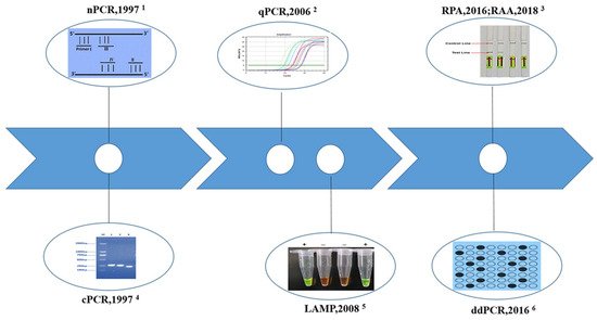
2.1. Conventional Polymerase Chain Reaction (cPCR)

cPCR emerged in the 1980s with a specific ability to amplify a small amount of target DNA and was the first nucleic acid amplification test used in schistosomiasis japonica diagnosis [53]. The character of amplification of microscale DNA of different samples greatly improves the analytical ability, simplifies the diagnostic process, and increases the sensitivity [14,16,51]. Another obvious advantage of this technology is that the amplified products can be visualized by gel electrophoresis and verified by sequencing. So far, many cPCR methods for the diagnosis of schistosomiasis japonica targeting various genes from chromosomes and mitochondria extracted from different samples have been established. One of the most important factors impacting the sensitivity of cPCR or other types of molecular detection assays is the abundance of the target sequences or biomarkers in the chromosome or mitochondrial genome [16,51]. The earliest reported PCR test for schistosomiasis diagnosis in China was based on the gene coding miracidium antigen named Sj5D [54,55,56]. Nowadays, the highly repetitive and conserved subunits of 18S rRNA [57,58,59,60], 28S rRNA [18,61,62,63], cytochrome c oxidase subunit 1 (COX I) [64,65], and the repetitive sequences SjR2 (an RTE-like, non-long terminal repeat retrotransposon from S. japonicum) [18,21,66,67,68,69] were the most common detection biomarkers. To evaluate the efficiency of established cPCR methods, several kinds of specimens, including genomic DNA (gDNA), pooled samples of snail and cercaria, and a mixture of eggs and feces, were employed. The lowest detection limit of cPCR established targeting a 607 base pair (bp) region of COX I can reach 10 fg of gDNA, which is less than that of 1 egg or 1 cercariae [64].
cPCR also provided a potential tool for the early detection and therapy evaluation of S. japonicum infection. The cPCR assay using a 230-bp sequence of SjR2 established by Xia could detect S. japonicum DNA in sera at the first week post-infection, and it became negative at 10 weeks post-treatment in a rabbit model infected by S. japonicum [21]. Similarly, schistosome DNA can be detected from one day post infection using pooled urine samples of mice by COX I-cPCR [65]. Moreover, the cPCR assay was always used as a reference method to assess the efficacy of other established methods, not only molecular methods but also serological methods [60,70,71]. The cPCR assay targeting 254-bp size of COX I gene was used in the detection of human samples in highly endemic areas of the Philippines, and the results showed that schistosome DNA in the serum and urine of KK-positive subjects could be detected by COX I -cPCR with 100% sensitivity [65]. However, few studies have reported on detecting field samples by the cPCR method, which may largely be due to its dependence on specific equipment and relatively complicated producers.

2.2. Nested PCR (nPCR)

The assay of nPCR, which can be considered a variant of cPCR, requires two rounds of PCR amplification using two sets of primers, commonly called the outer primers and the inner primers [17]. nPCR is more sensitive and specific than cPCR because the probability is extremely low if the first round amplification produces an erroneous fragment; primer pairing and amplification will occur in the second round amplification using the wrong fragment [51]. The repetitive sequences of SjR2 and SjCHGCS19 (a new 303-bp sequence from non-long terminal repeat (LTR) retrotransposon) are the common biomarkers in nPCR assays [73,74]. The published literature showed that the detection limit of SjR2-nPCR stabilized at fg level with 10fg of minimum limit [75]. Validated by the schistosome-infected mice model [76], rabbit model [74,77], and domestic animals (goat and buffalo) [78], the SjR2-nPCR or SjCHGCS19-nPCR could all be used for early diagnosis of schistosomiasis, even light infection, showing positive results at 3 days post-infection through testing sera samples. For samples from humans with chronic schistosomiasis, the detection rate of 230-bp SjR2-nPCR assay was 88.79% (95/107), significantly higher than that of the KK method (69.16%, 74/107) [79]. The sensitivity of the SjR2-nPCR method established by Zhang et al. using 14 and 28 days post-infection buffalo samples was 92.30% (36/39) and 100% (39/39), while the specificity was 97.60% (41/42) [78]. Moreover, the SjCHGCS19-nPCR assay demonstrated 97.67% sensitivity for 43 patient serum samples and 96.07% specificity for 51 serum samples from healthy individuals [74]. In addition, an nPCR assay targeting the 420-bp fragment of the Sjα1 gene, which is a short dispersal element retrotransposon gene with high copy and expression throughout the life cycle of schistosomes, could detect 0.1 fg of gDNA and distinguish the infection status of snail 4h post-infection [80]. However, the impressive performance of the detection efficacy of nPCR needed more verification.

2.3. Real-Time Quantitative PCR (qPCR)

The technique of the qPCR assay uses fluorescent labeled probes or double-stranded DNA-specific fluorescence dye to enable the continuous monitoring of amplicon (PCR product) formation throughout the reaction, thus allowing the quantification of PCR products by measuring fluorescence [81,82]. Compared with cPCR, qPCR has the following advantages: the amount of DNA in a sample can be measured using a standard curve, which can be determined by either spiking samples with a known amount of template or serial DNA dilutions; the qPCR procedures are streamlined with no need for an additional electrophoresis step to detect end-products of PCR, and the results can be preserved for a long time; and qPCR can utilize multiplex assays to detect multiple infections within a single clinical sample using specific probes and is preferred over cPCR in multiplex assays for having improved specificity by the use of probes [51,83].
The first reported qPCR assay for the detection of S. japonicum appeared in 2006, with mitochondrial NADH I as the target gene, and its detection limit could reach 1 egg per gram (EPG) fecal [84]. In the detection of 1,727 persons in field settings of Anhui Province, China, the prevalence (no. positive/no. examined) determined by NADH I-qPCR was 5.3%, significantly higher than those of the hatching test (3.2%) and Kato-Katz thick smear (3.0%) [85]. In a field evaluation of qPCR assay conducted in Hunan, Anhui, Hubei, and Jiangxi provinces of China, the qPCR assay exhibited a high level of sensitivity (100% for humans, 96.83% for bovines) and specificity (100%), and obtained a significantly higher prevalence in both the human (11.06% for qPCR, 0.93% for MHT) and bovine samples (24.73% for qPCR, 7.69% for MHT) [86]. The research conducted in the Philippines also showed similar trends, demonstrating that traditional copro-parasitological techniques underestimate the infection rate, signifying the advantages of qPCR for case finding and disease surveillance and monitoring [87,88,89,90,91]. Besides the gene of NADH I [85,86,87,88,89,90,91,92,93,94,95], several qPCR methods have been established targeting other genes, including COX I [96], 18S rRNA [97,98,99,100,101], ITS 2 [102], SjR2 [99,103,104], SjCHGCS20 [22], SjCHGC08270 [20,105], and Sjrh1.0 [99,106]. Those established qPCR methods have mostly completed the laboratory evaluation, but further validation should be conducted in field settings. Notably, the TaqMan qPCR assay targeting the COX I gene was developed to detect the environmental DNA (water samples) of S. japonicum, and its potential utility to schistosomiasis japonica surveillance in the Philippines was assessed. The results showed that the qPCR method could complement malacological surveys for monitoring schistosomes in endemic areas, especially those with a high risk of human infection [96].

2.4. Droplet Digital PCR (ddPCR)

The technique of ddPCR can still be considered a ‘new’ technology in parasitology, including schistosomiasis. Owing to its sensitivity and absolute quantitative characteristics, ddPCR is a potential candidate to become an appealing new method for parasite detection and quantitative analysis in the future [107]. Weerakoon et al. developed a ddPCR duplex assay targeting SjR2 and NADH I for the detection of S. japonicum, which provides improved detection sensitivity and specificity. The assay was able to detect as little as 0.05 fg of template DNA, and exhibited a high sensitivity for the detection of low levels of parasite DNA in stool, serum, urine, and saliva of mice model [108,109]. The ddPCR assay was also validated using clinical samples collected from 412 residents in a moderate-endemic area of schistosomiasis in the Philippines, proving its higher level of sensitivity obtained for human stool, serum, urine and saliva samples compared with the microscopy-based KK test [110,111]. Moreover, the capacity of ddPCR to quantify infection intensity has important public health implications for schistosomiasis control. Van Dorssen et al. determined the infection prevalence of S. japonicum in fecal samples of goats using the gene NADH I (46.4% ddPCR vs. 6.9% qPCR), showing that ddPCR was more sensitive than qPCR [93]. In general, the ddPCR technique with high sensitivity and specificity attracts increasing interest in its potential for clinical diagnosis and screening, and has the potential to be considered in schistosomiasis diagnosis as a complement to routine assays in schistosomiasis elimination programs.

2.5. Loop-Mediated Isothermal Amplification (LAMP)

The LAMP technique, which uses isothermal conditions to amplify DNA, is relatively simple, cost-effective, rapid, and more field-friendly compared with commonly used PCR-based methods [51,112]. Isothermal amplification does not require specific equipment, such as a thermocycler, electrophoresis apparatus, UV transilluminator, etc., while only a heating block or hot water bath is required for the reaction to progress [83,113,114]. The amplification results can be judged by precipitation turbidity of magnesium pyrophosphate or color reaction with the naked eye [115]. Hence, it is more suitable in resource-poor settings and grass-roots units. In addition, the four specific primers designed for six regions of target genes make the assay highly specific [113,114]. Of course, the LAMP technique has some shortcomings that need to be improved. It is complicated and time-consuming in the process of initial optimization with the use of multiple primers. Sometimes, the false-positive reaction is the fatal defect of the LAMP assay because of its high sensitivity [116,117]. Overall, the LAMP method is an extraordinary innovation trying to break through the restriction of equipment, and it has tremendous potential to apply in schistosomiasis control program for rapid screening, identification of transmission foci and environmental risk assessment.
A series of LAMP assays have been designed for schistosome-infected snail detection, schistosomiasis japonica diagnosis, and chemotherapy efficacy evaluation. The genes of CaBP (calcium-binding protein) [118,119], 28S rRNA [18,19,60], and SjR2 [120,121,122,123,124,125] were selected as the target sequence in the LAMP assay. Research has shown that the LAMP assay usually displays a higher detection rate than the conventional microscopy method for snail at different stages from 1 to 10 weeks post-infection [126]. 28S rRNA-LAMP was able to amplify the target band using DNA of a 1 day post-infection snail infected with one miracidium [18]. Nowadays, the LAMP assay has been applied to the surveillance of schistosoma infection of O. hupensis snail in national schistosomiasis sentinel sites in China [60,127]. In addition, a few studies have been conducted to evaluate the detection efficacy of LAMP for definitive hosts of S. japonicum. The SjR2-LAMP assay developed by Xu et al. was able to detect S. japonicum DNA in rabbit sera on the 3rd day post-infection. When LAMP was used to detect S. japonicum DNA in clinical serum samples (n = 152) from S. japonicum-infected patients and healthy persons, the sensitivity and specificity were 95.5% and 100%, respectively [71]. Moreover, for 47 patients after treatment 3 months, 6 months, and 9 months, the negative conversion rate of S. japonicum DNA in patient sera increased from 23.4% to 61.7% to 83.0%, respectively [123]. The above study demonstrated that the SjR2-LAMP method provides a useful and practical tool for the routine diagnosis and therapeutic evaluation of animals and human schistosomiasis.

2.6. Recombinase Polymerase Amplification (RPA)

Another isothermal amplification technology named RPA is a relatively new method that has experienced exponential growth in terms of publications, popularity, and applications since its first report in 2006 [128]. The central components of RPA mainly include DNA polymerase, DNA binding proteins, and recombinase. It is reported that RPA can operate at 37~42 °C and amplify as low as 1~10 copies of target DNA to detectable levels in less than 20 min. Therefore, the novel method is remarkable for its high sensitivity, simplicity, and extremely rapid amplification, as well as its operation at a low and constant temperature [129,130]. The RPA technique has been successfully integrated with different detection strategies, from end-point lateral flow strips to real-time fluorescent detection, among others, making this technique more user friendly, equipment-free, and facilitating the quantification of DNA [130]. In a meta-analysis of the diagnostic value of nucleic acid detection in schistosomiasis japonica, the isothermal amplification technique showed a relatively higher accuracy than the PCR-based amplification technique, and the sensitivity and specificity of the RPA method was higher than the LAMP assay [131]. However, RPA also has some disadvantages, such as the higher cost, carry-over contamination, and complicated optimization process [129,130]. Furthermore, due to the single source of RPA reagents or commercial kits, alternative products of the recombinase-aided isothermal amplification technique (RAA) have been developed in China [132,133,134,135,136,137].
The diagnostic method of RPA established for schistosomiasis japonica are concentrated after 2015, and the first retrievable literature was published in 2016 [23]. The visual detection method LFD-RPA (combination of RPA and lateral flow dipstick (LFD)) targeting SjR2 could detect 5 fg of S. japonicum DNA and showed no cross-reaction with other parasites. The reaction could be finished within 15~20 min at a wide temperature range (25–45 °C). Furthermore, the LFD-RPA assay performed 92.86% sensitivity (13/14), 100% of specificity (31/31) and excellent diagnostic agreement with the KK method (k = 0.947, Z = 6.36, p < 0.001), indicating that the LFD-RPA assay has a great potency in field application [35]. The real-time RPA (RT-RPA) targeting SjR2 gene performed 0.9 fg S. japonicum DNA detection limit, 100% sensitivity and specificity in detection of S. japonicum in stool samples from 30 infected patients and 30 healthy persons. The reaction could distinguish S. japonicum from other worms by measuring fluorescence using the TwistaTM incubator block [24]. Deng et al. tried to establish a detection method for S. japonicum using the SjR2 gene by RPA combined with electrochemical (EC) DNA biosensor. The RPA-EC combinational detection method also exhibited high sensitivity (0.01 fg detection limit), good specificity, and the ability to complete reaction within 30 min at 37 °C [138]. Afterwards, the RPA or LFD-RPA assay for different biomarkers of 28S rRNA and SjCHGCS19 was developed, which also proved that the technology was sensitive, specific, fast, and convenient [139,140].

This entry is adapted from the peer-reviewed paper 10.3390/pathogens11030287

This entry is offline, you can click here to edit this entry!
Video Production Service