Specific microRNAs Alter Autophagy and SCI Outcome: History
Please note this is an old version of this entry, which may differ significantly from the current revision.
Subjects: Neurosciences
Contributor:

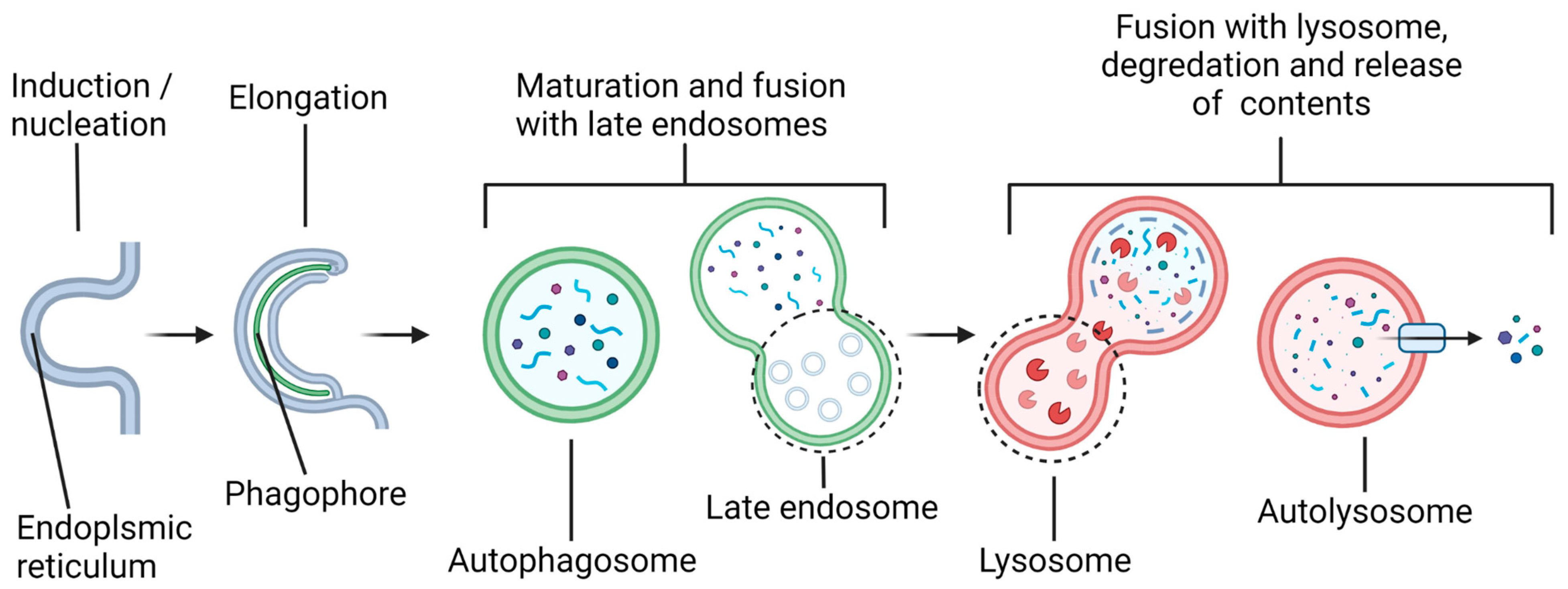
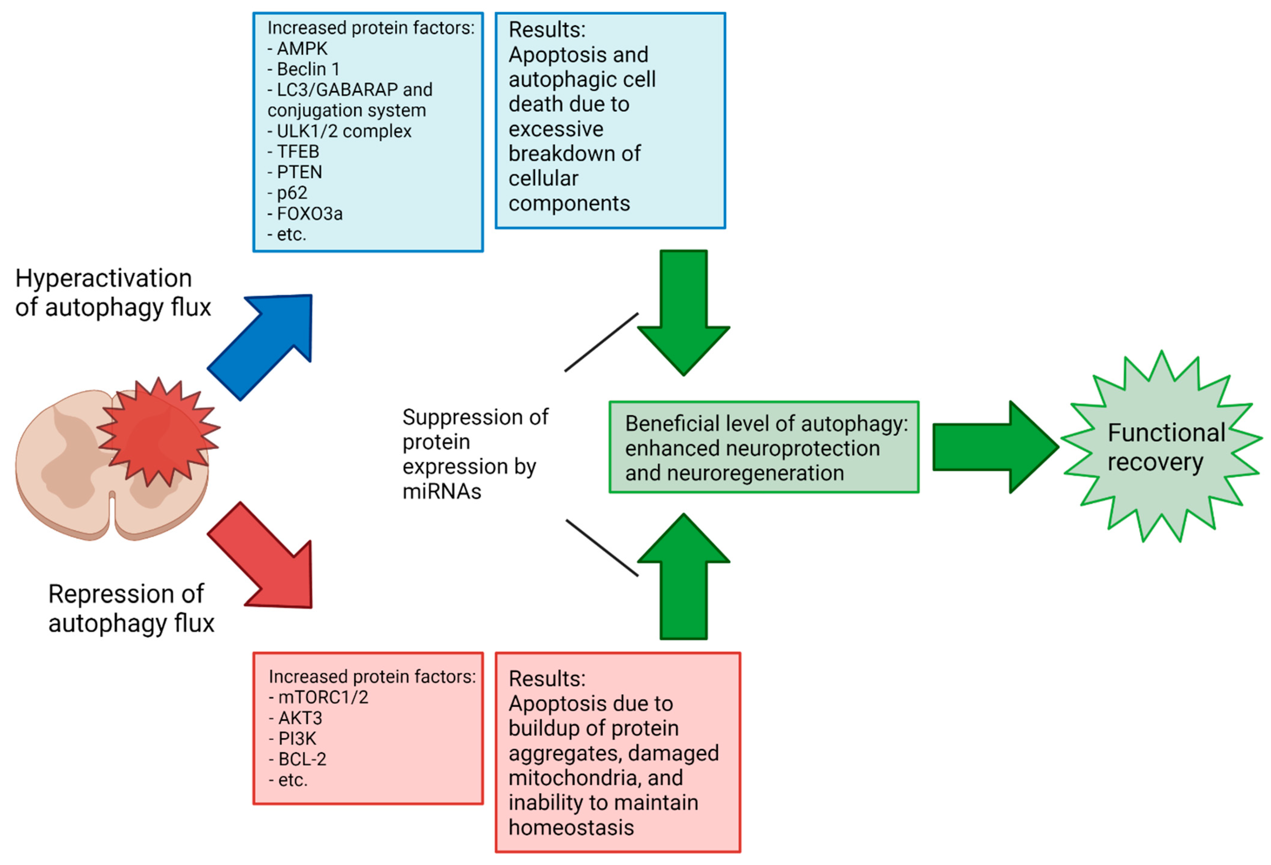
The treatment of spinal cord injury (SCI) is currently a major challenge, with a severe lack of effective therapies for yielding meaningful improvements in function. Therefore, there is a great opportunity for the development of novel treatment strategies for SCI. The modulation of autophagy, a process by which a cell degrades and recycles unnecessary or harmful components (protein aggregates, organelles, etc.) to maintain cellular homeostasis and respond to a changing microenvironment, is thought to have potential for treating many neurodegenerative conditions, including SCI. The discovery of microRNAs (miRNAs), which are short ribonucleotide transcripts for targeting of specific messenger RNAs (mRNAs) for silencing, shows prevention of the translation of mRNAs to the corresponding proteins affecting various cellular processes, including autophagy. 

  • spinal cord injury (SCI)
  • autophagy
  • neurodegeneration
  • miRNAs
  • miRNAs alter autophagy in SCI

1. Introduction

Alteration of the status of autophagy via microRNAs (miRNAs) may profoundly influence the course of pathogenesis and locomotor recovery in spinal cord injury (SCI). The increasing understanding of the biochemical mechanisms of autophagy and miRNAs and their interactions in preclinical models of SCI will open new therapeutic avenues for successful treatment of the patients who become the victims of this devastating neurological condition. Autophagy is a general term that can refer to several distinct cellular processes, including macroautophagy, chaperone-mediated autophagy (CMA), and microautophagy. Researchers focuses specifically on macroautophagy, which will hereafter be referred to as “autophagy”. Autophagy is utilized by a cell to break down and recycle its numerous substrates, either in “bulk autophagy”, targeting essentially random volumes of cytoplasmic contents, or in “selective autophagy”, targeting specific substrates, including protein aggregates, organelles such as mitochondria, parts of the nucleus, invading bacteria, proteasomes, peroxisomes, lysosomes, and others [1]. Over 40 proteins that make up the core autophagy machinery, known as autophagy-related (ATG) proteins, are highly conserved among eukaryotes [2][3] and have been most extensively studied in yeast, though analogs in several model organisms, as well as humans, have been identified [1]. Autophagy involves several steps (Figure 1), beginning with the formation of a double-membrane vesicle (called an autophagosome) from a nucleating membrane (called the phagophore or induction membrane) at sites on the endoplasmic reticulum known as omegasomes [4].
Figure 1. Overview of the autophagy pathway. Induction of autophagy begins with the formation of a cup-shaped domain of the endoplasmic reticulum called an omegasome. The nascent autophagosome, called a phagophore or induction membrane, is formed from the omegasome, and it grows until it has sealed around its cargo and produced a mature autophagosome. The autophagosome subsequently fuses with a late endosome, also known as multivesicular bodies (MVBs), ultimately merging with a lysosome or lysosomes. Acid hydrolases in the lysosome degrade the inner autophagosomal membrane and its contents into the cellular building blocks, which, afterwards, are released into the cytosol for recycling in the cell.

2. Specific miRNAs in Modulation of Autophagy in Preclinical Models of SCI

Autophagy at the basal levels is generally understood as promoting cell survival, but under certain conditions, changes in the autophagy flux or blockage of the autophagy pathway can promote cell death. Autophagy-dependent cell death in a strict sense appears to be a highly specific process primarily occurring during early development [5]. What occurs in many pathophysiological conditions like SCI is more accurately called autophagy-associated or autophagy-mediated cell death, wherein changes in the autophagy flux accompany and interact with the activation of other cell death pathways such as apoptosis. The crosstalk between autophagy and apoptosis is complex and an area of active research. There are several enlightening review papers already published on the subject [6][7][8]; however, some of the different ways autophagy and cell death are related will be briefly covered here for some context.
A buildup of autophagosomal bodies in cells has been shown to precede apoptosis in neurons and other cells [6][7][9]. Such a buildup may be the result of an increased production of autophagosomes and/or inhibition of autophagosome clearance. There is evidence that autophagosomal membranes act as scaffolds for the assembly of pro-apoptotic and pro-necrotic protein complexes [6]. An increased number of autophagosomes may therefore induce cell death by promoting the formation of these complexes. Defective autophagy can lead to the accumulation of protein aggregates as well as of damaged mitochondria, which subsequently can cause mitochondrial release of cytochrome c into the cytosol to initiate apoptosis [10]. Several ATG proteins are known to become pro-apoptotic factors when cleaved by calpain (ATG5) or caspases (ATG4 and Beclin 1) [6][7]. Selective autophagy may target either pro-death or pro-survival factors to influence the fate of a cell [11][12]. The exact nature of the interactions among the autophagy, apoptosis, and necrosis that determine cell death or survival are still not well understood.
Apoptotic activity following SCI varies significantly depending on the type and severity of the injury, as well as over the phases of SCI recovery [13][14]. However, a consistent difficulty in assessing the overall efficacy of autophagy activation/inhibition at a given phase in SCI recovery results from a lack of consensus in the literature on the results of autophagy activation or inhibition in a given injury context; a recent meta-analysis demonstrated that SCI recovery is improved by the modulation of autophagy but shows no significant difference in the Basso, Beattie, and Bresnahan (BBB) locomotor scores between upregulation and downregulation in mouse injury models [15]. In the previously referenced meta-analysis, the 33 included studies generally achieved a modulation of autophagy in SCI by chemical means (e.g., rapamycin and metformin) rather than via miRNAs. In comparison, the number of studies examining the modulation of autophagy in SCI by miRNAs specifically is small, and to the authors’ knowledge, no comparable meta-analysis exists. Notably, there is a lack of studies in the literature examining the miRNA-mediated upregulation of autophagy in SCI (miR-15a is the sole miRNA with sufficient previous research to be included in this entry that caused an increase in autophagy flux). As previously noted, the complexity and sensitivity of autophagy regulation pathways and the double-edged nature of autophagy’s relation to cell death suggest that this inconsistency in results may be due to small differences in the experimental procedure, and further research is needed to elucidate the points at which the neuroprotective or neurodegenerative properties of autophagy dominate (Figure 2 The effort toward full elucidation of the autophagy’s role in SCI across various injury types and phases might benefit from more standardized research methodologies regarding injury type and how long after the primary injury the intervention occurs. Due to the highly dynamic characteristics of SCI, comparisons of the results that arise from experimental conditions that are not highly similar are of limited value. Increased collaboration between research groups in the field to develop standardized methodologies to allow more meaningful comparisons of results could help resolve some of the seemingly conflicting conclusions so far obtained.
Figure 2. Potential role of miRNAs in promoting functional recovery in SCI. Autophagy may be hyperactivated or repressed to non-optimal levels at a given phase of the SCI secondary injury, leading to poorer recovery or even itself causing deleterious effects. An understanding of the effects that autophagy has on different SCI types and at different phases of secondary injury will be vital to developing effective therapies.
With that in mind, the following sections will explore the relationship between autophagy and SCI, with a particular focus on specific miRNAs that may modulate these interactions for neuroprotection in preclinical models of the SCI (Table 1). Most of the investigations in this field are currently focused on the fine tuning of interactions between specific miRNAs and the molecular components of autophagy, as researchers described above, to regulate the autophagy flux for functional neuroprotection in preclinical models of SCI with a hope to apply this approach to the SCI patients as soon as possible.

2.1. Autophagy in Neurons

Neuronal autophagy is critical for homeostasis in the central nervous system (CNS), as neurons are post-mitotic and therefore limited in their ability to deal with cellular waste. Additionally, their unique morphology creates the requirement for specialized autophagy processes not found in other cell types. Autophagosome formation is constitutively activated not just in the soma but occurs as far as the axon terminal, allowing for a rapid autophagy response along the axon [16]. Degradation of the autolysosomal contents takes place primarily in the soma, necessitating that the kinesin/dynein-mediated anterograde movement of lysosomes and retrograde movements of autophagosomes and autolysosomes along the cytoskeleton be tightly regulated due to the long distances involved [17][18]. The movement of autophagosomes through the axon appears to be strictly unidirectional; somatic autophagosomes are barred from entering the axon [19]. Finally, the LC3-II levels in neurons are relatively low in basal conditions; whether this indicates an innately lower rate of autophagosome formation or a more rapid flux rate in neurons is currently unclear [20].
Table 1. Specific miRNAs in modulating autophagy with prospect of influencing SCI recovery.
microRNA Molecular Target(s) Effect on Autophagy Flux Prospect of SCI Recovery References
miR-93-5p PTEN Decrease Beneficial [21][22][23]
ATG7
TLR4
miR-384-5p Beclin 1 Decrease Beneficial [24][25]
GRP78
miR-378 ATG12 Tissue dependent Beneficial [26][27][28][29]
GRB2
miR-27a FOXO3a Decrease Beneficial [30][31][32][33]
DRAM2
PINK1
miR-223 RPH1/KDM4A Decrease Beneficial [34][35]
miR-124 PI3K Decrease Beneficial [36][37][38]
AMPK
Bcl-2
p62
miR-212-3p PTEN Decrease Beneficial [39][40][41][42]
miR-15a Akt3 Increase Beneficial (in neuropathic pain model) [43][44]
Rictor
miR-384-5p Beclin 1 Decrease Beneficial [45]
miR-223 FOXO3a
ATG16L
Decrease Beneficial [46][47][48][49][50]
miR-30 Beclin 1 Tissue dependent Context dependent [51][52][53][54][55]
miR-30d Beclin 1 Increase or decrease Beneficial [56][57][58]
In rat retinal ganglion neurons, miR-93-5p was found to reduce autophagy-associated cell death after N-methyl-D-aspartate (NMDA)-induced excitotoxicity by downregulating the expression of phosphatase and tensin homolog (PTEN), which negatively regulates autophagy through the Akt/mTOR pathway [21]. ATG7 and Toll-like receptor 4 (TLR4, an inducer of neuronal autophagy and neuroinflammation [22]) have also been demonstrated to be targets of miR-93-5p, as the overexpression of miR-93-5p results in significantly reduced levels of those proteins, with an accompanying reduction in autophagy and inflammatory factors and increase in cell survival in rat myocardial tissue [23].
miR-384-5p is one of several miRNAs that target the expression of Beclin 1 (an inducer of autophagy), which contributes to functionality of the PI3K complex [24]. The suppression of Beclin 1 therefore halts autophagy at the point of PtdIns(3)P enrichment and prevents phagophore elongation. Specifically, the application of miR-384-5p was shown to significantly improve the BBB locomotor scores in rats with spinal cord compression injuries after 7 days and 21 days as compared to untreated rats [25]. The same study also reported that inhibition of ER stress via decreasing levels of glucose-regulating protein 78 (GRP78), an ER stress response-mediating protein that is also targeted by miR-384-5p, may have contributed to the lower level of autophagy seen in the study [25].
It has been demonstrated that miR-378 regulates the expression of several ATG proteins and appears to have a promising prospect for the modulation of autophagy in neurons. A report indicated that miR-378 negatively regulated ATG12 expression in rat neurons and reduced apoptosis when administered immediately after contusion SCI, with a corresponding improvement of the BBB locomotor scores 7 days post-injury compared to the sham group, though the study did not identify whether the improvement was necessarily due to autophagy impairment [26]. Studies on the long noncoding RNA (lncRNA)-based inhibition of miR-378 revealed that it acts to inhibit autophagy by targeting growth factor receptor-bound protein 2 (GRB2) and ATG12 [27][28]. Alternatively, one study has found that miR-378 activates autophagy in skeletal muscle, indicating that further research into how miR-378 functions in different cell types and what conditions are necessary [29].
Multiple studies have implicated miR-27a in modulating neurodegenerative processes through the modulation of autophagy. It has been reported that the overexpression of miR-27a downregulates autophagy in neurons and inhibits neurodegeneration by targeting FOXO3a (Forkhead bOX O3a, a transcription factor that promotes autophagy [30]) in mouse neurons after a traumatic brain injury [31]. Another study found that miR-27a also targets damage-regulated autophagy modulator 2 (DRAM2) and that the rno-circRNA_010705 (circLrp1b)-mediated repression of miR-27a can increase autophagy-associated neurodegeneration [32]. Additionally, miR-27a has been shown to downregulate mitophagy in HeLa cells by targeting PTEN-induced kinase 1 (PINK1), which is a mitochondrial serine/threonine kinase, to suggest that it may protect cells from excessive mitochondria loss in hyper-autophagic conditions [33].
RPH1 (Repressor of PHR1)/KDM4A (lysine- or K-specific DeMethylase 4A) is a DNA-binding protein that acts as a histone demethylase and negatively regulates the transcription of several ATG genes. It has been reported that miR-223 targets RPH1/KDM4A, subsequently reducing the autophagy levels in lipopolysaccharide-treated neuronal PC-12 cells and attenuating cell death [34]. Another study reported that, in yeast, RPH1/KDM4A acted as an autophagy inhibitor under nutrient-rich conditions and did not show any effect on translation in nitrogen-starved conditions [35]. Clearly, more research is necessary to clarify the functions of RPH1/KDM4A, specifically in mammalian cells, to rectify this difference.
The expression of miR-124, a key miRNA in neural development, has been linked to regulation of the autophagy pathway via its regulation of the Akt/AMPK/mTOR axis [36]. However, as in many other cases involving autophagy, there is conflicting evidence as to whether its overexpression or under-expression bestows neuroprotective and/or neuroregenerative effects in the spinal cord specifically. A study found that antagomiR-124 had a neuroprotective effect when introduced 5 days before spinal cord ischemia–reperfusion injury in rats, with a reduction in apoptosis and increase in the levels of mitochondrial LC3-II and Beclin 1, causing an increase in neuronal mitophagy [37]. Conversely, another report showed that antagomiR-124 administered 24 h before cerebral ischemia–reperfusion injury produces a neuroprotective effect by promoting the PI3K/Akt/mTOR pathway, with rats exhibiting an increase in the PI3K/Akt/mTOR levels and improvement in neurological function, in part, due to downregulation of autophagy by the PI3K/Akt/mTOR pathway [38]. To reconcile these conclusions, it should be noted that these studies involved neural tissue in different locations and quantified different proteins (PI3K/Akt/mTOR function upstream of LC3-II and Beclin 1 in the autophagy pathway) in different cell fractions (the whole cell vs. mitochondria). The conflicting results from these studies underline the need for more thorough research into the function of this specific miR-124 as it relates to pathophysiology of SCI before any potential therapy utilizing it is developed.
In a recent study, miR-212-3p has been shown to have a positive effect on recovery from SCI in rats [39]. PTEN is a possible target of miR-212-3p, and it is demonstrated that the silencing of PTEN may be the reason behind the improvement in the recovery in SCI rats treated with miR-212-3p. The molecular mechanism involved in this recovery process seems so far convincing. The silencing of PTEN leads to the activation of Akt and mTOR, suggesting that the beneficial effects of miR-212-3p may be due to the inhibition of autophagy. Indeed, other studies have linked miR-212-3p to autophagy regulation in cardiomyocytes, osteosarcoma cells, and prostate cancer cells [40][41][42]. However, it has yet to be conclusively shown that the neuroprotective effects of miR-212-3p in SCI are due to autophagy inhibition.

2.2. Autophagy in Glial Cells

A study shows that miR-15a positively regulates autophagy in rat microglial cells by reducing the Akt3 levels and increasing the expression of ATG proteins [43]. In addition, another study showed that miR-15a and miR-16 promote autophagy in HeLa cells by targeting the Rictor subunit of mTORC2, an upstream activator of Akt3 [44].
In addition to the previously mentioned role of miR-384-5p beneficially downregulating autophagy in neurons, there is evidence that it has anti-inflammatory effects in the brain through the inhibition of autophagy in macrophages [45]. The pro-inflammatory effects of autophagy in this case may be due to autophagy leading to an increase in the macrophage cell viability, which is negated by suppression of autophagy promoter Beclin 1 by overexpression of miR-384-5p that binds to 3’ UTR of Beclin 1 mRNA [45]. If so, this would highlight one of the ways that the modulation of autophagy in each direction might have significantly different effects (e.g., cell survival vs. cell death) in different cell types, including those that might be closely associated in the context of a particular injury.
As previously mentioned for its role in neural autophagy, miR-223 has also been implicated in reducing neuroinflammation in microglia by targeting ATG16L [46][47]. Interestingly, it has been found that FOXO3a is yet another autophagy-related target of miR-223, with a negative correlation between the miR-223 levels and FOXO3a expression reported in several cell types [48][49][50].
Various studies have clearly indicated that Beclin 1 in the CNS tissues is a potential target of several members of the miR-30 family [51][52][53][54][55]. Of these, the effects of miR-30d are the best characterized in relation to SCI. AntagomiRs of miR-30d promoted autophagy and reduced apoptosis in post-ischemia astrocytes [56] and neonatal rat neurons [57], while another study demonstrated that miR-30d decreased neuroinflammation by reducing autophagy in macrophages and subsequently promoting M2 macrophage polarization over M1 [58]. Additionally, the sponging of miR-30b and miR-30d by lncRNAs SNHG12 (small nucleolar RNA host gene 12) and C2dat2 (CAMK2D-associated transcript 2), respectively, was shown to increase autophagy and apoptosis after cerebral ischemia–reperfusion injury [52][59].

This entry is adapted from the peer-reviewed paper 10.3390/brainsci12020247

References

  1. Galluzzi, L.; Baehrecke, E.H.; Ballabio, A.; Boya, P.; Bravo-San Pedro, J.M.; Cecconi, F.; Choi, A.M.; Chu, C.T.; Codogno, P.; Colombo, M.I.; et al. Molecular definitions of autophagy and related processes. EMBO J. 2017, 36, 1811–1836.
  2. Reggiori, F.; Klionsky, D.J. Autophagy in the eukaryotic cell. Eukaryot. Cell 2002, 1, 11–21.
  3. Klionsky, D.J.; Cregg, J.M.; Dunn, W.A., Jr.; Emr, S.D.; Sakai, Y.; Sandoval, I.V.; Sibirny, A.; Subramani, S.; Thumm, M.; Veenhuis, M.; et al. A unified nomenclature for yeast autophagy-related genes. Dev. Cell 2003, 5, 539–545.
  4. Melia, T.J.; Lystad, A.H.; Simonsen, A. Autophagosome biogenesis: From membrane growth to closure. J. Cell Biol. 2020, 219, e202002085.
  5. Denton, D.; Kumar, S. Autophagy-dependent cell death. Cell Death Differ. 2019, 26, 605–616.
  6. Doherty, J.; Baehrecke, E.H. Life, death and autophagy. Nat. Cell Biol. 2018, 20, 1110–1117.
  7. Saleem, S. Apoptosis, autophagy, necrosis and their multi galore crosstalk in neurodegeneration. Neuroscience 2021, 469, 162–174.
  8. Chen, Q.; Kang, J.; Fu, C. The independence of and associations among apoptosis, autophagy, and necrosis. Signal Transduct. Target. Ther. 2018, 3, 18.
  9. Zhang, H.; Dong, X.; Zhao, R.; Zhang, R.; Xu, C.; Wang, X.; Liu, C.; Hu, X.; Huang, S.; Chen, L. Cadmium results in accumulation of autophagosomes-dependent apoptosis through activating Akt-impaired autophagic flux in neuronal cells. Cell Signal. 2019, 55, 26–39.
  10. Ghavami, S.; Shojaei, S.; Yeganeh, B.; Ande, S.R.; Jangamreddy, J.R.; Mehrpour, M.; Christoffersson, J.; Chaabane, W.; Moghadam, A.R.; Kashani, H.H.; et al. Autophagy and apoptosis dysfunction in neurodegenerative disorders. Prog. Neurobiol. 2014, 112, 24–49.
  11. Miller, D.R.; Cramer, S.D.; Thorburn, A. The interplay of autophagy and non-apoptotic cell death pathways. Int. Rev. Cell Mol. Biol. 2020, 352, 159–187.
  12. Ray, S.K. Modulation of autophagy for neuroprotection and functional recovery in traumatic spinal cord injury. Neural Regen. Res. 2020, 15, 1601–1612.
  13. Zhou, K.; Sansur, C.A.; Xu, H.; Jia, X. The temporal pattern, flux, and function of autophagy in spinal cord injury. Int. J. Mol. Sci. 2017, 18, 466.
  14. Fang, B.; Li, X.-Q.; Bao, N.-R.; Tan, W.-F.; Chen, F.-S.; Pi, X.-L.; Zhang, Y.; Ma, H. Role of autophagy in the bimodal stage after spinal cord ischemia reperfusion injury in rats. Neuroscience 2016, 328, 107–116.
  15. Zhang, D.; Zhu, D.; Wang, F.; Zhu, J.-C.; Zhai, X.; Yuan, Y.; Li, C.-X. Therapeutic effect of regulating autophagy in spinal cord injury: A network meta-analysis of direct and indirect comparisons. Neural Regen. Res. 2020, 15, 1120–1132.
  16. Valencia, M.; Kim, S.R.; Jang, Y.; Lee, S.H. Neuronal autophagy: Characteristic features and roles in neuronal pathophysiology. Biomol. Ther. 2021, 29, 605–614.
  17. Roney, J.C.; Li, S.; Farfel-Becker, T.; Huang, N.; Sun, T.; Xie, Y.; Cheng, X.-T.; Lin, M.-Y.; Platt, F.M.; Sheng, Z.-H. Lipid-mediated motor-adaptor sequestration impairs axonal lysosome delivery leading to autophagic stress and dystrophy in Niemann-Pick type, C. Dev. Cell 2021, 56, 1452–1468.e8.
  18. Kuijpers, M.; Azarnia Tehran, D.; Haucke, V.; Soykan, T. The axonal endolysosomal and autophagic systems. J. Neurochem. 2021, 158, 589–602.
  19. Maday, S.; Holzbaur, E.L.F. Compartment-specific regulation of autophagy in primary neurons. J. Neurosci. 2016, 36, 5933–5945.
  20. Ariosa, A.R.; Klionsky, D.J. Autophagy core machinery: Overcoming spatial barriers in neurons. J. Mol. Med. 2016, 94, 1217–1227.
  21. Li, R.; Jin, Y.; Li, Q.; Sun, X.; Zhu, H.; Cui, H. MiR-93-5p targeting PTEN regulates the NMDA-induced autophagy of retinal ganglion cells via AKT/mTOR pathway in glaucoma. Biomed. Pharmacother. 2018, 100, 1–7.
  22. Jiang, H.; Wang, Y.; Liang, X.; Xing, X.; Xu, X.; Zhou, C. Toll-like receptor 4 knockdown attenuates brain damage and neuroinflammation after traumatic brain injury via inhibiting neuronal autophagy and astrocyte activation. Cell Mol. Neurobiol. 2018, 38, 1009–1019.
  23. Liu, J.; Jiang, M.; Deng, S.; Lu, J.; Huang, H.; Zhang, Y.; Gong, P.; Shen, X.; Ruan, H.; Jin, M.; et al. miR-93-5p-Containing exosomes treatment attenuates acute myocardial infarction-induced myocardial damage. Mol. Ther. Nucleic Acids 2018, 11, 103–115.
  24. Wang, B.; Zhong, Y.; Huang, D.; Li, J. Macrophage autophagy regulated by miR-384-5p-mediated control of Beclin-1 plays a role in the development of atherosclerosis. Am. J. Transl. Res. 2016, 8, 606–614.
  25. Zhou, Z.; Hu, B.; Lyu, Q.; Xie, T.; Wang, J.; Cai, Q. miR-384-5p promotes spinal cord injury recovery in rats through suppressing of autophagy and endoplasmic reticulum stress. Neurosci. Lett. 2020, 727, 134937.
  26. Zhang, H.; Yu, H.; Yang, H.; Zhan, Y.; Liu, X. miR-378-3p alleviates contusion spinal cord injury by negatively regulating ATG12. Int. J. Exp. Pathol. 2021, 102, 200–208.
  27. Zhao, J.; Chen, F.; Ma, W.; Zhang, P. Suppression of long noncoding RNA NEAT1 attenuates hypoxia-induced cardiomyocytes injury by targeting miR-378a-3p. Gene 2020, 731, 144324.
  28. Luo, H.-C.; Yi, T.-Z.; Huang, F.-G.; Wei, Y.; Luo, X.-P.; Luo, Q.-S. Role of long noncoding RNA MEG3/miR-378/GRB2 axis in neuronal autophagy and neurological functional impairment in ischemic stroke. J. Biol. Chem. 2020, 295, 14125–14139.
  29. Li, Y.; Jiang, J.; Liu, W.; Wang, H.; Zhao, L.; Liu, S.; Li, P.; Zhang, S.; Sun, C.; Wu, Y.; et al. microRNA-378 promotes autophagy and inhibits apoptosis in skeletal muscle. Proc. Natl. Acad. Sci. USA 2018, 115, E10849–E10858.
  30. Zhou, J.; Liao, W.; Yang, J.; Ma, K.; Li, X.; Wang, Y.; Wang, D.; Wang, L.; Zhang, Y.; Yin, Y.; et al. FOXO3 induces FOXO1-dependent autophagy by activating the AKT1 signaling pathway. Autophagy 2012, 8, 1712–1723.
  31. Sun, L.; Zhao, M.; Wang, Y.; Liu, A.; Lv, M.; Li, Y.; Yang, X.; Wu, Z. Neuroprotective effects of miR-27a against traumatic brain injury via suppressing FoxO3a-mediated neuronal autophagy. Biochem. Biophys. Res. Commun. 2017, 482, 1141–1147.
  32. Li, H.; Lu, C.; Yao, W.; Xu, L.; Zhou, J.; Zheng, B. Dexmedetomidine inhibits inflammatory response and autophagy through the circLrp1b/miR-27a-3p/Dram2 pathway in a rat model of traumatic brain injury. Aging 2020, 12, 21687–21705.
  33. Kim, J.; Fiesel, F.C.; Belmonte, K.C.; Hudec, R.; Wang, W.-X.; Kim, C.; Nelson, P.T.; Springer, W.; Kim, J. miR-27a and miR-27b regulate autophagic clearance of damaged mitochondria by targeting PTEN-induced putative kinase 1 (PINK1). Mol. Neurodegener. 2016, 11, 55.
  34. Jia, D.; Niu, Y.; Li, D.; Zhang, Q. microRNA-223 alleviates lipopolysaccharide-induced PC-12 cells apoptosis and autophagy by targeting RPH1 in spinal cord injury. Int. J. Clin. Exp. Pathol. 2017, 10, 9223–9232.
  35. Bernard, A.; Jin, M.; González-Rodríguez, P.; Füllgrabe, J.; Delorme-Axford, E.; Backues, S.K.; Joseph, B.; Klionsky, D.J. Rph1/KDM4 mediates nutrient-limitation signaling that leads to the transcriptional induction of autophagy. Curr. Biol. 2015, 25, 546–555.
  36. Liu, X.; Feng, Z.; Du, L.; Huang, Y.; Ge, J.; Deng, Y.; Mei, Z. The potential role of microRNA-124 in cerebral ischemia injury. Int. J. Mol. Sci. 2019, 21, 120.
  37. Liu, K.; Yan, L.; Jiang, X.; Yu, Y.; Liu, H.; Gu, T.; Shi, E. Acquired inhibition of microRNA-124 protects against spinal cord ischemia-reperfusion injury partially through a mitophagy-dependent pathway. J. Thorac. Cardiovasc. Surg. 2017, 154, 1498–1508.
  38. Miao, W.; Yan, Y.; Bao, T.-H.; Jia, W.-J.; Yang, F.; Wang, Y.; Zhu, Y.; Yin, M.; Han, J. Ischemic postconditioning exerts neuroprotective effect through negatively regulating PI3K/Akt2 signaling pathway by microRNA-124. Biomed. Pharmacother. 2020, 126, 109786.
  39. Guan, C.; Luan, L.; Li, J.; Yang, L. miR-212-3p improves rat functional recovery and inhibits neurocyte apoptosis in spinal cord injury models via PTEN downregulation-mediated activation of AKT/mTOR pathway. Brain Res. 2021, 1768, 147576.
  40. Ucar, A.; Gupta, S.K.; Fiedler, J.; Erikci, E.; Kardasinski, M.; Batkai, S.; Dangwal, S.; Kumarswamy, R.; Bang, C.; Holzmann, A.; et al. The miRNA-212/132 family regulates both cardiac hypertrophy and cardiomyocyte autophagy. Nat. Commun. 2012, 3, 1078.
  41. Oh, J.Y.; Kim, E.H.; Lee, Y.-J.; Sai, S.; Lim, S.H.; Park, J.W.; Chung, H.K.; Kim, J.; Vares, G.; Takahashi, A.; et al. Synergistic autophagy effect of miR-212-3p in zoledronic acid-treated in vitro and orthotopic in vivo models and in patient-derived osteosarcoma cells. Cancers 2019, 11, 1812.
  42. Ramalinga, M.; Roy, A.; Srivastava, A.; Bhattarai, A.; Harish, V.; Suy, S.; Collins, S.; Kumar, D. microRNA-212 negatively regulates starvation induced autophagy in prostate cancer cells by inhibiting SIRT1 and is a modulator of angiogenesis and cellular senescence. Oncotarget 2015, 6, 34446–34457.
  43. Cai, L.; Liu, X.; Guo, Q.; Huang, Q.; Zhang, Q.; Cao, Z. miR-15a attenuates peripheral nerve injury-induced neuropathic pain by targeting AKT3 to regulate autophagy. Genes Genom. 2020, 42, 77–85.
  44. Huang, N.; Wu, J.; Qiu, W.; Lyu, Q.; He, J.; Xie, W.; Xu, N.; Zhang, Y. miR-15a and miR-16 induce autophagy and enhance chemosensitivity of camptothecin. Cancer Biol. Ther. 2015, 16, 941–948.
  45. Wang, B.; Huang, J.; Li, J.; Zhong, Y. Control of macrophage autophagy by miR-384-5p in the development of diabetic encephalopathy. Am. J. Transl. Res. 2018, 10, 511–518.
  46. Li, Y.; Zhou, D.; Ren, Y.; Zhang, Z.; Guo, X.; Ma, M.; Xue, Z.; Lv, J.; Liu, H.; Xi, Q.; et al. miR-223 restrains autophagy and promotes CNS inflammation by targeting ATG16L1. Autophagy 2019, 15, 478–492.
  47. He, Z.; Chen, H.; Zhong, Y.; Yang, Q.; Wang, X.; Chen, R.; Guo, Y. microRNA-223 targeting ATG16L1 affects microglial autophagy in the kainic acid model of temporal lobe epilepsy. Front. Neurol. 2021, 12, 704550.
  48. Hu, J.; Wang, X.; Cui, X.; Kuang, W.; Li, D.; Wang, J. Quercetin prevents isoprenaline-induced myocardial fibrosis by promoting autophagy via regulating miR-223-3p/FOXO3. Cell Cycle 2021, 20, 1253–1269.
  49. Long, C.; Cen, S.; Zhong, Z.; Zhou, C.; Zhong, G. FOXO3 is targeted by miR-223-3p and promotes osteogenic differentiation of bone marrow mesenchymal stem cells by enhancing autophagy. Hum. Cell. 2021, 34, 14–27.
  50. Zhou, Y.; Chen, E.; Tang, Y.; Mao, J.; Shen, J.; Zheng, X.; Xie, S.; Zhang, S.; Wu, Y.; Liu, H.; et al. miR-223 overexpression inhibits doxorubicin-induced autophagy by targeting FOXO3a and reverses chemoresistance in hepatocellular carcinoma cells. Cell Death Dis. 2019, 10, 843.
  51. Wang, P.; Liang, J.; Li, Y.; Li, J.; Yang, X.; Zhang, X.; Han, S.; Li, S.; Li, J. Down-regulation of miRNA-30a alleviates cerebral ischemic injury through enhancing beclin 1-mediated autophagy. Neurochem. Res. 2014, 39, 1279–1291.
  52. Sun, B.; Ou, H.; Ren, F.; Guan, Y.; Huan, Y.; Cai, H. Propofol protects against cerebral ischemia/reperfusion injury by down-regulating long noncoding RNA SNHG14. ACS Chem. Neurosci. 2021, 12, 3002–3014.
  53. Li, L.; Jiang, H.-K.; Li, Y.-P.; Guo, Y.-P. Hydrogen sulfide protects spinal cord and induces autophagy via miR-30c in a rat model of spinal cord ischemia-reperfusion injury. J. Biomed. Sci. 2015, 22, 50.
  54. Yang, X.; Zhong, X.; Tanyi, J.L.; Shen, J.; Xu, C.; Gao, P.; Zheng, T.M.; DeMichele, A.; Zhang, L. miR-30d Regulates multiple genes in the autophagy pathway and impairs autophagy process in human cancer cells. Biochem. Biophys. Res. Commun. 2013, 431, 617–622.
  55. Chakrabarti, M.; Klionsky, D.J.; Ray, S.K. miR-30e Blocks autophagy and acts synergistically with proanthocyanidin for inhibition of AVEN and BIRC6 to increase apoptosis in glioblastoma stem cells and glioblastoma SNB19 Cells. PLoS ONE 2016, 11, e0158537.
  56. Zhao, F.; Qu, Y.; Wang, H.; Huang, L.; Zhu, J.; Li, S.; Tong, Y.; Zhang, L.; Li, J.; Mu, D. The effect of miR-30d on apoptosis and autophagy in cultured astrocytes under oxygen-glucose deprivation. Brain Res. 2017, 1671, 67–76.
  57. Zhao, F.; Qu, Y.; Zhu, J.; Zhang, L.; Huang, L.; Liu, H.; Li, S.; Mu, D. miR-30d-5p Plays an important role in autophagy and apoptosis in developing rat brains after hypoxic-ischemic Injury. J. Neuropathol. Exp. Neurol. 2017, 76, 709–719.
  58. Jiang, M.; Wang, H.; Jin, M.; Yang, X.; Ji, H.; Jiang, Y.; Zhang, H.; Wu, F.; Wu, G.; Lai, X.; et al. Exosomes from miR-30d-5p-adscs reverse acute ischemic stroke-induced, autophagy-mediated brain injury by promoting M2 microglial/macrophage polarization. Cell Physiol. Biochem. 2018, 47, 864–878.
  59. Xu, Q.; Guohui, M.; Li, D.; Bai, F.; Fang, J.; Zhang, G.; Xing, Y.; Zhou, J.; Guo, Y.; Kan, Y. lncRNA C2dat2 facilitates autophagy and apoptosis via the miR-30d-5p/DDIT4/mTOR axis in cerebral ischemia-reperfusion injury. Aging 2021, 13, 11315–11335.
More
This entry is offline, you can click here to edit this entry!
Video Production Service