You're using an outdated browser. Please upgrade to a modern browser for the best experience.
Integrins in Modulating Smooth Muscle Cell Plasticity: History
Please note this is an old version of this entry, which may differ significantly from the current revision.
Contributor: Manish Jain

Smooth muscle cells (SMCs), present in the media layer of blood vessels, are crucial in maintaining vascular homeostasis. Upon vascular injury, SMCs show a high degree of plasticity, undergo a change from a “contractile” to a “synthetic” phenotype, and play an essential role in the pathophysiology of diseases including atherosclerosis and restenosis. Integrins are cell surface receptors, which are involved in cell-to-cell binding and cell-to-extracellular-matrix interactions. By binding to extracellular matrix components, integrins trigger intracellular signaling and regulate several of the SMC function, including proliferation, migration, and phenotypic switching. 

  • integrins
  • smooth muscle cell
  • phenotype switching
  • neointimal hyperplasia

1. Integrins: A Brief Overview

Integrins are transmembrane heterodimeric receptors that bind to cytoskeletal proteins of SMCs, including talin, vinculin, α-actinin, and filamin, and play a key role in SMC biology and in the development, maintenance, and remodeling of the vasculature [11,12,13]. The integrin family includes 18 alpha (α) and 8 beta (β) subunits that form 24 distinct αβ heterodimers. Each integrin heterodimer consists of a large extracellular domain region, two single-pass transmembrane helices (one in each subunit), and short cytoplasmic tails [14,15]. Integrins are known to adopt three central conformational states: inactive (low affinity, predominant state), active (high affinity, intermediate state), and ligand occupied (active state). Integrins can transmit signals from inside the cell to outside (inside-out signaling) and from outside to inside the cell (outside-in signaling). The process involves intracellular binding of ligands to the cytoplasmic domain, which causes a major change in the extracellular domain of the integrin receptor, leading to a high affinity for extracellular ligands [16,17]. Integrin outside-in signaling regulates cell growth, cell survival, and SMC-ECM interaction [2,16]. The activation of cell surface receptors, including growth factor receptors and cytokine receptors, also results in some conformational change in integrin receptors that, in turn, modulates its ligand-binding characteristics.
Depending on their ligand recognition pattern, integrins are classified as laminin-binding integrins (α3β1, α6β1, α7β1, and α6β4), collagen-binding integrins (α1β1, α2β1, α10β1, and α11β1), leukocyte-binding integrins, and Arg-Gly-Asp (RGD) binding integrins [14]. Laminin-binding integrins mediate the adhesion of cells to basement membranes; collagen-binding integrins mediate the adhesion of cells to collagen and chondroadherin; leucocyte-binding integrins bind intercellular adhesion molecule (ICAM) and plasma proteins. In contrast, RGD-binding integrins recognize three amino acid motifs, the ‘arginine-glycine-aspartic acid’ sequence commonly found in several ECM components, including vitronectin, fibronectin, fibrinogen, and von Willebrand factor [14]. Among the 24 human integrin subtypes known to date, eight integrin dimers recognize the tripeptide RGD motif within ECM proteins, namely: αvβ1, αvβ3, αvβ5, αvβ6, αvβ8, α5β1, α8β1, and αIIbβ3.

2. Role of Integrins in SMC Biology

Integrin signaling plays an essential role in SMC biology by regulating adhesion, migration, proliferation, contraction, and differentiation [10,16,18,19,20,21]. Several proteins such as integrin-associated protein, integrin-linked kinase, focal adhesion kinase (FAK), tetraspanin CD9, and urokinase-type plasminogen activator receptor modulate integrin-mediated cell motility and adhesion in SMCs [10,22,23,24,25]. Integrin signaling in SMC also involves growth factor receptors that crosstalk between signaling pathways [17,26]. Several studies suggest that synergism may occur between integrin and downstream signaling molecules [17,27]. For example, integrin-mediated adhesion to ECM can enhance growth factor signaling on its receptor. In some cases, interactions with ECM may aid in the effective presentation of the growth factors to their receptors [28]. Additionally, integrin activation includes receptor tyrosine phosphorylation [29]. For instance, integrin–ligand adhesion triggers FAK auto-phosphorylation at tyrosine (Tyr) 397, which prompts FAK association with steroid receptor coactivator (Src). Src then phosphorylates other tyrosine residues that contribute to the full activation of FAK [25]. The activated FAK/Src complex facilitates various key signaling cascades, including the activation of serine-threonine protein kinase (AKT), extracellular signal-regulated kinase (ERK), and p38 mitogen-activated protein kinase (MAPK) pathway [30,31], all of which are known to regulate SMC proliferation and migration. A schematic summary of the proposed mechanism is shown (Figure 1).
Figure 1. Schematic showing the signal transduction pathways regulated by integrins in smooth muscle cells (SMC). Depending on the type of integrin and its expression on SMCs, they can trigger signals promoting synthetic or paradoxically a contractile SMC phenotype. Many of the reported SMC-specific integrins promote synthetic SMC phenotype. For example, integrin binding to extracellular matrix (ECM) or activation of growth factor receptors (GFr) facilitates downstream signaling events via FAK-Src, Akt, or ERK pathway, resulting in SMC proliferation and migration and neointimal migration hyperplasia. Abbreviations: ERK: extracellular signal-regulated kinase; ECM: extracellular matrix; EDA: extra domain A; FAK: focal adhesion kinase, Fn: Fibronectin; IGFR: insulin-like growth factor receptor; LN: Laminin; OPN: Osteopontin; PDGFR: platelet-derived growth factor receptor; VEGFR: vascular endothelial growth factor receptor; VN: Vitronectin.
Integrin–ligand interactions play a crucial role in remodeling of the injured vessel wall during wound healing, arterial stent injury, and in maintaining typical vascular structure [32]. Several integrins contribute to SMC activation. The major α-integrin subunits present in SMC are α1, α3, α5, α8, and α9 [10,33], whereas β subunits are β1, β3, and β5. The expression of integrins is dynamic and varies dramatically in SMC with different phenotypes [21,33]. Few integrins are upregulated in activated SMC, while expression levels are very low or undetectable in differentiated quiescent SMCs [21]. For example, integrin α1β1 is a collagen-binding integrin that is highly expressed in resting SMCs, and its expression is significantly downregulated in culture conditions [34]. Similarly, integrin α8β1 is overexpressed in SMCs that display a contractile phenotype, and its expression is downregulated after vascular injury [35]. Studies have demonstrated that the downregulation of integrin α8β1 causes actin filaments (a hallmark feature of contractile SMC phenotype) to dissociate and subsequently disintegrate, favoring a synthetic SMC phenotype [12]. Other integrins, including α2β1, α5β1, α5β3, and α4β1, are often expressed on the surface of SMCs in a low-affinity ligand-binding conformation [18,19,36,37,38,39]. The α5β1, which is a receptor for fibronectin, is poorly expressed in quiescent vessels in vivo. Following injury, fibronectin and integrin α5β1 expression is upregulated [18]. Another integrin subunit β3 is also known to be upregulated in response to stimuli, such as mechanical injury and neointimal hyperplasia, whereas blocking β3 attenuates SMC migration [40]. Several other integrins, including α2β1, α5β1, α5β3, and α4β1, are known to contribute to SMC migration and synthetic phenotype [38,41], whereas α1β1 [42] and α7β1 [20] were shown to mediate the phenotypic switch of SMCs.

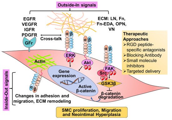
3. Role of Integrins in Neointimal Hyperplasia

Neointimal hyperplasia refers to post-intervention, pathological vascular remodeling due to the proliferation and migration of SMCs into the intimal layer, resulting in vascular wall thickening. During neointimal recruitment, SMCs are exposed to various ECM proteins, and integrin-ECM signaling has been shown to drive smooth muscle fibroproliferative remodeling. Several integrins are also known to promote neointimal hyperplasia, and evidence suggests that blocking integrins such as αIIbβ3 [43] and α4β1 [44,45] prevents neointimal hyperplasia. Besides these, the current literature strongly supports a role of signaling through αvβ3 in SMCs during neointimal hyperplasia [32]. In humans, αvβ3 is present in normal arteries and at the sites of SMC accumulation in atherosclerotic plaques. Several studies have shown that targeting αvβ3 integrin limits neointimal hyperplasia in small animal models of restenosis, including rat, rabbit, hamster, and guinea pig carotid angioplasty models [32,40,46,47]. In addition, an antibody to β3 integrin was demonstrated to prevent the development of intimal hyperplasia in wild-type diabetic mice [48]. Although β3-integrin blockade effectively reduces neointimal hyperplasia in animal models, the genetic ablation of β3 was found not to be effective for preventing intimal hyperplasia in animal models [49]. Therefore, it was speculated that the genetic loss of β3 might result in compensatory increases in the number and affinity of other adhesion receptors. In contrast, such compensation probably cannot occur with acute inhibition of αvβ3. In addition, the absence of β3 may affect signaling mediated by other integrins by decreased binding of intracellular proteins involved in signaling that ordinarily bind to the cytoplasmic domain of the missing integrin. Besides its detrimental role, some integrins are also known to prevent neointimal hyperplasia, such as α8β1 [50] and α7β1 [51]. The expression of different integrins on SMC, their ECM ligand, and their possible role in SMC function and neointimal hyperplasia are summarized in Table 1.
Table 1. Table representing the expression of different integrin subunits, their implication in smooth muscle cell (SMC) function and disease conditions such as atherosclerosis and neointimal hyperplasia, and integrin-directed drugs used in clinics. Collagen, Col; Laminin, LN; Fibronectin, Fn; Vascular cell adhesion molecule, VCAM; Osteopontin, OPN; Tenascin, TN; Vitronectin, VN; Fibrinogen, Fib; EDA, extra domain A.
Integrin ECM SMC Expression SMC Function Implication in Atherosclerosis/Restenosis Integrin-Targeting Agents in Clinics Reference
α1β1 Col 1-IV, LN High expression in resting SMCs. Downregulated in culture conditions and during neointimal hyperplasia Promotes SMC adhesion and contractile phenotype α1β1 deletion induces a stable plaque phenotype SAN-300 [10,34,42,52,53,54,55]
α2β1 Col 1 and IV, LN Undetectable levels in normal human SMCs, and high expression in cultured SMCs promote chemotaxis of arterial SMCs α2β1 deletion had no effect on atherosclerosis Vatelizu-mab [16,39,42,52,56]
α3β1 Col 1, Fn, and LN Detectable levels in normal human SMCs, and high expression in cultured SMCs No conclusive reports [10,33]
α4β1 Cellular-Fn, VCAM, OPN Undetectable levels in normal human SMCs, expressed in SMCs in culture and in intimal atherosclerotic thickening Induction of SMC differentiation blocking α4β1 prevents neointimal hyperplasia Natalizu-mab
AJM300
[38,44,45,52]
α5β1 Fn and LN Low levels in normal human SMCs, and high expression in cultured SMCs and during neointimal hyperplasia Promote SMC proliferation and migration Mediates early atherosclerosis Volocixi-mab
ATN61
[41,57,58]
α7β1 LN High levels in normal SMCs, and low expression in synthetic SMC Promotes contractile SMC phenotype α7 deletion promotes neointimal hyperplasia No conclusive reports [20,51,59,60]
α8β1 Fn, TN, VN Overexpressed in SMCs that display a contractile phenotype low expression in synthetic SMC phenotype and during neointimal hyperplasia Promotes contractile SMC phenotype. Prevents SMC proliferation and migration α8 deletion aggravates intimal thickening No conclusive reports [12,35,50,61]
α9β1 Fn-EDA, TN, VCAM Expression increases in synthetic SMC phenotype Promotes SMC proliferation, migration, and synthetic phenotype. α9 deletion prevents NH ASP5094 [21,62]
αvβ1 VN, Fn Weakly expressed in normal SMCs, and upregulated in SMCs cultured on fibronectin Inhibits contractility in SMC exposed to serum No conclusive reports PLN-74809
PLN-1474
[41,63,64]
αvβ3 VN, OPN, Fn Weakly expressed in normal SMCs, and upregulated in SMCs cultured on fibronectin and during neointimal hyperplasia Promotes SMC adhesion, proliferation and migration Promotes neointimal hyperplasia LM609, Abcixi-mab (c7E3Fab; ReoPro), Vitaxin, Intetumu-mab, Cillengitide [16,41,65,66,67,68]
αvβ5 Fib, Fn,
OPN
VN
highly abundant in cultured SMCs, upregulated upon vascular injury Promotes SMC adhesion and migration Promotes neointimal hyperplasia LM609
Intetumu-mab
[67,69]
 

This entry is adapted from the peer-reviewed paper 10.3390/cells11040646

This entry is offline, you can click here to edit this entry!
Academic Video Service