Gut Microbiota and Osteoarthritis: History
Please note this is an old version of this entry, which may differ significantly from the current revision.
Contributor:

Some say that all diseases begin in the gut. Interestingly, this concept is actually quite old, since it is attributed to the Ancient Greek physician Hippocrates, who proposed the hypothesis nearly 2500 years ago. The continuous breakthroughs in modern medicine have transformed our classic understanding of the gastrointestinal tract (GIT) and human health. Although the gut microbiota (GMB) has proven to be a core component of human health under standard metabolic conditions, there is now also a strong link connecting the composition and function of the GMB to the development of numerous diseases, especially the ones of musculoskeletal nature. The symbiotic microbes that reside in the gastrointestinal tract are very sensitive to biochemical stimuli and may respond in many different ways depending on the nature of these biological signals. Certain variables such as nutrition and physical modulation can either enhance or disrupt the equilibrium between the various species of gut microbes.

  • osteoarthritis
  • gut microbiota
  • metabolic syndrome
  • systemic inflammation

1. Introduction

Osteoarthritis (OA) has long been considered a degenerative disease that affects the hyaline cartilage alone. This orthopedic disorder still remains one of the most common degenerative and progressive joint diseases and a major cause of pain and disability in adults, affecting approximately 7% of the global population [1]. The Global Burden of Disease (GBD) 2019 study results revealed that the number of individuals affected by this condition increased globally by 48% between 1990 and 2019, classifying OA as the 15th highest cause of years lived with disability [2]. This significant increase is due to extrinsic factors such as the aging of the population as well as the indulgence in poor dietary habits [3][4][5]. Over the years, however, researchers started to notice that these pathological alterations were not exclusive to the chondral compartment. Adjacent structures including the subchondral bone, ligaments, synovium, and the joint capsule as a whole, are all involved in this pathological process albeit in varying degrees [6][7]. Many explanations have been proposed in attempts to fully elucidate the development of osteoarthritic alterations. Recent evidence indicates that OA progression is not exclusively attributed to biomechanical trauma but biochemical stressors as well, which may negatively affect regular activity of various cells and tissues [3]. Further research has shown that metabolic syndrome (MS), in particular, may in fact be one of the main culprits responsible for the development of OA. MS is a major health condition of modern-day society, and it only continues to expand and challenge public and clinical health on a global scale as a result of urbanization, increased calorie intake, the rise of obesity and sedentary life habits [8]. MS is connected to multiple physiological systems, being directly associated with the presence of four main clusters, which are: insulin resistance, obesity, vascular pathology, and dyslipidemia. MS paves the way for the progression of “meta-inflammation”, a state of persistent, low grade systemic inflammation triggered by metabolic stress [3]. This inflammatory stress disrupts cellular equilibrium and eventually aggravates systemic inflammation throughout the body [3]. By definition, meta-inflammation is a state of chronic inflammation mediated by macrophages present in multiple locations such as the liver, muscle, visceral fat, pancreas, colon and even the brain, for instance [9]. It is important to note that the state of chronic systemic inflammation also acts as a key mediator which drives the pathogenicity of OA by promoting harmful subchondral bone alterations in the onset of OA [10]. As a result, cartilage is also greatly affected by these alterations, aggravating inflammation even further, contributing to a shift towards a predominant pro-inflammatory and catabolic microenvironment in the joint [3]. Cytokines related to OA pathogenesis include tumor necrosis factor (TNF)-α, matrix metalloproteinases (MMPs), interleukin (IL)-1, IL-6, IL-2, IL-7, IL-15 and IL-21, and other chemokines which contribute to catabolic activity and detrimental effects [11][12]. These findings prove to be of particular significance since OA itself is influenced by the complex interplay between local, systemic and external factors, which consequently dictate disease progression and the manner in which patients respond to the treatment [13]. Despite its clinical and financial ramifications, conventional OA treatments still prove to be challenging. Conservative methods such as the administration of pharmacological agents only promote temporary alleviation of pain but do not address the etiological source of the disease and may, in some cases, cause serious adverse effects [7][14]. Pharmaceuticals may compromise the integrity of the gastrointestinal barrier, creating a state of hyper-permeability and inflammation [15]. As a matter of fact, long term administration of corticosteroids, for example, can increase the risk of serious side effects such as peptic ulcer disease, acute renal failure, and even myocardial infarction [16].

2. GBM-Derived Metabolites and Osteoarthritis (OA) Progression
Recent attention has been given to bacterial-derived LPS, specifically, as this microbial protein has been increasingly implicated in inflammatory disorders, namely OA. Researchers have revealed a correlation between elevated levels of circulatory inflammatory biomarkers (including LPS) with the severity of OA, therefore painting GMB-derived metabolites as pathogenic mediators responsible for driving inflammatory musculoskeletal disorders [17][18]. For instance, an animal study demonstrated that mice on a 28-week high-fat and high-sugar diet developed an obese phenotype and displayed increased cartilage damage, establishing a direct correlation between serum LPS levels and Mankin histological scores [17]. In this study the authors also examined GMB composition via 16S sequencing, detecting significant increases in Lactobacillus and Methanobrevibacter bacterial species, which indicated that MS promoted a strong dysbiotic shift in murine GMB with a strong predictive relationship with histological scores. In a similar study, Ulici et al. were able to demonstrate reduced severity of post-traumatic OA in germ-free mice, implying once again a causal role for the GMB in musculoskeletal pathogenesis [19]. Most of the animal studies evaluating the impact of GMB were performed on rodents due to the similarity of their GMB to that of the human gut microenvironment [20][21].
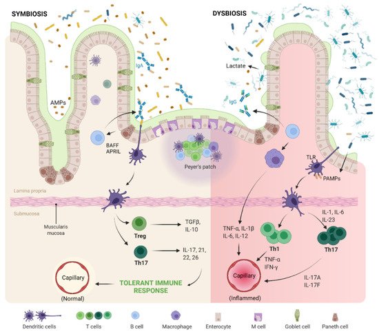
A similar pathogenic process occurs in humans. Dysbiosis of gut microbiome promotes excess porosity in the epithelial barrier of the gut and leakage of microbes and their by-products into the circulation, as shown in Figure 1 [22][23]. Stress involved in metabolic syndrome and pain involved in OA modulate gut microbiota through release of neurotransmitters and result in increased intestinal permeability [23][24].
Figure 1. Mechanism of regulation of immune response by gut microbiome. The native immune system is tolerant to the resident gut microbiome under the tight control of intestinal epithelial cells using mucosal barrier, secretory IgA and antimicrobial peptides (AMP). The native gut microbiome stimulates the intestinal epithelial cells, dendritic cells and macrophages to activate the T regulatory (T reg) cells and T helper 17 (Th17) cells. Upon activation of the intestinal epithelial cells with toll-like receptors (TLRs), B-cell activating factor (BAFF) and a proliferation inducing ligand (APRIL) are secreted which promotes the differentiation of IgA producing plasma cells, whereas in dysbiotic status of gut microbiome with the loss of barrier integrity and breach in the intestinal epithelial cell barrier, translocation of bacterial components, pathogen-associated molecular patterns (PAMPs), intestinal immune system is triggered through TLR activation. This results in an inflammatory cascade through hyperactivation of T helper 1 (Th1) and Th17 cells resulting in section of inflammatory cytokines.
The hypothesis behind the dysbiosis of gut and the development of OA are (a) low-grade intestinal inflammation [25][26], (b) elevated levels of microbial lipopolysaccharide (LPS) [27], (c) metabolic endotoxemia (interaction of gut-derived LPS and toll-like receptor (TLR)-4) [28][29], (d) meta-inflammation (metabolic inflammation mediated by macrophages present in multiple locations such as the liver, muscle, visceral fat, pancreas, colon and even the brain) [25][30][31], and (e) metabolic syndrome (abdominal obesity, dyslipidemia, hypertension, insulin resistance ± glucose intolerance, pro-inflammatory and prothrombotic states) [32][33]. The presence of inflammatory products and microbial genetic products in the joint pose a temporal association between gut microbiota and arthritis [34][35].

2.1. Gut–Joint Axis Distortion

“Gut–Joint” axis establishes the crosstalk between gut and joint [34][36][37][38][39]. Gut microbiota elaborates the wide range of metabolites, enzymes, and short chain fatty acids. These microbes produce lipopolysaccharides (LPS) which pave a way for increased intestinal permeability (“leaky gut”) and enter into systemic circulation to produce chronic low-grade intestinal inflammation. With respect to LPS, there exists an association with obesity and metabolic syndrome that are the potential risk factors for the development of OA. There is proven evidence of the role of LPS in OA pathogenesis [40]. Dunn et al. [41] revealed the identification of microbial DNA signatures in articular cartilage of rodents and humans. The authors performed 16S ribosomal RNA gene deep sequencing on eroded and intact cartilage samples from knee and hip OA patients, analyzing microbial DNA diversity and metagenomic profiles. The findings in human cartilage were compared to those in cartilage from OA-susceptible and OA-resistant mice. Result analysis indicated that alterations in microbial DNA signatures occur during OA progression. Although knee samples were microbiologically distinct from hip cartilage, microbial DNA in OA individuals was associated with increased Gram-negative constituents. This evidence shows that gut dysbiosis leads to the progression of the natural course of OA [28].
The role of MS on gut–joint instability in the absence of obesity has recently been investigated. In a mouse model of MS, Guss et al. [42] analyzed the effects of mechanically-induced OA on TLR5-deficient mice. Much like previous findings, histological evidence indicated that severe changes in cartilage were present in the high-fat diet mice groups, corresponding to GMB dysbiosis, increased body fat and systemic inflammation (as expected), only this time with an increased number of Firmicutes bacteria. Although metabolic irregularities were found in TLR5-deficient mice, the authors concluded that, in isolation, they could not have been solely responsible for the development of OA. Actually, the increased levels of LPS and the overgrowth of Firmicutes played a much more expressive role, here revealing a strong correlation between microbial components and OA progression.

2.2. Gut–Joint–Brain Axis Distortion

Turroni et al. established Gut–Joint–Brain (GJB) axis with OA pain [35]. The altered pain perception in OA cases is due to the modulation of the peripheral nociception and sensitization phenotype which results in the discrepancy in the results of the estimation of OA pain to the severity of radiological findings [43]. Increased intestinal permeability allows microbial metabolites to prime macrophages and exacerbate the joint inflammation, resulting in pain [44]. With the existing “Gut–Joint” axis, the exposure of stress and pain alter the interactions between the brain and the intestine, resulting in distorted quorum sensing signals and microbial gene expression, altered GI secretion, increased gut permeability and mobility, and dysbiotic gut. All this disequilibrium between gut microbiota and pain perception results in joint and systemic inflammation [45][46]. Understanding the temporal relationship among the pain perception in OA, gut microbiota, and joint inflammation leads to improved therapeutic strategies in the management of patient health in OA.

2.3. Evidences on Pathogenesis of OA

In a study involving 25 patients with knee OA, researchers were able to establish a link between serum and synovial fluid levels of LPS with known hallmark features of OA: the presence of activated macrophages (M1—pro-inflammatory) in the knee joint capsule and synovium; joint space narrowing; osteophyte formation; and high WOMAC (The Western Ontario and McMaster Universities Arthritis Index) scores, indicative of severe pain [18]. This also lies in parallel with a larger cohort study in the Dutch population [47]. The Rotterdam study-III recruited 1444 patients with hip and/or knee OA, where a solid association between increased WOMAC scores and abundance of Streptococcus bacteria with pro-inflammatory profile was found. For these reasons, physicians have been prompted to view the GMB from a new perspective, as a patient’s GMB must also be accounted for in consideration of possible dysbiotic shifts and secondary pathogenic effects.
Coulson et al. compared 3000 mg/day of green-lipped mussel (GLM) and 3000 mg/day of glucosamine (GS) in OA patients for 12 weeks and evaluated therapeutic efficacy on gut microbiota. In the GLM group, increased Bifidobacterium and decreased Enterococcus and yeasts were observed whereas in the GS group decreased Bacteroides and increased yeasts and coliforms, most notably Escherichia coli, were observed. Clostridia was reduced in both the groups, which is a potent immunomodulatory that decreases inflammation, improved WOMAC and GSRS scores and improved OA symptoms in response with colonic Th17 and CD4+ regulatory T cells [48].
Boer et al. evaluated gut microbiome and joint pain and inflammation. They demonstrated a spurious association between increased amounts of Streptococcus spp. and higher OA-related knee pain, but the causal association needs to be established. The possible hypothesis for OA-related knee pain and Streptococcus spp. is due to the production of microcellular vesicles by Streptococcus spp. in the GI tract. With the above findings, by reversing the gut dysbiosis through diet interventions, OA-related knee pain can be reversed. The causal association between Streptococcus spp. and OA has to be establish before translating into clinical practice [44].
Huang et al. demonstrated the role of LPS, a pro-inflammatory mediator from Gram-negative microbes, in accelerating the severity of OA [40]. They established a correlation between the presence of LPS in synovial fluid in the knee with the increased activated macrophages in the knee and clinical and radiographic severity of OA knee [49].

This entry is adapted from the peer-reviewed paper 10.3390/ijms23031494

References

  1. Hunter, D.J.; March, L.; Chew, M. Osteoarthritis in 2020 and beyond: A Lancet Commission. Lancet 2020, 396, 1711–1712.
  2. Cui, A.; Li, H.; Wang, D.; Zhong, J.; Chen, Y.; Lu, H. Global, Regional Prevalence, Incidence and Risk Factors of Knee Osteoarthritis in Population-Based Studies. EClinicalMedicine 2020, 29, 100587.
  3. Azzini, G.O.M.; Santos, G.S.; Visoni, S.B.C.; Azzini, V.O.M.; Dos Santos, R.G.; Huber, S.C.; Lana, J.F. Metabolic Syndrome and Subchondral Bone Alterations: The Rise of Osteoarthritis—A Review. J. Clin. Orthop. Trauma 2020, 11, S849–S855.
  4. Chen, D.; Shen, J.; Zhao, W.; Wang, T.; Han, L.; Hamilton, J.L.; Im, H.-J. Osteoarthritis: Toward a Comprehensive Understanding of Pathological Mechanism. Bone Res. 2017, 5, 16044.
  5. Zhang, Y.; Jordan, J.M. Epidemiology of Osteoarthritis. Clin. Geriatr. Med. 2010, 26, 355–369.
  6. Lana, J.F.; Macedo, A.; Ingrao, I.L.G.; Huber, S.C.; Santos, G.S.; Santana, M.H.A. Leukocyte-Rich PRP for Knee Osteoarthritis: Current Concepts. J. Clin. Orthop. Trauma 2019, 10, S179–S182.
  7. Setti, T.; Arab, M.G.L.; Santos, G.S.; Alkass, N.; Andrade, M.A.P.; Lana, J.F.S.D. The Protective Role of Glutathione in Osteoarthritis. J. Clin. Orthop. Trauma 2021, 15, 145–151.
  8. Alberti, K.G.M.M.; Eckel, R.H.; Grundy, S.M.; Zimmet, P.Z.; Cleeman, J.I.; Donato, K.A.; Fruchart, J.-C.; James, W.P.T.; Loria, C.M.; Smith, S.C.; et al. Harmonizing the Metabolic Syndrome: A Joint Interim Statement of the International Diabetes Federation Task Force on Epidemiology and Prevention; National Heart, Lung, and Blood Institute; American Heart Association; World Heart Federation; International Atherosclerosis Society; and International Association for the Study of Obesity. Circulation 2009, 120, 1640–1645.
  9. Li, C.; Xu, M.M.; Wang, K.; Adler, A.J.; Vella, A.T.; Zhou, B. Macrophage Polarization and Meta-Inflammation. Transl. Res. J. Lab. Clin. Med. 2018, 191, 29–44.
  10. Robinson, W.H.; Lepus, C.M.; Wang, Q.; Raghu, H.; Mao, R.; Lindstrom, T.M.; Sokolove, J. Low-Grade Inflammation as a Key Mediator of the Pathogenesis of Osteoarthritis. Nat. Rev. Rheumatol. 2016, 12, 580–592.
  11. Kapoor, M.; Martel-Pelletier, J.; Lajeunesse, D.; Pelletier, J.-P.; Fahmi, H. Role of Proinflammatory Cytokines in the Pathophysiology of Osteoarthritis. Nat. Rev. Rheumatol. 2011, 7, 33–42.
  12. Sellam, J.; Berenbaum, F. The Role of Synovitis in Pathophysiology and Clinical Symptoms of Osteoarthritis. Nat. Rev. Rheumatol. 2010, 6, 625–635.
  13. Mora, J.C.; Przkora, R.; Cruz-Almeida, Y. Knee Osteoarthritis: Pathophysiology and Current Treatment Modalities. J. Pain Res. 2018, 11, 2189–2196.
  14. Hafsi, K.; McKay, J.; Li, J.; Lana, J.F.; Macedo, A.; Santos, G.S.; Murrell, W.D. Nutritional, Metabolic and Genetic Considerations to Optimise Regenerative Medicine Outcome for Knee Osteoarthritis. J. Clin. Orthop. Trauma 2019, 10, 2–8.
  15. Vitetta, L.; Coulson, S.; Linnane, A.W.; Butt, H. The Gastrointestinal Microbiome and Musculoskeletal Diseases: A Beneficial Role for Probiotics and Prebiotics. Pathogens 2013, 2, 606–626.
  16. Marcum, Z.A.; Hanlon, J.T. Recognizing the Risks of Chronic Nonsteroidal Anti-Inflammatory Drug Use in Older Adults. Ann. Long-Term Care Off. J. Am. Med. Dir. Assoc. 2010, 18, 24.
  17. Collins, K.; Paul, H.; Reimer, R.; Seerattan, R.; Hart, D.; Herzog, W. Relationship between inflammation, the gut microbiota, and metabolic osteoarthritis development: Studies in a rat model. Osteoarthr. Cartil. 2015, 23, 1989–1998.
  18. Huang, Z.; Stabler, T.; Pei, F.; Kraus, V. Both systemic and local lipopolysaccharide (LPS) burden are associated with knee OA severity and inflammation. Osteoarthr. Cartil. 2016, 24, 1769–1775.
  19. Ulici, V.; Kelley, K.; Azcarate-Peril, M.; Cleveland, R.; Sartor, R.; Schwartz, T.; Loeser, R. Osteoarthritis induced by destabilization of the medial meniscus is reduced in germ-free mice. Osteoarthr. Cartil. 2018, 26, 1098–1109.
  20. Nguyen, T.L.A.; Vieira-Silva, S.; Liston, A.; Raes, J. How informative is the mouse for human gut microbiota research? Dis. Model. Mech. 2015, 8, 1–16.
  21. Wos-Oxley, M.L.; Bleich, A.; Oxley, A.P.; Kahl, S.; Janus, L.M.; Smoczek, A.; Nahrstedt, H.; Pils, M.C.; Taudien, S.; Platzer, M.; et al. Comparative evaluation of establishing a human gut microbial community within rodent models. Gut Microbes 2012, 3, 234–249.
  22. Szychlinska, M.A.; Di Rosa, M.; Castorina, A.; Mobasheri, A.; Musumeci, G. A correlation between intestinal microbiota dysbiosis and osteoarthritis. Heliyon 2019, 5, e01134.
  23. Romero, E.S.; Oliva, E.M.; Pérez, J.A.; Pérez, S.M.; Turroni, S.; Marchese, L.; Villafañe, J. Relationship between the Gut Microbiome and Osteoarthritis Pain: Review of the Literature. Nutrients 2021, 13, 716.
  24. Collins, S.M.; Surette, M.; Bercik, P. The interplay between the intestinal microbiota and the brain. Nat. Rev. Microbiol. 2012, 10, 735–742.
  25. Wang, X.; Hunter, D.; Xu, J.; Ding, C. Metabolic triggered inflammation in osteoarthritis. Osteoarthr. Cartil. 2015, 23, 22–30.
  26. Liu, Y.; Ding, W.; Wang, H.; Dai, L.; Zong, W.; Wang, Y.; Bi, J.; Han, W.; Dong, G. Gut microbiota and obesity-associated osteoarthritis. Osteoarthr. Cartil. 2019, 27, 1257–1265.
  27. Wang, J.; Gu, X.; Yang, J.; Wei, Y.; Zhao, Y. Gut Microbiota Dysbiosis and Increased Plasma LPS and TMAO Levels in Patients with Preeclampsia. Front. Cell. Infect. Microbiol. 2019, 9, 409.
  28. Hao, X.; Shang, X.; Liu, J.; Chi, R.; Zhang, J.; Xu, T. The gut microbiota in osteoarthritis: Where do we stand and what can we do? Arthritis Res. Ther. 2021, 23, 42.
  29. Boutagy, N.E.; McMillan, R.P.; Frisard, M.I.; Hulver, M.W. Metabolic endotoxemia with obesity: Is it real and is it relevant? Biochimie 2016, 124, 11–20.
  30. Berenbaum, F. Deep phenotyping of osteoarthritis: A step forward. Ann. Rheum. Dis. 2019, 78, 3–5.
  31. Chadha, R. Revealed aspect of metabolic osteoarthritis. J. Orthop. 2016, 13, 347–351.
  32. Sellam, J.; Berenbaum, F. Is osteoarthritis a metabolic disease? Jt. Bone Spine 2013, 80, 568–573.
  33. Berenbaum, F.; Griffin, T.M.; Liu-Bryan, R. Review: Metabolic Regulation of Inflammation in Osteoarthritis. Arthritis Rheumatol. Hoboken NJ 2017, 69, 9–21.
  34. Favazzo, L.J.; Hendesi, H.; Villani, D.A.; Soniwala, S.; Dar, Q.-A.; Schott, E.M.; Gill, S.R.; Zuscik, M.J. The Gut Microbiome-Joint Connection: Implications in Osteoarthritis. Curr. Opin. Rheumatol. 2020, 32, 92–101.
  35. Turroni, S.; Pedersini, P.; Villafañe, J.H. The Human Gut Microbiome and Its Relationship with Osteoarthritis Pain. Pain Med. Malden Mass 2021, 22, 1467–1469.
  36. de Sire, A.; de Sire, R.; Petito, V.; Masi, L.; Cisari, C.; Gasbarrini, A.; Scaldaferri, F.; Invernizzi, M. Gut–Joint Axis: The Role of Physical Exercise on Gut Microbiota Modulation in Older People with Osteoarthritis. Nutrients 2020, 12, 574.
  37. Gracey, E.; Vereecke, L.; McGovern, D.; Fröhling, M.; Schett, G.; Danese, S.; De Vos, M.; Bosch, F.V.D.; Elewaut, D. Revisiting the gut–joint axis: Links between gut inflammation and spondyloarthritis. Nat. Rev. Rheumatol. 2020, 16, 415–433.
  38. Zaiss, M.M.; Wu, H.-J.J.; Mauro, D.; Schett, G.; Ciccia, F. The gut–joint axis in rheumatoid arthritis. Nat. Rev. Rheumatol. 2021, 17, 224–237.
  39. Qaiyum, Z.; Lim, M.; Inman, R.D. The gut-joint axis in spondyloarthritis: Immunological, microbial, and clinical insights. Semin. Immunopathol. 2021, 43, 173–192.
  40. Huang, Z.; Kraus, V.B. Does lipopolysaccharide-mediated inflammation have a role in OA? Nat Rev Rheumatol. 2016, 12, 123–129.
  41. Dunn, C.M.; Velasco, C.; Rivas, A.; Andrews, M.; Garman, C.; Jacob, P.B.; Jeffries, M.A. Identification of Cartilage Microbial DNA Signatures and Associations with Knee and Hip Osteoarthritis. Arthritis Rheumatol. 2020, 72, 1111–1122.
  42. Guss, J.D.; Ziemian, S.N.; Luna, M.; Sandoval, T.N.; Holyoak, D.T.; Guisado, G.G.; Roubert, S.; Callahan, R.L.; Brito, I.L.; van der Meulen, M.C.; et al. The effects of metabolic syndrome, obesity, and the gut microbiome on load-induced osteoarthritis. Osteoarthr. Cartil. 2019, 27, 129–139.
  43. King, C.; Sibille, K.; Goodin, B.; Cruz-Almeida, Y.; Glover, T.; Bartley, E.; Riley, J.; Herbert, M.; Sotolongo, A.; Schmidt, J.; et al. Experimental pain sensitivity differs as a function of clinical pain severity in symptomatic knee osteoarthritis. Osteoarthr. Cartil. OARS Osteoarthr. Res. Soc. 2013, 21, 1243–1252.
  44. Boer, C.G.; Radjabzadeh, D.; Medina-Gomez, C.; Garmaeva, S.; Schiphof, D.; Arp, P.; Koet, T.; Kurilshikov, A.; Fu, J.; Ikram, M.A.; et al. Intestinal microbiome composition and its relation to joint pain and inflammation. Nat. Commun. 2019, 10, 4881.
  45. Mukhtar, K.; Nawaz, H.; Abid, S. Functional gastrointestinal disorders and gut-brain axis: What does the future hold? World J. Gastroenterol. 2019, 25, 552–566.
  46. Silva, Y.P.; Bernardi, A.; Frozza, R.L. The Role of Short-Chain Fatty Acids from Gut Microbiota in Gut-Brain Communication. Front. Endocrinol. 2020, 11, 25.
  47. Boer, C.; Radjabzadeh, D.; Uitterlinden, A.; Kraaij, R.; van Meurs, J. The role of the gut microbiome in osteoarthritis and joint pain. Osteoarthr. Cartil. 2017, 25, S10.
  48. Coulson, S.; Butt, H.; Vecchio, P.; Gramotnev, H.; Vitetta, L. Green-lipped mussel extract (Perna canaliculus) and glucosamine sulphate in patients with knee osteoarthritis: Therapeutic efficacy and effects on gastrointestinal microbiota profiles. Inflammopharmacology 2013, 21, 79–90.
  49. Kundu, P.; Blacher, E.; Elinav, E.; Pettersson, S. Our Gut Microbiome: The Evolving Inner Self. Cell 2017, 171, 1481–1493.
More
This entry is offline, you can click here to edit this entry!
ScholarVision Creations