Antioxidants in Alzheimer’s Disease: History
Please note this is an old version of this entry, which may differ significantly from the current revision.
Subjects: Biology
Contributor:

Alzheimer’s disease (AD) is the most common neurodegenerative disease, intensifying impairments in cognition, behavior, and memory. Histopathological AD variations include extracellular senile plaques’ formation, tangling of intracellular neurofibrils, and synaptic and neuronal loss in the brain. Multiple evidence directly indicates that oxidative stress participates in an early phase of AD before cytopathology. Oxidative stress plays a crucial role in activating and causing various cell signaling pathways that result in lesion formations of toxic substances, which advances the disease. Antioxidants are widely preferred to combat oxidative stress, and those derived from natural sources, which are often incorporated into dietary habits, can play an important role in delaying the onset as well as reducing the progression of AD. However, this approach has not been extensively explored yet. Moreover, a combination of antioxidants in conjugation with a nutrient-rich diet might be more effective in tackling AD pathogenesis.

  • Alzheimer’s disease
  • antioxidants
  • oxidative stress
  • reactive oxygen species
  • therapeutics

1. Role of Antioxidant-Rich Diet in Alzheimer’s Disease

It is well established that diet affects both mental and physical health. Food sources have a significant role in treating AD, as reported in many studies [88,89,90]. Vitamin C, E, carotenoids, flavonoids, polyphenols, and others are included in natural dietary antioxidants [91]. Moreover, it is suggested by literature and clinical reports that a proper diet including vitamins, proteins, and minerals will surely complement the medicine for the treatment of AD [89,90,91]. Apple cider has been reported to increase the activity of SOD, CAT, and glutathione peroxidase (GPx) to reduce lipid peroxidation [92,93].
Dietary potassium helps reduce ROS, alters the Aβ aggregation pattern, and helps in improving cognitive abilities [94]. Furthermore, increasing dietary potassium (to the optimum concentration) benefits individuals by preventing or delaying age and diet-related neurodegenerative diseases [95]. Garlic and its components show an admirable effect on brain function and neuronal physiology, leading to pharmacotherapy for AD [96]. Citrus fruits containing flavanone glycoside may be responsible for the conformational change of the beta-amyloid precursor protein cleaving enzyme 1 [97]. It has been found that supplementation of soaked almonds in the AD animal models of C57Bl/6 mice and adults of Sprague–Dawley rats, after overnight fasting, enhances memory due to the enrichment of vitamin E [97]. Despite under-conducted research, the cross-sectional studies of the overall role of diet and its patterns in AD are still questionable. Thus, research in this area is also needed. Moreover, people’s apprehension of the quantity and quality of individual bioactive components present in food items is insufficient for significant neuroprotection. Different classes of antioxidants with potential therapeutic applications against AD are displayed in Figure 2.
Figure 2. Various classes of antioxidants are documented for having the potential to counter AD.

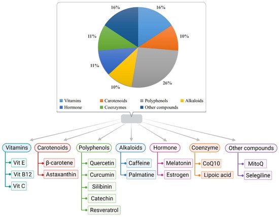
2. Role of Antioxidants in Alzheimer’s Disease

2.1. Vitamin E

Vitamin E is the most promising antioxidant for peroxyl radicals [98]. It can act on lipid-soluble membrane lipoproteins and low-density lipoproteins [99]. It has the potential to inhibit and delay neuronal death caused by inflammation. Moreover, it eliminates free radicals present in the red blood cell membrane and inhibits the spread of lipoperoxidation [100]. Furthermore, α-tocopherol is the most abundant form of vitamin E with high bioavailability in human tissue [101,102]. Vitamin E can be helpful in overcoming the increased expression of alpha-tocopherol transfer protein (α-TTP) in the patient’s brain suffering from AD [103]. A meta-analysis report of AD patients shows a reduced level of vitamin E in the blood plasma [104]. In one of the clinical trials, vitamin E and Ginkgo biloba extract were potentially significant in improving cognitive function of the brain [11]. Additionally, in another meta-analysis, it has been reported that a low concentration of serum vitamin E is associated with AD [105]. Moreover, substantial evidence suggested that vitamin E successfully suppresses tau-induced neurotoxicity in Drosophila [106,107,108]. In one of the recent studies, it is proposed that vitamin E has significantly reduced oxidative and nitrosative damage in AD [109]. However, the positive effect being evaluated of vitamin E in AD is still in the ongoing phases of various clinical trials.

2.2. Glutathione

Glutathione also plays a significant role in protein and DNA synthesis, cell cycle regulation, and storage and transport of cysteine. It has the potential to scavenge lipid peroxidation products like acrolein, 4-hydroxy-2-nonenal (HNE), and others [110]. It is used to maintain the thiol redox of cells, detox electrophiles, and metals, and protect from oxidative stress. It also can form metal complexes that reduce the toxicity of the metals and facilitate their further excretion from the body [110,111,112]. Recently, it was reviewed that cholesterol-mediated depletion of mitochondrial glutathione is linked with increased Aβ-induced oxidative stress in mitochondria [113]. The introduction of glutathione ethyl ester in transgenic mice featuring a high expression of sterol regulatory element-binding protein-2 (SREBP-2) has been shown to prevent neuroinflammation and neuronal damage [114]. Further, one recent study reveals the redox pathway of glutathione antioxidant responsible for regulating mitochondrial dynamics in axons [115]. However, the mechanistic overview of the exclusive role of glutathione in AD is still unclear.

2.3. Molecular Hydrogen

Molecular hydrogen is also an antioxidant that can modulate the Keap1-Nrf2-ARE signaling pathway and reduce inflammation [116]. It has a potential role in the selective reduction of hydroxyl radicals involved in the demolishing of proteins, nucleic acid and leads to lipid peroxidation, which is also a reported feature in AD [117]. It has been reported that molecular hydrogen administration increases short-lived Drosophila’s survival and life span [118]. At the same time, it is found that the hydrogen-rich water causes the increment in the level of glutathione and SOD [52]. Having both an indirect and direct role, the application of molecular hydrogen shows satisfying results for AD. However, more human trials are required for solid suggestions and recommendations.

2.4. Monoamine Oxidase-b Inhibitor

Monoamine oxidase catalyzes the oxidative deamination of xenobiotic and biogenic amines. In peripheral tissue and the central nervous system, they play an important role in the metabolism and control of vasoactive and neuroactive amines. In cerebral blood arteries, a monoamine oxidase-b inhibitor can rapidly produce the vasodilator nitric oxide [119]. By blocking oxidative deamination, it shields the vascular endothelium from the effects of Aβ and improves the survival and function of nigral neurons [120,121]. It is also reported to decrease the progression of AD by reducing neuronal damage [122]. L-deprenyl, a monoamine oxidase-b inhibitor, enhances nitric oxide production accompanied by vasodilation; however, the study also suggests that L-deprenyl may involve other pathways for its effectivity [119].

2.5. Melatonin

Melatonin, a mammalian hormone synthesized in the pineal gland, can scavenge oxygen and nitrogen-based reactants. It performs by stimulating and promoting the activity and expression of NO synthase, SOD, and GPx [123]. It has a significant role in reducing oxidative damage of cells [124]. In recent literature, it has been reported that antioxidant melatonin can mitigate tau hyperphosphorylation [125,126,127,128,129] and inhibit the toxicity induced by Aβ [130].

2.6. Ascorbyl Palmitate

It is a lipid-soluble form of vitamin C. It maintains all the vitamin C activity without creating problems associated with ascorbic acids, such as less recycling capacity of α-tocopherol in the lipid bilayer, reduced viability in-vivo, and others [122]. Additionally, it is reported that the demand for vitamin C can be better fulfilled with lipophilic form rather than hydrophilic form [131]. Ascorbyl palmitate can successfully cross the blood-brain barrier (BBB) [132] and is reported for its significant role in treating AD [133]. As ascorbyl palmitate resides in the cell membrane, it can accelerate the production of vitamin E. However, the protective role of vitamin C is still in debate as it is not yet clear whether vitamin C is acting alone or in combination for treating AD.

2.7. Curcumin

Multiple desirable features reside in curcumin for a neuroprotective drug, including antioxidant, anti-protein aggregates, and anti-inflammatory activities [134]. It has been studied that curcumin reduces inflammation, oxidative damage, and cognitive deficits in rats where Aβ toxicity has affected their central nervous system. Curcumin possesses substantial free radical scavenging properties, whereby it targets NO-based radicals to scavenge them, which helps inhibit lipid peroxidation [135]. Curcumin has also been reported to bind with metal ions, which prevents them from causing aggregation of Aβ and reduces oxidative stress [136]. Moreover, curcumin was also found to restore glutathione levels in brain tissue and reduce oxidized proteins in mice models with AD [137,138]. However, in one of the clinical trials, curcumin’s beneficial effect in AD couldn’t be determined; this may be due to highly poor pharmacokinetics and pharmacodynamics properties [139].

2.8. Coenzyme Q and SK-PC-B70M

Coenzyme Q is currently studied for its role in Parkinson’s disease and amyotrophic lateral sclerosis [106]. Moreover, it helps in the generation of ATP. It is the only lipid synthesized directly within the body and can maintain a redox function [140]. Coenzyme Q has the potential to neutralize free radicals and stabilize the optimal functioning of the cell membrane. The contribution of coenzyme Q in AD treatment must be explored as there is a high possibility that it might play an influential, protective, and preventing role in AD. SK-PC-B70M, an oleanolic-glycoside saponin enriched fraction, is derived from Pulsatilla Korean. Currently, it has been reported for its neuroprotective activity against the cytotoxicity effect induced by Aβ in SK-N-SH [141].

2.9. Estrogen, Astaxanthin, and Quercetin

Estrogen protects neurons against the toxicity of Aβ by acting as an antioxidant [142]. It appears to have a neuroprotective effect [52] without improving function or cognition in people with AD [142]. Astaxanthin is a powerful carotenoid that can prevent apoptosis, oxidative stress, inflammation, memory loss, and protect against Aβ’s neurotoxic effects [94,142,143,144]. Quercetin is the most prominent and significant dietary antioxidant effective on health as it protects against severe diseases like lung cancer, cardiovascular disease, osteoporosis, and others [145]. There are ongoing clinical trials for estimating its accurate effect on AD [146].

2.10. Lipoic Acid

The medicinal antioxidant lipoic acid (α-lipoic acid) is found in the mitochondria. Pyruvate dehydrogenase and α-ketoglutarate dehydrogenase both use it as a cofactor. However, it is also involved in the recycling of other antioxidants such as vitamin C and E, as well as glutathione, in order to boost ACh production [53]. Lipoic acid is also implicated in some redox-active chelating metals, which helps to prevent lipid peroxidation from building up [147]. When used in combination with acetylcarnitine, lipoic acid was found to protect neuronal cells through cell-signaling pathways, including specific extracellular kinase pathways, mainly the Ras-MAPK pathway that were dysregulated in AD [148]. Studies undertaken on the brain of control and AD mouse models showed that lipoic acid reduced the expression of F2 isoprostanes and neuroprostanes, which are oxidative stress markers [149]. Lipoic acid also induces the transcription factor Nrf2, which regulates a number of different antioxidant enzymes involved in protection from oxidative stress [150]. Lipoic acid improved memory and reversed oxidative stress indices in the senescence-accelerated mouse-prone 8 (SAMP8) models [151]. Lipoic acid is a potent antioxidant as it can traverse the BBB, making it ideal for therapeutic applications in AD [152].

2.11. Resveratrol

Resveratrol (3, 5, 4′-trihydroxy-trans-stilbene) is a polyphenolic compound found in a number of plants, like red grapes, blueberries, dark chocolate, and peanut butter. Resveratrol has been reported to possess antioxidant properties and was found to diminish malondialdehyde and nitrite levels and restore glutathione levels [84]. Studies in a number of cell lines expressing mutant AβPP695 reported that resveratrol exhibited anti-amyloidogenic activity through reduction in secreted intracellular Aβ peptide levels [153]. Levels of intracellular antioxidant enzymes SOD, CAT, GPx, and HO-1 were increased by resveratrol while simultaneously reducing lipid peroxidation [79]. Another essential function of resveratrol was diminishing ROS production in brain tissue by preventing disruption in the mitochondrial membrane potential [154]. The binding of metal ions to Aβ and NFTs enhances their aggregation and increases ROS production. Resveratrol counteracts this through dysregulation of the metal ion balance [84]. Along with antioxidant properties, resveratrol has been reported to promote an anti-inflammatory response, reduce levels of tau protein phosphorylation and increase the activity of SIRT-1 [154]. This makes resveratrol an interesting natural antioxidant in combating AD pathogenesis.

2.12. MitoQ

MitoQ is an antioxidant that targets the mitochondria in AD. MitoQ is made by adding the lipophilic triphenylphosphonium (TPP+) cation to ubiquinone, a component of the mitochondrial electron transport chain, via a ten-carbon chain [155]. TPP+ facilitates entry of ubiquinone into the mitochondrial matrix, where the complex II reduces ubiquinone to ubiquinol, the active antioxidant form, decreasing lipid peroxidation, which reduces oxidative damage [156]. MitoQ is able to traverse the BBB rapidly and has been found to accumulate several hundred folds in the mitochondrial membrane. The uptake of MitoQ in the mitochondria is driven by the high membrane potential of the inner mitochondrial membrane [157]. MitoQ has been found to reduce free radicals and oxidative damage while helping to regulate mitochondrial functions of the cells [158]. MitoQ was found to lower Aβ peptide levels, minimize synaptic loss and astrogliosis and improve cognitive functions in AD mouse model studies wherein the administration of MitoQ was initiated at a young age [155,159]. MitoQ was also reported to enhance neurite outgrowth in neurons and protection against Aβ peptide toxicity in cells of AD mouse models [160].

2.13. Catechins

Catechins are the bioactive components found in tea—most abundant in green tea (green tea catechins or GTC)—which includes four different types of catechins: viz. epicatechin (EC), epicatechin gallate (ECG), epigallocatechin (EGC), and epigallocatechin gallate (EGCG) [153]. Catechins exhibit antioxidative effects by scavenging ROS and chelating metal ions like copper, iron, and zinc, thereby reducing their accumulation in the brain of AD patients [86]. EGCG was reported to reduce caspase levels and oxidative stress along with reducing lipid peroxidation in the hippocampus of the rat model [161]. A long-term study on male Wistar rats revealed that the administration of 0.5% GTC in water resulted in counteracting Aβ-induced cognitive impairment, along with reduced levels of plasma lipid peroxide and ROS levels [162]. In addition toantioxidant properties, catechins were also reported to exhibit anti-inflammatory properties, along with inhibition of acetylcholinesterase (AChE) activity. At the same time, EGCG was found to directly interact with Aβ peptides and prevent the formation of aggregates [86,163,164]. Furthermore, catechins are BBB permeable, as found in rodent models, making them a potential therapeutic candidate for AD treatment [86].

2.14. Silibinin

Silibinin, an antioxidant flavonolignan obtained from Silybum marianum, can boost the amount of newly formed microglia, astrocytes, neurons, and neural precursor cells in the brain [165]. In one study, silibinin was found to be a dual inhibitor of AChE and Aβ peptide aggregation, implying a therapeutic method for treating Alzheimer’s disease [165]. It can potentially prevent the injuries caused by Aβ1-42-indued oxidative stress by lowering the production of H2O2 in Aβ1-42-stressed neurons [166]. Another study reported that streptozotocin-induced tau hyperphosphorylation (ser404) in the hippocampus was substantially reduced by silibinin [167]. Though these results indicate that silibinin may be a novel therapeutic agent for treating AD, no clinical trials are on board.

2.15. Palmatine

Palmatine, an isoquinoline alkaloid, acts against Aβ induced neurotoxicity [168]. It is reported that palmatine activated the Nfr2 knockdown and AMPK pathway [168]. It is reported for having anti-inflammatory, antioxidative and antiproliferative effects [169]. Another study reported the combined impact of palmatine and berberine on the inhibition of AChE [169,170]. Though it is reported in several in-silico and in-vivo studies, there is still a massive absence of its proper application in AD. Moreover, the mode of action underlying their neuroprotective effect is poorly characterized in vivo.

2.16. Serotonin

Serotonin, an indoleamine neurotransmitter, can disassemble performed Aβ fibrils [171]. Ample evidence reflects that a combination of disturbances in serotonergic and cholinergic function may possess a vital role in cognitive impairment in AD [172]. In one study, it is indicated that alterations of the serotonergic system contribute to neuropsychiatric symptoms in AD as their results suggest that a decline in neurons expressing 5-HT2A plays a role in the etiopathology of neuropsychiatric symptoms in AD [173]. Furthermore, while many of these compounds will likely be used as adjuvant therapy in the treatment of AD symptoms, there are currently just a few pharmacological entities with activity against serotonin receptors that have the potential to slow the illness’s progression.

2.17. Gintonin

Gintonin, a glycol-lipoprotein, can help in maintaining the integrity of BBB [174]. It can suppress the activated inflammatory mediators and microglial cells in the brains of Aβ-injected mice [175]. Recent findings suggest that treatment with gintonin in AD results in improved synaptic and memory functions in the brain [176]. It reflects an emerging role as a modulator of neurogenesis and synaptic transmission, and it has the potential to regulate autophagy in primary cortical astrocytes [176,177]. Moreover, as a novel agonist of lysophosphatidic acid receptors, gintonin regulated several GPCR, including GPR55 and GPR40 [177]. Nevertheless, further exploration is still required to understand gintonin’s underlying mode of action in AD.

3. Role of Other Nutrients in Alzheimer’s Disease

Apart from antioxidant activities, natural products have exhibited other vital properties to combat AD progression through anti-inflammatory response, prevention of Aβ aggregation, accumulation of tau protein, and the promotion of cholinergic signaling [178]. Alkaloids, such as cryptolepine and tetrandrine, have been reported to be involved in the inhibition of NF-κB, thereby acting as anti-inflammatory agents [179]. Flavonoids, owing to their characteristic property of inhibiting inflammatory response, have shown potential for working against AD progression [180]. Studies in animal models of AD have reported terpenoids, such as artemisinin, parthenolide, and carnosol can inhibit NF-κB and p38 MAPK pathways [181,182,183]. Ginsenoside Rg1, a compound obtained from the roots of the Ginseng plant, has been reported to cause a significant drop in levels of Aβ peptide levels in AD mice [184]. Natural plant products like crocin, α-cyperone, chrysophanol, and aloe-emodin have been found to exhibit properties that inhibit tau protein formation and reduce AD progression [185,186,187]. Caffeine, one of the most widely consumed alkaloids, has been found to inhibit Aβ deposition in vitro [188]. It was also found to reduce ROS production and enhance SOD levels in human neuroblastoma cells cultured with Aβ [189]. Caffeine has also been shown to exhibit anti-neuroinflammatory properties as well as decreasing tau protein phosphorylation in the hippocampus [190]. In low to moderate doses, caffeine inhibits AChE, thereby improving cognitive actions and reducing the progression of AD [191]. Eugenol, found in cloves, has been reported to reduce amyloid plagues and increase memory in rat models induced with Aβ peptides [192]. Dietary patterns have also been found to impact the onset and progression of AD. A Western diet characterized by higher meat intake was associated with an increased risk of AD [193]. In contrast to this, the Mediterranean diet, characterized by higher consumption of fruits, vegetables, and fish with lower meat intake, was found to reduce the risk of AD in the population [194].

4. Conclusions

With a long asymptomatic period, AD is a chronic neurodegenerative condition. Multiple literature and evidence infer that oxidative damage or stress plays a significant role in the pathogenesis of AD through various mechanisms and pathways. Thus, new treatment strategies are required to either prevent or reduce oxidative damage and may provide therapeutic efficacy against AD. Natural bioactive, often incorporated into the diet, can become a widely adopted approach to avoid the onset of AD. Moreover, this approach can be conjugated with approved drugs for patients with progressive AD. The integrated system of antioxidants with multiple drugs may provide higher effectiveness. Some antioxidants have proven positive effectors on AD, but some still need attention and work. Moreover, there is limited data on the role of antioxidants in AD from human clinical trials and epidemiological studies. Additionally, some antioxidants show significant effects on an animal model but exhibit diminished efficacy on humans during clinical trials. Due to this, there is a lot of skepticism about the success of antioxidant therapy for AD. It is quite necessary to explore a more definitive and precise approach integrated with antioxidants for lowering or inhibiting the progression of AD. The link between inflammation and AD is unavoidable, so antioxidants’ integrated role in decreasing inflammation must be considered. Thus, further advanced studies and human clinical trials are necessary to determine and estimate the antioxidants potential for AD.

This entry is adapted from the peer-reviewed paper 10.3390/biology11020212

This entry is offline, you can click here to edit this entry!
Video Production Service