You're using an outdated browser. Please upgrade to a modern browser for the best experience.
Breast Cancer Metastases Development: History
Please note this is an old version of this entry, which may differ significantly from the current revision.
Subjects: Oncology
Contributor: Barbara Ciastek

Breast cancer is one of the main causes of morbidity and mortality in women. Early breast cancer has a relatively good prognosis, in contrast to metastatic disease with rather poor outcomes. Metastasis formation in distant organs is a complex process requiring cooperation of numerous cells, growth factors, cytokines, and chemokines. Tumor growth, invasion, and finally systemic spread are driven by processes of angiogenesis, vasculogenesis, chemotaxis, and coagulation.

  • breast cancer
  • metastasis
  • angiogenesis
  • vasculogenesis
  • chemotaxis
  • coagulation

1. Introduction

1.1. General Information Related to Breast Cancer

Breast cancer (BC) is in first place in terms of the most commonly diagnosed malignancies in women worldwide, with almost 2.3 million new cases and 685 thousand deaths in 2020 alone [1]. As the incidence of BC in recent years has increased, the prognosis for patients has improved as a result of progress in early diagnosis and treatment approaches, leading to a drop in mortality from BC in North America and the European Union [2,3]. The rise in the number of new cases in developing countries is attributed mostly to lifestyle changes, diet, environment alterations, older gestational ages, alcohol consumption, and the use of menopausal hormone therapy [2]. The established BC risk factors include being female, early age at menarche and older age at menopause, nulliparity, no breastfeeding, higher body mass index, family history of BC, alcohol use, and use of contraceptives or menopausal hormone therapy. However, as BC is a complex disease, for particular BC subtypes, the presence of some of these factors may be not associated with an increased risk of the disease, or they could be even considered protective (Figure 1) [4].
Figure 1. Breast cancer molecular subtypes and their risk factors [4]. Abbreviations: BMI, body mass index; * there is insufficient or inconsistent evidence of an association between the luminal B breast cancer subtype and greater parity, older age at first birth, older age at menopause, greater BMI, alcohol use, use of oral contraceptives, or menopausal hormone therapy use; ** there is insufficient or inconsistent evidence of an association between HER2-overexpressing breast cancer and younger age at menarche, greater parity, older age at first birth, breastfeeding, older age at menopause, greater BMI, or menopausal hormone therapy use.

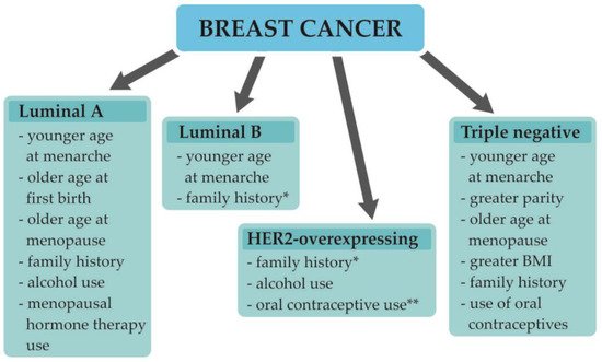
1.2. Understanding Breast Cancer Heterogeneity

BC is a complex disease with miscellaneous morphologic and biological features and, hence, various clinical courses and prognoses [5]. The traditional classification of BC is based on tumor stage and grade. The TNM staging system by the American Joint Committee on Cancer encompasses both clinical and pathologic data on tumor size (T), the status of regional lymph nodes (N), and distant metastases (M); subsequently, based on these data, the disease is established as being in one of five stages (0–IV) [5]. Tumor grade brings information on cell differentiation—it includes evaluation of the particular features of the cell, which include tubule formation, size and shape of the nucleus in tumor cells, and mitotic rate [5]. With more than 20 different histologic types; BC can be classified based on clinicopathologic features such as cell type of the tumor, extracellular secretion, architectural features, or immunohistochemical profile. The most common type is invasive ductal carcinoma not otherwise specified (IDC-NOS), which is found in 70–80% of cases. It encompasses adenocarcinomas that cannot be classified as one of the special types [5,6]. The most common among special types of BC is invasive lobular carcinoma, accounting for about 10% of cases. Other less common special types include: tubular, cribiform, mucinous, papillary, apocrine, medullary, metaplastic, or mixed type carcinomas [5,6]. In recent decades, there has been meaningful development in the understanding of BC biology, which has led to the conclusion that the previous classifications do not display the heterogeneity of the disease—both response to therapy and prognosis are defined by the biological features of a tumor [5,7]. Routine immunohistochemical analysis of estrogen receptor (ER), progesterone receptor (PR), human epidermal growth factor receptor 2 (HER2), and Ki-67 expression (proliferation index) is currently used to determine the four basic molecular subtypes of BC: Luminal A-like, luminal B-like, HER2-overexpresing, and triple-negative BCs. This classification (approved by the St Gallen Consensus Conference) facilitates choosing the right therapeutic approach and allows to determine a patient’s future outcome [5,8]. Apart from the molecular subtype of BC, the TNM stage and the patient’s preferences, as well as many other factors, play a role in choosing the most personalized therapy. The clinico-prognostic characteristics of particular subtypes are shown in Table 1. The first-line therapy for the early BC combines surgery and postoperative radiation. Main surgical approaches include mastectomy or an excision followed by radiation. Breast cancer surgery may also include dissection of axillary lymph nodes if there are indications for the procedure. Furthermore a pre- (neoadjuvant) or postoperative (adjuvant) systemic therapy may be administered, which includes chemotherapy, endocrine therapy, or trastuzumab-based therapy. In metastatic disease, the major objectives are prolongation and maintaining quality of life. The systemic therapy encompasses chemotherapy, endocrine therapy, trastuzumab-based therapy, or other targeted therapies such as CDK4/6 inhibitor or PARP inhibitor [9]. In some patients, breast surgery may be implemented as it could improve local progression free survival; however, it may worsen distant progression free survival. There is no evidence that surgical treatment of the primary tumor improves overall survival in metastatic disease [10].
Table 1. The clinico-prognostic characteristics of the molecular subtypes of breast cancer [5,11].
  Luminal A-Like Luminal B-Like HER2-
Overexpressing
Triple-Negative
ER + + - -
PR ≥20% <20% 1 - -
HER2 - + 1 + -
Ki-67 <20% ≥20% 1 all all
Grade Low Low/High High High
Frequency 30–40% 20–30% 12–20% 15–20%
Local–Regional
Recurrence
0.8–8% 1.5–8.7% 1.7–9.4% 3–17%
Prognosis Favorable Intermediate Unfavorable Unfavorable/Poor

Abbreviations: ER, estrogen receptor; PR, progesterone receptor; HER2, human epidermal growth factor 2; Ki-67, proliferation index; 1 and/or.

1.3. Breast Cancer Development, Progression, and Metastasis Formation

BC development is a complex and intricate process that depends on genetic and epigenetic alterations, as well as on the tumor microenvironment. Cancer cells are surrounded by a modified stroma, which creates the tumor microenvironment (TME). It comprises miscellaneous stromal cells, encompassing fibroblasts, immune cells, inflammatory cells, endothelial cells (ECs), pericytes, adipocytes, and bone marrow-derived cells [2,12,13]. Both tumor and stromal cells interact mutually through secreted proteins, cytokines, chemokines, and growth factors, which lead to BC growth, progression, and metastasis formation [14,15,16].
Early diagnosis and targeted therapy are the main factors that, in recent decades, have allowed for improvement in the overall survival of BC patients. As non-metastatic BC is considered to be generally curable with a five-year overall survival greater than 80%, in metastatic disease, only palliative treatment is applicable, with a reduction in the five-year overall survival rate of only 25% [15,16,17].
Metastasis formation is a multi-step and complex process, which implies cooperation between angiogenesis, vasculogenesis, chemotaxis, and coagulation. During this action, cancer cells acquire proangiogenic phenotype and are able to escape from their initial location, leading to remodeling the extracellular matrix (ECM), intravasate into the blood, survive in the circulation, and extravasate from blood vessels to colonize new organs. In recent years, it has been speculated that BC cells spread systematically during the early disease stage, regardless of the primary tumor size [18]. During cancer development, numerous cell types continuously liberate signaling molecules into the microenvironment, leading to the formation of a sophisticated web of communication that attracts new cell types to remodel the tumor microenvironment [19,20].

2. Angiogenesis

2.1. The Role of Angiogenesis in Tumor Invasion

Angiogenesis is the process of new blood vessels development from preexisting vasculature. This process occurs during physiological processes such as wound healing or endometrial growth during the menstrual cycle, and can also be part of a disease; for instance, in cancer transformation, when tumor diameter exceeds 1–2 mm3, it requires its own blood supply. Angiogenesis also plays an important role in cancer dissemination and the formation of metastases [21,22]. The initiation of angiogenesis—the so-called angiogenic switch—is the point in tumor development where proangiogenic factors surpass antiangiogenic factors and a progressive tumor growth is initiated. The angiogenic switch is set off either by genetic mutations of tumor cells, resulting in increased proliferation and hypoxia or the expression of proangiogenic factors, or by tumor-associated inflammation and the recruitment of immune cells [23,24].

2.2. The Role of Angiogenesis in BC

Hypoxia plays a major role in the stimulation of angiogenesis in BC. Endothelial cells (ECs) start expressing hypoxia-inducible factor-1 (HIF-1) as a result of low intratumoral oxygen levels. Furthermore, overexpression of HER2 and expression of estrogen receptor on BC cells is associated with increased HIF-1 levels. High HIF-1 levels have been also found in triple-negative breast cancer, which is probably a result of loss of p53, PTEN mutations, and EGFR overexpression [22]. HIF-1 upregulation leads to the transcription of genes involved in adaptation to the hypoxic BC environment [21,22,25]. Hypoxia, via HIF-1, stimulates cancer cells to secrete large amounts of vascular endothelial growth factor A (VEGF-A), which binds to its receptor on the surface of ECs. This leads to the activation, growth, and migration of ECs toward the tumor [26]. The migrating cells start to proliferate and subsequently form the lumen of the new vessel, which supplies cancer cells with oxygen and nutrients [21]. This process is supported by various cytokines, enzymes, and growth factors (Table 2). Furthermore, as VEGF-A has the ability to mobilize endothelial progenitor cells (EPCs) from bone marrow, it facilitates formation of new blood vessels de novo via vasculogenesis [27].
Table 2. Involvement of various cytokines in angiogenesis [23,28,29,30,31,32,33,34,35,36].
Cytokines and Growth Factors Involved in
Angiogenesis
Role/Action
EGFR
-
upregulates of VEGF-A and other proangiogenic factors;
-
increases tumor cells proliferation and migration through EGFR-Ras/Raf/MEK/ERK and EGFR-PI3K/AKT pathways.
bFGF
-
promotes tumor progression and metastases;
-
increases motility and invasiveness of BC cells;
-
stimulates proliferation, migration, and differentiation of endothelial cells;
-
increases production of proteases;
-
promotes of integrin and cadherin receptor expression;
-
supports communication across intercellular gap junctions.
IL-8
-
stimulates the proliferation and survival of ECs;
-
upregulates MMPs in ECs;
-
supports recruitment of neutrophils into the tumor tissue;
-
promotes modification of expression of cells-adhesion molecules in BC cells and neutrophils.
VEGF-A
-
the most potent proangiogenic molecule;
-
promotes proliferation and migration of ECs;
-
increases vascular permeability;
-
stimulates actin rearrangement;
-
influences gap junction;
-
enhances tumor cells extravasation;
TNF- α
-
increases the expression of angiogenic factors, such as VEGF-A, bFGF, IL-8 in ECs;
-
proinflammatory cytokine;
-
induces cells survival, proliferation, and metastasis.
MMPs
-
degrade and remodel ECM;
-
enhance ECs migration;
-
promote initiation of angiogenesis.

Abbreviations: bFGF, basic fibroblast growth factor; EGFR, epidermal growth factor receptor; VEGF-A, vascular endothelial growth factor A; IL-8, interleukin 8; MMPs, matrix metalloproteases; ECM, extracellular matrix; ECs, endothelial cells; BC, breast cancer; TNF-α, tumor necrosis factor-α.

2.3. Interactions of VEGF with Tumor and Tumor Microenviroment

Elevated levels of VEGF-A in BC are linked to aggressive tumor behavior and poor prognosis [26,37]. Tumors overexpressing HER2 also over-release VEGF-A. The poor prognosis of patients with HER2-overexpressing tumors is linked to excessive angiogenesis [38,39]. Close cooperation between proangiogenic factors (including VEGF-A), their receptors, and the components of the ECM matrix is demanded in angiogenesis in BC [22]. There are numerous factors that stimulate angiogenesis. These include estradiol and progesterone. Estradiol upregulates VEGF-A expression by vascular epithelium and supports EC proliferation and migration [40]. Cancer-associated fibroblasts (CAFs) are one of the main components of the TME and boost cancer growth and metastasis formation. They secrete tumor-promoting mediators, causing remodeling of the ECM, thus supporting the invasion of cancer [41]. Another component of the TME in BC is tumor-associated macrophages (TAMs), which promote angiogenesis and tumor progression via releasing a proangiogenic factor, CCL18 [28].

2.4. Contribution of VEGF to the Metastatic Spread of BC

VEGF-A binds to its receptors on ECs—either to VEGFR1 or VEGFR2. VEGFR2 is the major receptor for VEGF-A, and its activation leads to the proliferation of ECs, invasion, migration, and survival (via the ERK and PI3K/Akt pathways) [24]. VEGFR1 is found not only on ECs, but also on several other cell types, including monocytes or macrophages [42]. In BC it activates MAPK/ERK and PI3K/Akt pathways, resulting in tumor growth and induction of epithelial-to-mesenchymal transition (EMT) [43]. PI3K/Akt may trigger nuclear factor-kappa B (NF-κB) to activate migration pathway. Furthermore, AKT pathway stimulates permeability factors and inactivates proapoptotic factors [44]. Presence of receptors for VEGF on BC cells is linked to cancer cells proliferation, survival, migration, adhesion, and invasion as a result of autocrine signaling pathway activation [45]. Interestingly, in small metastatic lesions the extensive activation of proangiogenic factors—the angiogenic switch—is necessary for the progression of small avascular micrometastases to macrometastases and thus escaping of cancer cells from dormancy [46]. The soluble form of VEGFR1 (sVEGFR1), which is a product of the short mRNA transcript of the VEGFR1 gene, impairs angiogenesis—it binds to VEGF-A, thus preventing it from binding to VEGFR1 or VEGFR2 on ECs and inhibiting signal transduction [40,42].

2.5. Structure of Tumoral Vessels

Newly formed blood vessels may serve as a gate for cancer cells to enter the systemic circulation, as they are fairly distinct from normal blood vessels [25]. Partially, it is caused by the structure of tumor-associated capillaries—they are abnormally formed, being circuitous or twisted, instead of bifurcated hierarchical figures found in regular capillaries. The ECs in these vessels are morphologically immature and irregular, with cytoplasmic extensions to the lumen. Tumor blood vessels are fenestrated, with endothelial gaps, thus making them highly permeable. Moreover, most of these capillaries lack a pericyte layer or their endothelial cells are loosely bound to the pericyte layer, different than in normal vessels [21,47]. All of this favors cancer cell extravasation, dissemination, and metastasis formation.

3. Vasculogenesis

3.1. Alternative Method of Tumor Vessel Formation

In embryos, the vasculature is formed in a process of vasculogenesis. This requires the involvement of endothelial progenitor cells (EPCs) derived from bone marrow, which differentiate into ECs to form a primary capillary network [24,48]. EPCs can also support physiological and pathological vessel formation in adults [48]. EPCs constitute 0.002% of the total mononuclear cells in the peripheral blood of healthy individuals [49].
Neovascularization involves the development of all types of new blood vessels during the entire postnatal lifetime [50]. The formation of new vessels within a tumor comprises angiogenesis and vasculogenesis—the process of the de novo formation of blood vessels involving EPCs [40,50,51]. EPCs are a subtype of stem cells that are involved in some processes of tissue repair such as myocardial ischemia and infarction, limb ischemia, and wound healing [49]. Furthermore, low oxygen levels in sites such as fetal liver, umbilical cord, and tumor tissues can trigger EPCs mobilization [49]. Circulating EPCs can be generally divided into two subpopulations: early and late. Early EPCs promote new vessels formation by paracrine secretion of a number of growth factors and cytokines such as VEGF, CXCL12, insulin-like growth factor 1, but late ones are able to mature into functional endothelial cells [52].

3.2. Role of EPCs in Breast Cancer

EPCs are a subtype of stem cells that have the ability to self-renew, proliferate, mediate neovascularization, and rebuild endothelial tissue, and are suspected to support early stages of BC development [53]. The number of EPCs is increased in the blood of BC patients [50]. Patients with advanced BC are characterized by an increased number of EPCs compared to those with an early-stage disease [54]. The amount of EPCs is suspected to be a prognostic factor for BC patients; however, the results of such studies remain contradictory [55,56]. It was found that patients with good response to systemic therapy had greater drop of circulating EPCs [57]. Some cytotoxic drugs could mobilize EPCs from the bone marrow to the tumor site, enhancing formation of new blood vessels [58]. This could also happen during anti-angiogenic therapy, which might be a possible explanation of resistance to anti-angiogenic therapy in some cases [59]. There are various sources of EPCs, which include hematopoietic stem cells, myeloid cells, circulating mature ECs, and other circulating progenitor cells [24]. In BC, EPCs are mobilized from the bone marrow and move to the tumor site, stimulated by chemotaxis [40]. The main surface markers of EPCs are CD34, CD309 (VEGFR2/KDR), and CD133 [49], but they can also express VE-cadherin, CXCR4, CD31, CD105, CD144, CD106, and CD117 [60].

3.3. Role of Hypoxia and Inflammation in EPCs Mobilization and Homing

Hypoxia is the main driver of EPCs homing at the tumor site—this induces, via HIF-1α expression, numerous growth factors, cytokines, and chemokines, including VEGF-A, CXCL12, and CXCR4 [40]. These molecules, along with hypoxia and chronic inflammation, engage EPCs [40].
Other factors such as chemokines CCL2 or CCL5 and adiponectin also contribute to EPC mobilization [24]. Furthermore, VEGF-A shifts EPCs to the tumor site by binding the VEGFR1 and VEGFR2 found on EPCs [60]. ECs, together with immune cells, release paracrine factors, favoring EPCs at the tumor site. EPCs are attracted by the CXCL12 gradient into a tumor’s hypoxic microenvironment [49]. Moreover, CXCL12 increases vascular permeability and reduces endothelial tight junction proteins, facilitating their recruitment from bone marrow [19].
Thus far, both CXCL12/CXCR4 and VEGF/VEGFR are the dominant pathways of EPC mobilization from bone marrow in cancer development [60]. Chronic inflammation at the tumor site also attracts EPCs and pericyte progenitors from bone marrow to the inflammation site [49].

3.4. Interactions of EPCs with Tumor and Tumor Microenviroment

Recruitment of EPCs from circulation to the tumor bed is an essential step in the formation of new vessels in the process of neoplastic vasculogenesis. Circulating EPCs follow a CXCL12 gradient towards the tumor bed and then integrate into the nascent vessels [52]. After homing, EPCs secrete proangiogenic factors and inflammatory cytokines, leading to exacerbation of angiogenesis and inflammation processes [40]. EPCs lead to tumor progression through increasing production of VEGF-A, CXCL12, or CXCR4 [49]. On the other hand, VEGF-A enhances proliferation of EPCs via activation of PI3K/Akt/eNOS pathway, leading to an increase of NO level, subsequently resulting migration and proliferation of EPCs and tubule formation [49]. Undoubtedly, all those reactions intensify the vicious cycle between vasculogenesis, angiogenesis, and chemotaxis, which lead to uncontrolled cancer cells proliferation and enhanced tumor mass. Furthermore, EPCs might be activated on particular sites before arrival of tumor cells, contributing to formation of pre-metastatic niche [49].

This entry is adapted from the peer-reviewed paper 10.3390/biomedicines10020300

This entry is offline, you can click here to edit this entry!
Academic Video Service