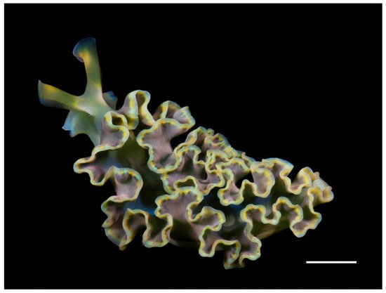
Some species of sacoglossan sea slugs are able to steal chloroplasts from the algae they feed on and maintain them functional for several months, a process termed “kleptoplasty”. One of these photosynthetic slugs is Elysia crispata, found in coral reefs of the Gulf of Mexico. This sacoglossan inhabits different depths (0–25 m), being exposed to different food sources and contrasting light conditions.
- Mollusca
- Heterobranchia
- kleptoplasty
- lipidomics
- phospholipids
- glycolipids
1. Introduction
2. Sample Collection

3. Photosynthetic Pigment Analysis
4. Lipid Analysis
Ultrapure water was added (800 µL) and centrifuged at 2000 rpm for 10 min to recover the organic phase. An additional volume of 800 µL of dichloromethane was added to the aqueous phase, centrifuged at 2000 rpm for 10 min and the organic phase was recovered. Organic phases were dried under a nitrogen stream and preserved at –20 °C for further analysis. Total lipid extract weight was estimated by gravimetry.
Glycolipid (GL) quantification in total lipid extracts was performed using the orcinol colorimetric method [43]. Briefly, 200 µg of total lipid extract were transferred to a glass tube and 1 mL of orcinol solution (0.2% in 70% H2SO4) was added after dying dichloromethane under a nitrogen flow. Tubes were then incubated for 20 min at 80 °C. D-Glucose standards of 2-50 µg (standard solution of D-glucose 2.0 mg mL−1) were used to prepare the calibration curve. At room temperature, the absorbance of standards and samples was measured at 505 nm using a microplate UV-Vis spectrophotometer. The conversion factor 100/35 (ca. 2.8) was used to estimate the total glycolipid content in total lipid extracts [44].
Phospholipid (PL) content from total lipid extract was quantified through the phosphorus assay, according to Bartlett and Lewis [45]. Lipid extracts were re-suspended in 300 µL of dichloromethane and 10 µL of each sample were transferred to a glass tube washed with 5% nitric acid. After drying under a nitrogen flow, 125 µL of perchloric acid (70%) was added and samples were incubated for 1 h at 180 °C in a heating block. A total of 825 µL of ultrapure water, 125 µL of NaMoO4∙H2O (2.5%), and 125 µL of ascorbic acid (10%) was added to each sample, with the mixture being homogenized in a vortex following each addition. Tubes were then incubated for 10 min at 100 °C in a water bath. Standards of 0.1-2 µg phosphate (standard solution of NaH2PO4∙2H2O, 100 µg of phosphorus mL-1) underwent the same treatment as samples, without the heating block step. At room temperature, absorbance of standards and samples was measured at 797 nm, using a microplate UV-Vis spectrophotometer. The conversion factor 775/31 (25) was used to estimate the total phospholipid content in total lipid extracts.
FA profile of E. crispata was analyzed by gas chromatography-mass spectrometry (GC-MS). FA methyl esters (FAME) were prepared using 30 µg of total lipid extract, 1 mL of the internal standard 19:0 (0.5 µg mL−1 in n-hexane, CAS number 1731-94-8, Merck) and 200 µL of a methanolic solution of potassium hydroxide (2 M) [46]. After homogenization of this mixture, 2 mL of an aqueous solution of sodium chloride (10 mg mL−1) were added. Sample was centrifuged at 2000 rpm for 5 min to separate the phases. The organic phase containing the FAME was transferred to a microtube and dried under a nitrogen stream. FAME were then dissolved in 40 µL of n-hexane, and 2 µL of this solution were injected on an Agilent Technologies 6890 N Network chromatograph equipped with a DB-FFAP column with 30 m length, an internal diameter of 0.32 mm, and a film thickness of 0.25 µm (J&W Scientific, Folsom, CA, USA). The GC was connected to an Agilent 5973 Network Mass Selective Detector operating with an electron impact mode at 70 eV and scanning the mass range m/z 50−550 in 1 s cycle in a full scan mode acquisition. The initial oven temperature was 80 °C, staying at this temperature for 3 min and increasing linearly to 160 °C at 25 °C min−1, followed by linear increases to 210 °C at 2 °C min−1 and 250 °C at 30 °C min−1. Temperature was maintained at 250 °C for 10 min. The injector and detector temperatures were 220 °C and 250 °C, respectively. Helium was used as carrier gas at a flow rate of 1.4 mL min−1. FAME present in the sample were identified by comparing their retention time and mass spectra with a commercial FAME standard mixture (Supelco 37 Component FAME Mix, ref. 47885-U, Sigma-Aldrich) and confirmed by comparison with the spectral library from ‘The Lipid Web’ [47]. FAME were quantified by using calibration curves of FAME standards acquired under the same instrumental conditions [48].
5. Conclusions
Authors provided a characterization of pigments, FA and lipid classes (PL and GL) from E. crispata sampled at different depths, which helps to fill a knowledge gap of one of the animal models most commonly employed to study kleptoplasty. To our knowledge, this is the first effort assessing differences in depth for a population of a sacoglossan sea slug, and the first work in the most western distribution recorded for this species.
The heterogeneity recorded in the profile of photosynthetic pigments and FA composition of E. crispata was not related with the habitat depth at the coral reef where they were sampled in Southern Gulf of Mexico. The total lipid, PL and GL contents found in this work were similar for specimens collected at shallow (0-4 m) and deeper (8-12 m) habitats. The conserved heterogeneity of their photosynthetic pigment profiles, as well as the high content of molecules exclusive of chloroplasts recorded on E. crispata, such as Chl a and GL after a month of food deprivation confirms that these sea slugs retain chloroplasts in good condition for long periods of time after stealing them from macroalgae.
This entry is adapted from the peer-reviewed paper 10.3390/ani11113157
References
- Händeler, K.; Grzymbowski, Y.P.; Krug, P.J.; Wägele, H. Functional chloroplasts in metazoan cells—A unique evolutionary strategy in animal life. Front. Zool. 2009, 6, 1–18.
- Van Steenkiste, N.W.L.; Stephenson, I.; Herranz, M.; Husnik, F.; Keeling, P.J.; Leander, B.S. A new case of kleptoplasty in animals: Marine flatworms steal functional plastids from diatoms. Sci. Adv. 2019, 5, 1–9.
- Baumgartner, F.A.; Pavia, H.; Toth, G.B. Individual specialization to non-optimal hosts in a polyphagous marine invertebrate herbivore. PLoS ONE 2014, 9.
- de Vries, J.; Rauch, C.; Christa, G.; Gould, S.B. A sea slug’s guide to plastid symbiosis. Acta Soc. Bot. Pol. 2014, 83, 415–421.
- Rauch, C.; Tielens, A.G.M.; Serôdio, J.; Gould, S.B.; Christa, G. The ability to incorporate functional plastids by the sea slug Elysia viridis is governed by its food source. Mar. Biol. 2018, 165, 1–13.
- Christa, G.; Händeler, K.; Kück, P.; Vleugels, M.; Franken, J.; Karmeinski, D.; Wägele, H. Phylogenetic evidence for multiple independent origins of functional kleptoplasty in Sacoglossa (Heterobranchia, Gastropoda). Org. Divers. Evol. 2015, 15, 23–36.
- Vieira, S.; Calado, R.; Coelho, H.; Serôdio, J. Effects of light exposure on the retention of kleptoplastic photosynthetic activity in the sacoglossan mollusc Elysia viridis. Mar. Biol. 2009, 156, 1007–1020.
- Laetz, E.M.J.; Wägele, H. How does temperature affect functional kleptoplasty? Comparing populations of the solar-powered sister-species Elysia timida Risso, 1818 and Elysia cornigera Nuttall, 1989 (Gastropoda: Sacoglossa). Front. Zool. 2018, 15, 1–13.
- Dionísio, G.; Faleiro, F.; Bispo, R.; Lopes, A.R.; Cruz, S.; Paula, J.R.; Repolho, T.; Calado, R.; Rosa, R. Distinct bleaching resilience of photosynthetic plastid-bearing mollusks under thermal stress and high CO2 conditions. Front. Physiol. 2018, 9, 1–11.
- Green, B.J.; Li, W.-Y.; Manhart, J.R.; Fox, T.C.; Summer, E.J.; Kennedy, R.A.; Pierce, S.K.; Rumpho, M.E. Mollusc-algal chloroplast endosymbiosis. Photosynthesis, thylakoid protein maintenance, and chloroplast gene expression continue for many months in the absence of the algal nucleus. Plant Physiol. 2000, 124, 331–342.
- Costa, J.; Giménez-Casalduero, F.; Melo, R.; Jesus, B. Colour morphotypes of Elysia timida (Sacoglossa, Gastropoda) are determined by light acclimation in food algae. Aquat. Biol. 2012, 17, 81–89.
- Middlebrooks, M.L.; Curtis, N.E.; Pierce, S.K. Algal sources of sequestered chloroplasts in the sacoglossan sea slug Elysia crispata vary by location and ecotype. Biol. Bull. 2019, 236, 88–96.
- Curtis, N.E.; Massey, S.E.; Pierce, S.K. The symbiotic chloroplasts in the sacoglossan Elysia clarki are from several algal species. Invertebr. Biol. 2006, 125, 336–345.
- Clark, K.B.; Busacca, M. Feeding specificity and chloroplast retention in four tropical Ascoglossa, with a discussion of the extent of chloroplast symbiosis and the evolution of the order. J. Molluscan Stud. 1978, 44, 272–282.
- Jensen, K.R. A review of sacoglossan diets with comparative notes on radular and buccal anatomy. Malacol. Rev. 1980, 13, 55–77.
- Middlebrooks, M.L.; Pierce, S.K.; Bell, S.S. Foraging behavior under starvation conditions is altered via photosynthesis by the marine gastropod, Elysia clarki. PLoS ONE 2011, 6, e22162.
- Curtis, N.E.; Middlebrooks, M.L.; Schwartz, J.A.; Pierce, S.K. Kleptoplastic sacoglossan species have very different capacities for plastid maintenance despite utilizing the same algal donors. Symbiosis 2015, 65, 23–31.
- Curtis, N.E.; Schwartz, J.A.; Pierce, S.K. Ultrastructure of sequestered chloroplasts in sacoglossan gastropods with differing abilities for plastid uptake and maintenance. Invertebr. Biol. 2010, 129, 297–308.
- Terashima, I.; Fujita, T.; Inoue, T.; Chow, W.S.; Oguchi, R. Green light drives leaf photosynthesis more efficiently than red light in strong white light: Revisiting the enigmatic question of why leaves are green. Plant Cell Physiol. 2009, 50, 684–697.
- Wilhelm, C.; Jungandreas, A.; Jakob, T.; Goss, R. Light acclimation in diatoms: From phenomenology to mechanisms. Mar. Genom. 2014, 16, 5–15.
- Cruz, S.; Cartaxana, P.; Newcomer, R.; Dionísio, G.; Calado, R.; Serôdio, J.; Pelletreau, K.N.; Rumpho, M.E. Photoprotection in sequestered plastids of sea slugs and respective algal sources. Sci. Rep. 2015, 5, 1–8.
- Cartaxana, P.; Morelli, L.; Quintaneiro, C.; Calado, G.; Calado, R.; Cruz, S. Kleptoplasts photoacclimation state modulates the photobehaviour of the solar- powered sea slug Elysia viridis. J. Exp. Biol. 2018, 221.
- Cartaxana, P.; Morelli, L.; Jesus, B.; Calado, G.; Calado, R.; Cruz, S. The photon menace: Kleptoplast protection in the photosynthetic sea slug Elysia timida. J. Exp. Biol. 2019, 222, 3–6.
- Jesus, B.; Ventura, P.; Calado, G. Behaviour and a functional xanthophyll cycle enhance photo-regulation mechanisms in the solar-powered sea slug Elysia timida (Risso, 1818). J. Exp. Mar. Bio. Ecol. 2010, 395, 98–105.
- Pierce, S.K.; Curtis, N.E.; Schwartz, J.A. Chlorophyll a synthesis by an animal using transferred algal nucleargenes. Symbiosis 2009, 49, 121–131.
- Middlebrooks, M.L.; Bell, S.S.; Pierce, S.K. The kleptoplastic sea slug Elysia clarki prolongs photosynthesis by synthesizing chlorophyll a and b. Symbiosis 2012, 57, 127–132.
- Trench, R.K.; Smith, D.C. Synthesis of pigment in symbiotic chloroplasts. Nature 1970, 227, 196–197.
- Ventura, P.; Calado, G.; Jesus, B. Photosynthetic efficiency and kleptoplast pigment diversity in the sea slug Thuridilla hopei (Vérany, 1853). J. Exp. Mar. Bio. Ecol. 2013, 441, 105–109.
- Cruz, S.; Calado, R.; Serôdio, J.; Jesus, B.; Cartaxana, P. Pigment profile in the photosynthetic sea slug Elysia viridis (Montagu, 1804). J. Molluscan Stud. 2014, 80, 475–481.
- Parrish, C.C. Lipids in Marine Ecosystems. ISRN Oceanogr. 2013, 2013, 1–16.
- Voogt, P.A. Lipids: Their Distribution and Metabolism. In The Mollusca. Metabolic Biochemistry and Molecular Biomechanics; Hochachka, P.W., Ed.; Academic Press, Inc.: New York, NY, USA, 1983; Volume 1, pp. 329–370. ISBN 0127514015.
- Joseph, J.D. Lipid composition of marine and estuarine invertebrates. Part II: Mollusca. Prog. Lipid Res. 1982, 21, 109–153.
- Pelletreau, K.N.; Weber, A.P.M.; Weber, K.L.; Rumpho, M.E. Lipid accumulation during the establishment of kleptoplasty in Elysia chlorotica. PLoS ONE 2014, 9, 1–16.
- Trench, R.K.; Boyle, J.E.; Smith, D.C. The association between chloroplasts of Codium fragile and the mollusc Elysia viridis. II. Chloroplast ultrastructure and photosynthetic carbon fixation in E. viridis. Proc. R. Soc. Lond.-Biol. Sci. 1973, 184, 63–81.
- Rey, F.; Da Costa, E.; Campos, A.M.; Cartaxana, P.; MacIel, E.; Domingues, P.; Domingues, M.R.M.; Calado, R.; Cruz, S. Kleptoplasty does not promote major shifts in the lipidome of macroalgal chloroplasts sequestered by the sacoglossan sea slug Elysia viridis. Sci. Rep. 2017, 7, 1–10.
- Rey, F.; Melo, T.; Cartaxana, P.; Calado, R.; Domingues, P.; Cruz, S.; Domingues, M.R.M. Coping with starvation: Contrasting lipidomic dynamics in the cells of two sacoglossan sea slugs incorporating stolen plastids from the same macroalga. Integr. Comp. Biol. 2020, 60, 43–56.
- Clark, K.B. Ascoglossan (=Sacoglossa) molluscs in the Florida Keys: Rare marine invertebrates at special risk. Bull. Mar. Sci. 1994, 54, 900–916.
- Camacho-García, Y.E.; Pola, M.; Carmona, L.; Padula, V.; Villani, G.; Cervera, J.L. Diversity and distribution of the heterobranch sea slug fauna on the Caribbean of Costa Rica. Cah. Biol. Mar. 2014, 55, 109–127.
- Lalli, C.M.; Parsons, T.R. Biological Oceanography, An Introduction, 2nd ed.; The Open University: Burlington, MA, USA, 1997.
- Krug, P.J.; Vendetti, J.E.; Valdés, Á. Molecular and morphological systematics of Elysia Risso, 1818 (Heterobranchia: Sacoglossa) from the Caribbean region. Zootaxa 2016, 4148, 1–137.
- Mendes, C.R.; Cartaxana, P.; Brotas, V. HPLC determination of phytoplankton and microphytobenthos pigments: Comparing resolution and sensitivity of a C18 and a C8 method. Limnol. Oceanogr. Methods 2007, 5, 363–370.
- Bligh, E.G.; Dyer, W.J. A rapid method of total lipid extraction and purification. Can. J. Biochem. Physiol. 1959, 37, 911–917.
- Leray, C. CyberLipid. Available online: http://cyberlipid.gerli.com/techniques-of-analysis/analysis-of-complex-lipids/glycoglycerolipid-analysis/quantitative-estimation/ (accessed on 13 May 2021).
- Bell, B.M.; Daniels, D.G.H.; Fearn, T.; Stewart, B.A. Lipid compositions, baking qualities and other characteristics of wheat varieties grown in the U.K. J. Cereal Sci. 1987, 5, 277–286.
- Bartlett, E.M.; Lewis, D.H. Spectrophotometric determination of phosphate esters in the presence and absence of orthophosphate. Anal. Biochem. 1970, 36, 159–167.
- Aued-Pimentel, S.; Lago, J.H.G.; Chaves, M.H.; Kumagai, E.E. Evaluation of a methylation procedure to determine cyclopropenoids fatty acids from Sterculia striata St. Hil. Et Nauds seed oil. J. Chromatogr. A 2004, 1054, 235–239.
- Christie, W.W. The Lipid Web. Available online: www.lipidmaps.org/resources/lipidweb (accessed on 4 December 2019).
- Cartaxana, P.; Rey, F.; Ribeiro, M.; Moreira, A.S.P.; Rosário, M.; Domingues, M.; Calado, R.; Cruz, S. Nutritional state determines reproductive investment in the mixotrophic sea slug Elysia viridis. Mar. Ecol. Prog. Ser. 2019, 611, 167–177.
