Livestock-Associated Zoonoses: History
Please note this is an old version of this entry, which may differ significantly from the current revision.
Contributor:

Human and animal health are intimately connected. This idea has been known for more than a century but now it has gained special importance because of the increasing threat from zoonoses. Zoonosis is defined as any infection naturally transmissible from vertebrate animals to humans. As the frequency and prevalence of zoonotic diseases increase worldwide, they become a real threat to public health. In addition, many of the newly discovered diseases have a zoonotic origin. Due to globalization and urbanization, some of these diseases have already spread all over the world, caused by the international flow of goods, people, and animals. However, special attention should be paid to farm animals since, apart from the direct contact, humans consume their products, such as meat, eggs, and milk. Therefore, zoonoses such as salmonellosis, campylobacteriosis, tuberculosis, swine and avian influenza, Q fever, brucellosis, Shiga-toxic Escherichia coli (STEC) infections, and listeriosis are crucial for both veterinary and human medicine. Consequently, in the suspicion of any zoonoses outbreak, the medical and veterinary services should closely cooperate to protect the public health. 

  • One Health
  • zoonotic pathogens
  • foodborne diseases

1. Background

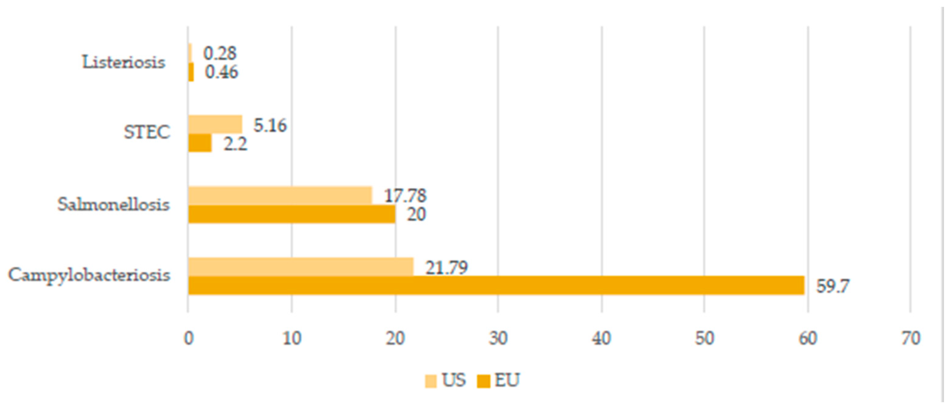
The twenty-first century is the age of globalization and urbanization, and is characterized by more and more free flows of people, animals, and goods around the world. Therefore, the conception of One Health gains importance like never before. The main assumption behind this idea is that the environment and human as well as animal health are intimately connected and interdependent. Any infection naturally transmissible from vertebrate animals to humans is called zoonosis. The pathogen transmission from animal to human is not only associated with the direct contact but also may occur via vectors or consuming animal products such as milk, meat, or eggs (foodborne diseases). Zoonotic diseases, particularly those associated with livestock and poultry, are becoming an increasing threat for public health due to different reasons. For example, the predictions suggest that the global human population will constantly increase and reach almost 10 billion by 2050 [1]. Consequently, it will result in a higher food demand. Thus, the livestock population is also expected to increase in order to cover the need for food, in particular regarding the high nutritional value of, for example, dairy or meat products. In 2020, the world meat and milk production was estimated at 337.2 and 906 million tonnes, respectively [2]. However, according to FAO, more than 70% of additional animal protein will be needed to feed the world by 2050, which suggests that animal production worldwide will be expected to grow. In turn, this potentially increases the risk of transmitting pathogens from animals to humans. The World Organization for Animal Health (OIE) suggests that 60% of pathogens that cause human diseases originate from domestic animals or wildlife [3]. Another fact is that 75% of emerging human pathogens are of animal origin [3]. For example, it has been suspected from the beginning that the original outbreak of SARS-CoV-2 was of zoonotic origin, possibly linked to a market in Wuhan, which sold a variety of animals including wild birds, poultry, fish, shellfish, and other exotic species [4]. It is important to note that the significance of particular zoonotic diseases differs within the continent and/or country mainly due to different zoohygienic conditions, human dietary habits, dominant livestock species, and legal environments. For example, according to the European Food Safety Authority (EFSA), the most frequent zoonoses in 2020 in the EU were campylobacteriosis, salmonellosis, Shiga-toxic Escherichia coli (STEC) infections, yersiniosis, listeriosis, and tularaemia, each reaching more than 1000 human cases requiring hospitalization [5]. Meanwhile, the Centre for Diseases Control (CDC) suggests prioritizing in the USA the following diseases and pathogens with zoonotic properties: influenza (zoonotic influenza A viruses), Salmonellosis (Salmonella species), West Nile virus, Plague (Yersinia pestis), emerging coronaviruses (Coronaviridae; i.e., severe acute respiratory syndrome (SARS-CoV) and Middle East respiratory syndrome (MERS-CoV)), Rabies (Rhabdoviridae, Lyssavirus), Brucellosis (Brucella species), and Lyme disease (Borrelia burgdorferi) [6]. A brief comparison of the chosen zoonoses incidences in the UE and US in 2019 is given in Figure 1 [5][7]. On the other hand, research from East Africa [8] revealed that the greatest concern regarding zoonoses is reserved for trypanosomiasis and brucellosis in this part of the world. From the Chinese point of view, major emerging zoonoses include SARS, Highly Pathogenic Avian Influenza (HPAI), rabies, Japanese encephalitis, brucellosis, and schistosomiasis japonica [9].
A brief summary of zoonotic threats from livestock is given in Table 1. It is important to note that decent knowledge of these diseases and their transmission is crucial since it enables people to take action, including introducing proper risk assessment models. It involves the application of new technologies such as metagenomics, which is now the main method used to identify novel viruses and thus plays a central role in studies aimed at assessing zoonotic risk [10]. From a global point of view, the key reference regarding risk assessment models is the tripartite guide addressing zoonotic diseases, which was developed by the experts from the Food and Agriculture Organization of the United Nations (FAO), World Health Organization (WHO), and World Organization for Animal Health (OIE). Besides, constant epidemiological surveillance and report systems must be timely and efficient since surveillance in animals and humans is critical for the early identification and possible prediction of future outbreaks, allowing for preemptive action [11]. Similarly, timely, accurate, and reliable laboratory tests are critical for identifying etiologies of disease and to monitor both endemic and emerging zoonotic diseases in humans and livestock, which allows for implementing proper prevention as well as detection and response strategies [11]. However, it is important to note that animals other than livestock can also be a risk for human health including, e.g., dogs and cats (rabies), as well as wildlife (rabies, tularemia, and Lyme disease) [12][13]. Another important perspective regards climate change and the possible detrimental influence on vector-borne diseases, which may in the future expand and/or alter the geographical ranges of biological vectors and consequently the zoonotic diseases transmitted by them [14].
Idr 14 00008 g001
Figure 1. Selected livestock-associated zoonoses: comparison of the number of cases in United States (US) and European Union (EU) in 2019 (cases per 100,000 population). Created based on data from [5][7].
Table 1. A brief summary of the most significant livestock-associated zoonoses.
Disease Aetiological Agent Human Symptoms Transmission Route Epidemiology References
Q fever Coxiellaburnetti Self-limited febrile illness, pneumonia, hepatitis, and endocarditis Inhalation of aerosolized bacteria, ingestion, transfusion of blood, and sexual transmission EU—950 human cases in 2019
USA—178 human cases in 2019
[5]
[6]
[15]
Brucellosis Brucellaabortus,
B. melitensis,
B. canis,
B. suis
Systematic syndrome (fever, sweat, chills, and fatigue), located presentations (epididymoorchitis and spondylodiscitis), neurobrucellosis, and endocarditis Contaminated food and dairy products, occupational contact, and inhalation World—around 500,000
human cases per year
EU—310 human cases in 2019
USA—80–120 cases annually
[5]
[6]
[16]
Tuberculosis Mycobacterium bovis
M. caprae
Generalized symptoms (fever, fatigue, arthralgia, and muscle pain), respiratory and cardiac complications, hepatitis, osteoarthritis, central nervous system dysfunction, and orchitis/epididymitis Inhalation of aerosol, infected milk, dairy products, and meat EU—147 human cases in 2019
USA—7174 human cases in 2020
[5]
[17]
[18]
Trichinellosis Trichinella sp. Diarrhea, abdominal pain at first, fever, myalgia, myocarditis, facial oedemas, and encephalitis Ingestion of raw or undercooked muscle tissue containing encysted larvae EU—96 human cases in 2019
USA—90 human cases during 2008–2012
[5]
[6]
[19]
Yersiniosis Yersinia enterocolitica,
Y. pseudotuberculosis
Fever, vomiting, abdominal pain, and bloody diarrhea Eating raw or undercooked pork; ingestion of dairy products, seafood, and vegetables; or drinking contaminated water EU—6961 human cases
USA—nearly 117,000 illnesses per year
[5]
[6]
[20]
Swine
influenza
Swine influenza
virus (SIV)
Sneezing, coughing, difficult breathing, fever, lethargy, and decreased appetite Contact with respiratory discharges or inhalation of exhalated aerosol by sick pig No specific epidemiological
data available, spread worldwide
[21]
[22]
[23]
Salmonellosis Salmonella sp. Acute enterocolitis accompanied by inflammatory diarrhea, abdominal pain, fever, nausea, and vomiting Ingestion of uncooked contaminated foods (eggs, milk, and meat), drinking contaminated water, direct contact with infected animals, their feces and environment, and human-to-human transmission through fecal–oral route EU—87,923 human cases in 2019
USA—about 1.35 million human illnesses per year
Sub-Saharan Africa—535,500 cases of non-typhoidal salmonellosis in 2019
[5]
[6]
[24]
[25]

2. Q Fever

Q fever is a severe, zoonotic worldwide disease caused by Coxiella burnetii. This disease was first described by Derrick in 1937 following an epidemic fever outbreak among employees at a slaughterhouse in Brisbane (Australia) [26]. Coxiella burnetii is an obligate intracellular bacterium. Its cell wall is similar to that of Gram-negative bacteria but is not stainable with the Gram technique [15]. Coxiella burnetii is a microorganism with a very high infection capacity; a single germ is capable of causing infection [27]. This bacterium presents a phenomenon called “antigenic phase variation”. It is a molecular phenomenon that is produced by modification in the complexity of the membrane LPS, which will cause a difference in virulence [28]. The main reservoirs of C. burnetii are cattle, sheep, and goats, but infections were detected in other animals such as domestic mammals, marine mammals, reptiles, birds, and ticks. Coxiella burnetii is most abundant in birth products and in the urine, feces and milk of infected animals. Transmission to humans most commonly occurs through inhalation of aerosolized bacteria from the placenta (delivery or abortion), feces, or urine of infected animals. Human-to-human transmission is extremely rare [15][28][29]. Other routes of transmission of the disease are oral transmission after the ingestion of contaminated raw dairy products, transfusion of blood products, sexual transmission, and professional exposure, as in the case of pathologists or microbiologists [30][31]. In humans, the average incubation time is 18 days (between 7 and 32 days) [29]. Infected animals are usually asymptomatic. Spontaneous abortions, endometritis, mastitis, and infertility are the only signs that can be observed [15]. In humans, Q fever can manifest as an acute disease usually as a self-limited febrile illness, pneumonia, or hepatitis. It may also occur as a persistent focalized infection with endocarditis [32]. In humans, the diagnosis of Q fever is mainly made by serology, microbiological cultures, or PCR tests [28]. The European Union (EU) One Health Zoonoses Report indicates that 950 human cases of Q fever have been reported in the EU in 2019 [5]. According to CDC, a total of 212 cases of Q fever have been reported in the US in 2019, including 178 human cases of acute illness and 34 cases of chronic Q fever disease [6]. In Africa, seroprevalence rates in humans varied from 1% in Chad to 16% in Egypt [15]. Between 2007 and 2010, there was an outbreak of Q fever in the Netherlands, with more than 4000 reported human cases and an estimation of probably more than 40,000 total human cases [15]. The highest prevalence recorded is in Cayenne, French Guiana, where C. burnetii causes 24% of community-acquired pneumonia [33].

3. Brucellosis

Brucellosis is caused by the intracellular pathogens from genus Brucella [34]. Brucella spp. can multiply within phagocytic cells with human beings as end hosts [35]. Four species of Brucella can infect humans: B. abortus, B. canis, B. melitensis, and B. suis. Of these species, B. melitensis is the most commonly isolated from ruminants [16]. In sheep and goats, which are the main hosts of B. melitensis, the bacterium causes impaired fertility and abortions [36]. Brucellosis in cattle is caused by B. abortus, which can be easily transmitted to humans. Brucellosis may be transmitted to humans through contaminated food and dairy products, occupational contact, or inhalation of infected aerosols [16]. Another important route of infection is the contamination of mucous membranes or open wounds with fetal fluids, making veterinarians, farmers, and abattoir workers the most vulnerable to infection. In other cases, transmission from animals to humans is mainly associated with drinking contaminated milk [37]. Human-to-human transmission takes place through lactation, sexual intercourse, and tissues such as blood transfusion and bone marrow transplantation [38]. Brucellosis in humans has several, often non-specific, presentations, including a systemic syndrome (fever, sweat, chills, and fatigue), but also some located presentations (epididymoorchitis and spondylodiscitis). Severe forms of this disease are neurobrucellosis and endocarditis [38]. Brucellosis is one of the most frequent zoonosis in many parts of the world. However, this disease mainly affects humans in developing countries, as it is effectively controlled in developed countries [16]. Brucellosis is an endemic zoonosis for the Middle East, the Mediterranean rim, Asia, Africa, and South and Central America. These are regions with a very high consumption of dairy products and insufficient animal health care [37]. Around 500,000 cases of brucellosis in humans are reported worldwide each year [36]. However, true incidence is estimated to be 5,000,000 to 12,500,000 cases annually [39]. Seroprevalence by country in sub-Saharan Africa is 24.1% and 31.82% in Nigeria, 17% in Uganda, 7.7% in Tanzania, 3.8% in Chad, and 3.3% in the Central African Republic [40]. Brucellosis is a major economic problem in African countries such as Nigeria. In this country, high losses are generated for cattle producers due to stillbirths, reduced calving percentages, medical costs, births of weak calves, culls due to infertility, and the loss of man-hours in infected people. Furthermore, wastes in meat and dairy production are estimated at USD 224 million per year. To compare, in the Republic of South Africa, the losses due to brucellosis are USD 37.5 million and in the USA, they equal to USD 800 million per year [41]. According to the EU One Health Zoonoses Report, 310 human cases of brucellosis have been reported in the EU in 2019 [5]. As reported by the CDC, areas currently listed as high risk of brucellosis are the Mediterranean Basin (Portugal, Spain, Southern France, Italy, Greece, Turkey, and North Africa), Mexico, South and Central America, Eastern Europe, Asia, Africa, the Caribbean, and the Middle East [6]. In the United States, brucellosis is a rare disease, with 80–120 cases reported annually [6]. Syria has the highest number of human brucellosis with 1603.4 cases per 1,000,000 individuals. This is followed by Mongolia (3910), Iraq (268.8), Tajikistan (211.9), Saudi Arabia (149.5), and Iran (141.6) [39]. In China, brucellosis is also an important public health threat. In 2014, 4.2 cases/100,000 people were reported [42].

4. Tuberculosis Caused by Mycobacterium bovis and Mycobacterium caprae
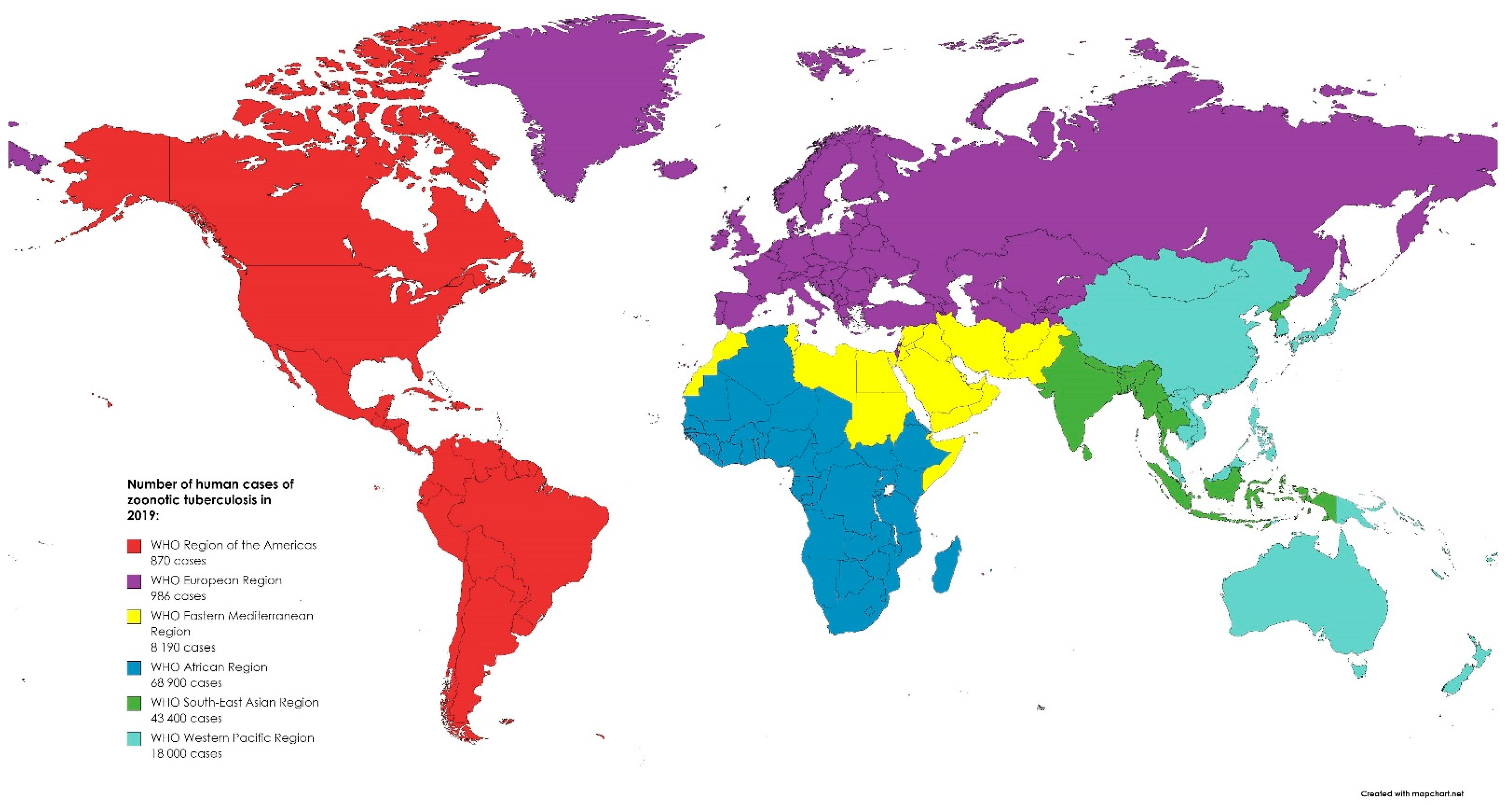
Mycobacterium caprae and Mycobacterium bovis are members of the Mycobacterium tuberculosis complex and cause tuberculosis (TB) in animals and humans. M. caprae is isolated not only from goats but also from sheep, red deer, cattle, wild boar, the Siberian tiger, camel, bison, and humans [17]. M. caprae causes lesions and diseases like that of M. bovis but occurs only in a low proportion of human TB cases. Moreover, M. caprae is evolutionarily older than its epidemiological twin, M. bovis. This bacterium is not globally distributed but primarily restricted to European countries [43]. On the other hand, the most common host of M. bovis is cattle, but other mammals, such as marsupials, carnivores, pinnipeds, lagomorphs, rodents, and some avian species, could be also infected [44]. The main route of TB transmission in animals is via aerosol by the droplet nuclei generated during coughing and sneezing. Humans may be also infected through milk, dairy products, and by eating meat from infected animals [45]. The disease is manifested in humans by fever, fatigue, arthralgia, and muscle pain, and variety of other symptoms depending on the part of the body affected by the disease. WHO reported that in 2016, there were 147,000 new cases and 12,500 people died due to TB, but with no information of the potential zoonotic origin [18]. M. bovis is responsible only for 3.1% cases, with the exception for Tanzania, in which it reached 16% of TB in humans possibly due to poor zoohygienic conditions [46]. In 2019, most of the zoonotic TB human cases occurred in Africa (50%) and South-East Asia (31%). Globally, there were 140,000 human cases of zoonotic TB. However, the uncertainty level is estimated to be 69,800 to 235,000 [47]. While in Europe, TB is a rare infection with 147 confirmed cases in humans reported in 2019 in the EU. Between 2015 and 2019, 918 cases of TB were confirmed in the EU, including 54 caused by M. caprae [5]. The global distribution of zoonotic TB human cases in 2019 is presented in Figure 2. The most effective way to eliminate TB in farm animals is through implementation of eradication programs. In developed countries, infection with M. bovis is not common in cattle. This is related to compulsory tuberculin testing, the pasteurization of milk, and the removal of positive reactors [48]. Before the routine application of milk pasteurization in the United Kingdom (UK), M. bovis was isolated from 8% of churn milk samples from 3000-gallon tankers in 1945 [49]. In the 21st century, only 315 cases of human TB have been reported in the UK over a 10-year period [50]. However, it is important to note that cattle can become infected from wild mammals. This can also have an impact on the eradication of the disease. To date, the following have been identified as reservoirs of the mycobacteria around the world: brushtail possum and badger, European bison, African buffalo, wild boar, and white-tailed deer, among many others [51]. Research shows that Michigan deer may have infected surrounding cattle. Data indicate that while recording cases of the disease in wild deer between 1975 and 1994, infected animals were found in sixteen domestic cattle herds in four counties in the north-western part of the state [52]. Transmission from humans to cattle is also possible. M. bovis is usually transmitted directly by inhalation but also indirectly by hay and bedding contaminated with urine. In the Netherlands, humans were the source of transmission for 50 cattle herds [53]. Zoonotic M. bovis infections are mainly a problem in undeveloped countries. In the developing countries, due to the lack of control of zoonotic products, poor production hygiene, and outbreaks of other diseases (e.g., AIDS), the pathogen will continue to persist and remain a real challenge for public health in the future.
Idr 14 00008 g002
Figure 2. The global distribution of zoonotic tuberculosis human cases in 2019. Created based on data from [47].

5. Salmonellosis

Salmonella is a large, ubiquitous genus of Gram-negative, rod-shaped, facultative anaerobic bacteria belonging to the family of Enterobacteriaceae and is responsible for zoonotic infections of global significance. It can be persistent in dry environments as well as in water for months [54]. There are two main species distinguished: S. enterica (which includes more than 2600 known serovars) and S. bongori. The majority of variants of S. enterica are motile by the means of flagella but the most important virulence factors are invasion and intracellular replication [54]. Salmonella was first isolated in 1884 by an American bacteriologist, D. E. Salmon, from porcine intestine [24] and in the 1980s, the first pandemic of S. enterica s. Enteritidis emerged due to contaminated poultry products [55]. Salmonella sp. may cause clinical disease in livestock or subclinical infections in asymptomatic animals (carriers), such as dogs and cats, which transmit and contaminate the environment of food-producing animals. A very important role is played by vertical transmission, especially in the poultry and bovine reproduction sector, but pests are also a significant vector of the germ [25][54]. An infection in humans can occur after drinking contaminated water or ingesting uncooked contaminated eggs, milk, and meat originating from poultry, cattle, or swine, although there have been reports about other foods, including vegetables contaminated by manure and ready-to-eat foods that caused infection. Human-to-human transmission through the fecal–oral route and infection after direct contact with infected animals, their feces, and the environment are less common, although still significant. What is a concern is that Salmonella can pass through the entire food chain, starting from animal feed contaminated by manure and primary production to the table in households, food services, and institutions (farm-to-fork continuum) [6][56]. EFSA reported that salmonellosis after campylobacteriosis was the second most often reported gastrointestinal infection in humans. In 2019, 87,923 cases were confirmed in the EU [5]. According to OIE, salmonellosis qualifies as one of the most common foodborne bacterial diseases in the world. Human infections caused by Salmonella species are most frequently caused by S. Enteritidis and S. Typhimurium, which are normally found in the intestines of humans and animals, as they are the main reservoir of these bacteria [3][24]. A distinction is made between three major diseases caused by Salmonella in humans, namely non-invasive non-typhoidal salmonellosis, invasive non-typhoidal salmonellosis, and typhoid fever, but in general, salmonellosis manifests with acute enterocolitis accompanied by inflammatory diarrhea, abdominal pain, fever, nausea, and vomiting in humans [57]. Most cases of the disease are underdiagnosed, turning salmonellosis into a disease that contributes to the deaths of thousands of people worldwide, especially in economically underdeveloped countries [54].

6. Conclusions

Human and animals’ health are intimately connected since they share a communal environment. Consequently, pathogen transmission is possible and it may occur via direct and/or indirect contact, including consuming products of animal origin. In the case of confirmed infection derived from animals or animal products, a comprehensive approach should be applied. Thus, in the suspicion of any zoonoses, the medical and veterinary doctors should closely cooperate to protect public health and work in accordance with the One Health conception.

This entry is adapted from the peer-reviewed paper 10.3390/idr14010008

References

  1. United Nations, Department of Economic and Social Affairs, Population Division. World Population Prospects 2019: Highlights (ST/ESA/SER.A/423); United Nations: New York, NY, USA, 2019.
  2. FAO. World Food and Agriculture—Statistical Yearbook 2021; FAO: Rome, Italy, 2021.
  3. World Organization for Animal Health Official Website. Available online: https://www.oie.int/en/what-we-do/global-initiatives/one-health/ (accessed on 25 November 2021).
  4. Pomorska-Mól, M.; Włodarek, J.; Gogulski, M.; Rybska, M. Review: SARS-CoV-2 Infection in farmed minks—An overview of current knowledge on occurrence, disease and epidemiology. Animal 2021, 15, 100272.
  5. European Food Safety Authority; European Centre for Disease Prevention and Control. The European Union One Health 2019 Zoonoses Report; EFS2; 2021; Volume 19, pp. 1–286. Available online: https://efsa.onlinelibrary.wiley.com/doi/full/10.2903/j.efsa.2021.6406 (accessed on 5 December 2021).
  6. Centers for Disease Control and Prevention Official Website. Available online: https://www.cdc.gov/onehealth/pdfs/us-ohzdp-report-508.pdf (accessed on 25 November 2021).
  7. Centers for Disease Control and Prevention. National Notifiable Diseases Surveillance System, 2019 Annual Tables of Infectious Disease Data; CDC Division of Health Informatics and Surveillance: Atlanta, GA, USA, 2021.
  8. Kemunto, N.; Mogoa, E.; Osoro, E.; Bitek, A.; Kariuki Njenga, M.; Thumbi, S.M. Zoonotic disease research in East Africa. BMC Infect. Dis. 2018, 18, 545.
  9. Liu, Q.; Cao, L.; Zhu, X.-Q. Major emerging and re-emerging zoonoses in China: A matter of global health and socioeconomic development for 1.3 billion. Int. J. Infect. Dis. 2014, 25, 65–72.
  10. Wille, M.; Geoghegan, J.L.; Holmes, E.C. How accurately can we assess zoonotic risk? PLoS Biol. 2021, 19, e3001135.
  11. Belay, E.D.; Kile, J.C.; Hall, A.J.; Barton-Behravesh, C.; Parsons, M.B.; Salyer, S.; Walke, H. Zoonotic disease programs for enhancing global health security. Emerg. Infect. Dis. 2017, 23, S65.
  12. Acharya, K.P.; Acharya, N.; Phuyal, S.; Upadhyaya, M.; Lasee, S. One-Health approach: A best possible way to control rabies. One Health 2020, 10, 100161.
  13. Kruse, H.; Kirkemo, A.-M.; Handeland, K. Wildlife as source of zoonotic infections. Emerg. Infect. Dis. 2004, 10, 2067–2072.
  14. Rocklöv, J.; Dubrow, R. Climate change: An enduring challenge for vector-borne disease prevention and control. Nat. Immunol. 2020, 21, 479–483.
  15. Eldin, C.; Mélenotte, C.; Mediannikov, O.; Ghigo, E.; Million, M.; Edouard, S.; Mege, J.-L.; Maurin, M.; Raoult, D. From Q fever to Coxiella burnetii infection: A paradigm change. Clin. Microbiol. Rev. 2017, 30, 115–190.
  16. Amjadi, O.; Rafiei, A.; Mardani, M.; Zafari, P.; Zarifian, A. A review of the immunopathogenesis of brucellosis. Infect. Dis. 2019, 51, 321–333.
  17. Kozińska, M.; Krajewska-Wędzina, M.; Augustynowicz-Kopeć, E. Mycobacterium caprae—the first case of the human infection in Poland. Ann. Agric. Environ. Med. 2020, 27, 151–153.
  18. World Health Organization; Food and Agriculture Organization of the United Nations. World Organisation for Animal Health Roadmap for Zoonotic Tuberculosis; World Health Organization: Geneva, Switzerland, 2017; ISBN 978-92-4-151304-3.
  19. Bilska-Zając, E.; Różycki, M.; Korpysa-Dzirba, W.; Bełcik, A.; Ziętek-Barszcz, A.; Włodarczyk-Ramus, M.; Gontarczyk, A.; Cencek, T. Trichinella outbreaks on pig farms in Poland in 2012–2020. Pathogens 2021, 10, 1504.
  20. Laukkanen-Ninios, R.; Fredriksson-Ahomaa, M.; Maijala, R.; Korkeala, H. High prevalence of pathogenic Yersinia enterocolitica in pig cheeks. Food Microbiol. 2014, 43, 50–52.
  21. Smith, G.J.D.; Vijaykrishna, D.; Bahl, J.; Lycett, S.J.; Worobey, M.; Pybus, O.G.; Ma, S.K.; Cheung, C.L.; Raghwani, J.; Bhatt, S.; et al. Origins and evolutionary genomics of the 2009 swine-origin H1N1 influenza A epidemic. Nature 2009, 459, 1122–1125.
  22. Ito, T.; Couceiro, J.N.S.S.; Kelm, S.; Baum, L.G.; Krauss, S.; Castrucci, M.R.; Donatelli, I.; Kida, H.; Paulson, J.C.; Webster, R.G.; et al. Molecular basis for the generation in pigs of influenza A viruses with pandemic potential. J. Virol. 1998, 72, 7367–7373.
  23. Myers, K.P.; Olsen, C.W.; Setterquist, S.F.; Capuano, A.W.; Donham, K.J.; Thacker, E.L.; Merchant, J.A.; Gray, G.C. Are swine workers in the United States at increased risk of infection with zoonotic influenza virus? Clin. Infect. Dis. 2006, 42, 14–20.
  24. Popa, G.L.; Popa, M.I. Salmonella spp. infectio—A continuous threat worldwide. Germs 2021, 11, 88–96.
  25. Roadmap to Reducing Salmonella Driving Change through Science-Based Policy; Food Safety the U.S. Department of Agriculture: Washington, DC, USA, 2020.
  26. Derrick, E.H. “Q” fever, a new fever entity: Clinical features, diagnosis and laboratory investigation. Med. J. Aust. 1937, 2, 281–299.
  27. Miller, H.K.; Priestley, R.A.; Kersh, G.J. Transmission of Coxiella burnetii by ingestion in mice. Epidemiol. Infect. 2020, 148, 1–8.
  28. Pechstein, J.; Schulze-Luehrmann, J.; Lührmann, A. Coxiella burnetii as a useful tool to investigate bacteria-friendly host cell compartments. Int. J. Med. Microbiol. Suppl. 2018, 308, 77–83.
  29. España, P.P.; Uranga, A.; Cillóniz, C.; Torres, A. Q fever (Coxiella burnetii). Semin. Respir. Crit. Care. Med. 2020, 41, 509–521.
  30. Kersh, G.J.; Priestley, R.; Massung, R.F. Stability of Coxiella burnetii in stored human blood: C. burnetii stability in blood. Transfusion 2013, 53, 1493–1496.
  31. Signs, K.A.; Stobierski, M.G.; Gandhi, T.N. Q fever cluster among raw milk drinkers in Michigan, 2011. Clin. Infect. Dis. 2012, 55, 1387–1389.
  32. Angelakis, E.; Raoult, D. Q fever. Vet. Microbiol. 2010, 140, 297–309.
  33. Epelboin, L.; Chesnais, C.; Boulle, C.; Drogoul, A.-S.; Raoult, D.; Djossou, F.; Mahamat, A. Q fever pneumonia in French Guiana: Prevalence, Risk Factors, and Prognostic Score. Clin. Infect. Dis. 2012, 55, 67–74.
  34. Galińska, E.M.; Zagórski, J. Brucellosis in humans—Etiology, diagnostics, clinical forms. Ann. Agric. Environ. Med. 2013, 20, 6.
  35. Franco, M.P.; Mulder, M.; Gilman, R.H.; Smits, H.L. Human brucellosis. Lancet Infect. Dis. 2007, 7, 775–786.
  36. Głowacka, P.; Żakowska, D.; Naylor, K.; Niemcewicz, M.; Bielawska-Drózd, A. Brucella—virulence factors, pathogenesis and treatment. Pol. J. Microbiol. 2018, 67, 151–161.
  37. Rubach, M.P.; Halliday, J.E.B.; Cleaveland, S.; Crump, J.A. Brucellosis in low-income and middle-income countries. Curr. Opin. Infect. Dis. 2013, 26, 404–412.
  38. Tuon, F.F.; Gondolfo, R.B.; Cerchiari, N. Human-to-human transmission of Brucella—A systematic review. Trop. Med. Int. Health 2017, 22, 539–546.
  39. Hull, N.C.; Schumaker, B.A. Comparisons of Brucellosis between human and veterinary medicine. Infect. Ecol. Epidemiol. 2018, 8, 1500846.
  40. Awah-Ndukum, J.; Mouiche, M.M.M.; Kouonmo-Ngnoyum, L.; Bayang, H.N.; Manchang, T.K.; Poueme, R.S.N.; Kouamo, J.; Ngu-Ngwa, V.; Assana, E.; Feussom, K.J.M.; et al. Seroprevalence and risk factors of brucellosis among slaughtered indigenous cattle, abattoir personnel and pregnant women in Ngaoundéré, Cameroon. BMC Infect. Dis. 2018, 18, 611.
  41. Mai, H.M.; Irons, P.C.; Kabir, J.; Thompson, P.N. A large seroprevalence survey of brucellosis in cattle herds under diverse production systems in Northern Nigeria. BMC Vet. Res. 2012, 8, 144.
  42. Zheng, R.; Xie, S.; Lu, X.; Sun, L.; Zhou, Y.; Zhang, Y.; Wang, K. A systematic review and meta-analysis of epidemiology and clinical manifestations of human brucellosis in China. BioMed Res. Int. 2018, 2018, 5712920.
  43. Prodinger, W.M.; Indra, A.; Koksalan, O.K.; Kilicaslan, Z.; Richter, E. Mycobacterium caprae infection in humans. Expert Rev. Anti Infect. Ther. 2014, 12, 1501–1513.
  44. Kaneene, J.B.; Miller, R.; de Kantor, I.N.; Thoen, C.O. Tuberculosis in wild animals. Int. J. Tuberc. Lung Dis. 2010, 14, 1508–1512.
  45. Khan, M.; Islam, M.M.; Ferdous, J.; Alam, M. An overview on epidemiology of tuberculosis. Mymensingh Med. J. 2019, 28, 259–266.
  46. El-Sayed, A.; El-Shannat, S.; Kamel, M.; Castañeda-Vazquez, M.A.; Castañeda-Vazquez, H. Molecular epidemiology of Mycobacterium bovis in humans and cattle. Zoonoses Public Health 2016, 63, 251–264.
  47. World Health Organization. Global Tuberculosis Report 2019; World Health Organization: Geneva, Switzerland, 2019.
  48. Michel, A.L.; Müller, B.; van Helden, P.D. Mycobacterium bovis at the animal–human interface: A problem, or not? Vet. Microbiol. 2010, 140, 371–381.
  49. Grange, J.M. Mycobacterium bovis infection in human beings. Tuberculosis 2001, 81, 71–77.
  50. Jalava, K.; Jones, J.A.; Goodchild, T.; Clifton-Hadley, R.; Mitchell, A.; Story, A.; Watson, J.M. No increase in human cases of Mycobacterium bovis disease despite resurgence of infections in cattle in the United Kingdom. Epidemiol. Infect. 2007, 135, 40–45.
  51. Palmer, M.V. Mycobacterium bovis: Characteristics of wildlife reservoir hosts. Transbound. Emerg. Dis. 2013, 60, 1–13.
  52. De Lisle, G.W.; Schlundt, J.; Schmitt, S.M.; O’Brien, D.J. Tuberculosis in free-ranging wildlife: Detection, diagnosis and management. Rev. Sci. Tech. OIE 2002, 21, 317–334.
  53. O’Reilly, L.M.; Daborn, C.J. The epidemiology of Mycobacterium bovis infections in animals and man: A review. Tuberc. Lung Dis. 1995, 76, 1–46.
  54. Jajere, S.M. A review of Salmonella enterica with particular focus on the pathogenicity and virulence factors, host specificity and antimicrobial resistance including multidrug resistance. Vet. World 2019, 12, 504–521.
  55. Li, S.; He, Y.; Mann, D.A.; Deng, X. Global spread of Salmonella enteritidis via centralized sourcing and international trade of poultry breeding stocks. Nat. Commun. 2021, 12, 5109.
  56. World Health Organization Official Website. Influenza (Avian and Other Zoonotic). Available online: https://www.who.int/news-room/fact-sheets/detail/influenza-(avian-and-other-zoonotic) (accessed on 25 November 2021).
  57. Kurtz, J.R.; Goggins, J.A.; McLachlan, J.B. Salmonella infection: Interplay between the bacteria and host immune system. Immunol. Lett. 2017, 190, 42–50.
More
This entry is offline, you can click here to edit this entry!
ScholarVision Creations