You're using an outdated browser. Please upgrade to a modern browser for the best experience.
Fungal Pathogens on Bast Fiber Crops: History
Please note this is an old version of this entry, which may differ significantly from the current revision.
Subjects: Mycology | Agronomy
Contributor: Jianping Xu

Bast fiber crops are an important group of economic crops for the purpose of harvesting fibers from stems. These fibers are sclerenchyma fibers associated with the phloem of plants. They arise either with primary tissues from the apical meristem, or with secondary tissues produced by the lateral meristem. Fungal diseases have become an important factor limiting their yield and quality, causing devastating consequences for the production of bast fiber crops in many parts of the world.

  • bast fiber crops
  • molecular identification
  • fungal disease
  • DNA barcode
  • PCR assay

1. Introduction

Plant infectious diseases are among the most important constraints on the quality and yield of crops. It is estimated that plant diseases cause losses of 10%–15% of the world’s major crops, with direct economic losses of up to hundreds of billions of dollars each year. About 70%–80% of crop diseases are caused by fungal pathogens and the damage can be very serious, significantly reducing the yield and quality of many staple food crops and economic crops like fruits, vegetables, and fiber crops [1]. In addition, several fungal pathogens can secrete a variety of toxins and metabolites harmful to humans and animals, posing a great threat to the safety of agricultural products [2]. At present, most control measures against plant fungal pathogens rely on the applications of broad-spectrum fungicides. However, such fungicides not only increase production costs, but also can bring problems such as environmental pollution, fungicide resistance, and persistent residues on foods and other consumer goods with further implications for human health. In order to minimize the damage to crops caused by fungal diseases, as well as to maximize productivity and ensure agricultural sustainability, early detection and quantification of fungal pathogens is essential for disease prevention and control. However, conventional protocols based on morphological and physiological methods are time-consuming, require significant experience, and may not be sensitive and specific for individual pathogens [3]. Moreover, many fungal pathogens can remain latent in “sub-infection” stages with no obvious symptoms and/or in low numbers, making them difficult to detect, and causing confusion with regard to their roles in diseases. These issues can contribute to delayed or wrong control measures.
During the last three decades, to overcome these problems and minimize crop losses caused by fungal diseases, a diversity of DNA molecule-based tools has been developed for the detection and identification of fungal pathogens. These techniques include conventional polymerase chain reaction (PCR) [4], quantitative PCR (qPCR) [5,6], immunocapture-PCR (IC-PCR) [7,8], droplet digital PCR (dd-PCR) [9], loop-mediated isothermal amplification (LAMP) [10], multiplex tandem PCR [11], fluorescence in situ hybridization (FISH) [12], and DNA microarrays [3]. These methods are typically faster and more accurate than those based on colony morphology, microscopic features, and/or physiological/biochemical characters of pure fungal cultures. Indeed, methods targeting DNA sequences have been applied to detect pathogens during crops’ growth, harvest and post-harvest processing stages [13]. Moreover, they have also enabled a deeper understanding of microbial populations and communities associated with crops, especially the microorganisms that are difficult or impossible to cultivate in the lab. Together, technological advances and developments in DNA molecule-based methods have allowed fast and accurate detection and quantification of several fungal pathogens simultaneously in many important crops [14,15]. Information resulting from such work has been used to improve disease control and prevention with more rational decisions about the choice of fungicides to use, the appropriate cultivar(s) to plant, and necessary sanitary measures to apply during various stages of the crop production and processing cycle [16,17,18,19].

2. Bast Fiber Crops

Bast fiber crops are an important group of economic crops for the purpose of harvesting fibers from stems [20]. These fibers are sclerenchyma fibers associated with the phloem of plants. They arise either with primary tissues from the apical meristem, or with secondary tissues produced by the lateral meristem. Bast fiber is one of four major types of natural plant fibers, with the other three being leaf fiber (e.g., banana and pineapple fibers), fruit and seed fiber (e.g., cotton and coconut fiber), and stalk fiber (e.g., straw fiber from rice, wheat, and bamboo). Bast fiber crops comprise six main species (flax, hemp, ramie, kenaf, jute, and sunn hemp) that are broadly cultivated (Table 1) as well as a few others (kudzu, linden, milkweed, nettle, okra, and paper mulberry) with more limited fiber production [21]. Table 1 summarizes the main bast fiber crops, including their geographic distributions, habitats, commercial use, and main fungal diseases.
Table 1. Major types of bast fiber crops and their distributions around the world [20,21,22].

Crop

Main Distribution

Main Characters of Growth Habitat

Main Applications

Main Fungal Diseases

Flax (Linum usitatissimum Linnaeus)

France, Russia, Netherlands, Belarus, Belgium, Canada, Kazakhstan, China, India

Well-drained loam and cool, moist, temperate climates

Linen, flax yarn, flax seed, linseed oil

flax wilt, flax blight, flax anthracnose

Hemp

(Cannabis sativa Linnaeus)

China, Canada, USA, Europe, East Asia, Nepal

Grows at 16–27 °C, sufficient rain at the first six weeks of growth, short day length.

Textiles, hempseed oil, prescription drugs

hemp powdery mildew, hemp leaf spot disease, hemp blight, hemp root and crown rot wilt, hemp charcoal rot

Jute

(Corchorus capsularis Linnaeus)

India, Bangladesh, Burma, China

Tropical lowland areas, humidity of 60% to 90%, rain-fed crop

Textiles, medicine

jute anthracnose, jute brown wilt, jute leaf spot

Kenaf

(Hibiscus cannabinus Linnaeus)

India, Bangladesh, China, Malaysia, Thailand

Sandy loam and warm, humid subtropical, or tropical climates, few heavy rains or strong winds, at least 12 h light each day

Textiles

kenaf anthracnose, kenaf lack rot, kenaf sooty mold

Ramie (Boehmeria nivea Linnaeus) Gaudich

China, Brazil, Philippines, India, Vietnam, Laos, Cambodia

Sandy soil and warm, wet climates, rainfall averaging at least 75 to 130 mm per month

Textiles, soil and water conservation, medicine

ramie anthracnose, ramie powdery mildew, ramie black leaf spot, ramie blight

Sunn Hemp

(Crotalaria juncea Linnaeus)

India, USA, China

Wide variety of soil condition, altitude from 100 to 1000 m, temperatures above 28 °C, photoperiod-sensitive

Cover crop or green manure, forage producer

sunn hemp fusarium wilt, sunn hemp root rot, sunn hemp powdery mildew

3. Fungal Pathogens of Bast Fiber Crops

As shown in Table 1, most bast fiber crops can grow in a diversity of geographic regions and ecological niches. However, some of them have relatively limited geographic and/or ecological distributions and can’t grow well in certain environments. As a result, the types of land used to cultivate certain bast fiber crops may be limited and the same fields may be used to grow the same crop over many years. Even for bast fiber crops with broad ecological adaptability, the limited agricultural land in certain regions and the drive to seek high commercial benefits often mean that only certain types of fields are used for growing each specific crop. In these fields, fungal infectious diseases often increase over time, leading to large yield loss, or even total destruction of the harvest. Fungal pathogens occurring on bast fiber crops are taxonomically very broad (Table 2). Below we describe the major genera and species of fungal pathogens impacting bast fiber crops.
Table 2. List of fungal pathogens on bast fiber crops identified using molecular method.

Pathogen

Disease

Method

Marker

Host Plant

Geographic Region(s)

Reference

Alternaria


A. alternata

Hemp leaf spot

Conventional PCR

ITS

Cannabis sativa

Shanxi, China

[46]

A. alternata

Ramie black leaf spot

Conventional PCR

ITS, GAPDH

Boehmeria nivea

Hunan, Hubei, China

[47]

Cercospora


Cercospora cf. flagellaris

Hemp leaf spot disease

Not mentioned

ITS, EF-1α, CAL, H3, actin

Cannabis sativa

Kentucky, USA

[48]

Colletotrichum


C. corchorum capsularis

Jute anthracnose

Conventional PCR

ACT, TUB2, CAL, GAPDH, GS, and ITS

Corchorus capsularis L.

Zhejiang, Fujian, Guangxi, and Henan, China

[27]

C. fructicola

Jute anthracnose

Conventional PCR

ACT, TUB2, CAL, GAPDH, GS, and ITS

Corchorus capsularis L.

Zhejiang, Fujian, Guangxi, and Henan, China

[26]

C. fructicola

Jute anthracnose

Conventional PCR

ACT, TUB2, CAL, GAPDH, GS, and ITS

Corchorus capsularis L.

Zhejiang, Fujian, Guangxi, and Henan, China

[27]

C. gloeosporioides

Ramie anthracnose

Conventional PCR

ITS

Boehmeria nivea

HuBei, HuNan, JiangXi, and SiChuan, China

[30]

C. higginsianum

Ramie anthracnose

Conventional PCR

ITS

Boehmeria nivea

HuBei, China

[29]

C. phormii

New Zealand flax anthracnose

Conventional PCR

ITS

Phormium tenax

California, USA

[24]

C. phormii

New Zealand flax anthracnose

Conventional PCR

ITS

Phormium tenax

Perth, Australia

[25]

C. siamense

Jute anthracnose

Conventional PCR

ACT, TUB2, CAL, GAPDH, GS, and ITS

Corchorus capsularis L.

Zhejiang, Fujian, Guangxi, and Henan, China

[26]

Colletotrichum sp.

Kenaf anthracnose

Conventional PCR

ITS

Corchorus olitorius

South Korea

[28]

Curvularia


C. cymbopogonis

Hemp leaf spot

Conventional PCR

25S

Cannabis sativa

USA

[52]

Exserohilum


E. rostratum

Hemp floral blight

Not mentioned

ITS, RPB2

Cannabis sativa

North Carolina, USA

[49]

Fusarium


F. oxysporum

Hemp roots and crown rot

Conventional PCR

ITS, EF-1α

Cannabis sativa

Canada

[32]

F. oxysporum

Jute brown wilt

Conventional PCR

ITS

Corchorus olitorius

Dhaka, Manikgonj, Kishorgonj, Rangpur, and Monirampur, Bangladesh

[40]

F. oxysporum

Hemp wilt

Conventional PCR

ITS, EF-1α

Cannabis sativa

California, USA

[34]

F. solani

Hemp crown root

Conventional PCR

ITS, EF-1α

Cannabis sativa

Canada

[32]

F. solani

Hemp wilt

Conventional PCR

ITS, EF-1α

Cannabis sativa

California, USA

[34]

F. solani

Sunn hemp root rot and wilt

Conventional PCR

ITS, EF-1α

Crotalaria juncea

Ceará, Brazil

[41]

F. brachygibbosum

Hemp wilt

Conventional PCR

ITS, EF-1α

Cannabis sativa

California, USA

[34]

F. udum f. sp. crotalariae

Sunn hemp fusarium wilt

Conventional PCR

EF-1α, β-tubulin

Crotalaria juncea

Tainan, China

[42]

Glomus


G. mosseae

Hemp root rot

Conventional PCR

25S

Cannabis sativa

USA

[52]

Golovinomyces


G. spadiceus

Hemp powdery mildew

Not mentioned

ITS, 28S

Cannabis sativa

Kentucky, USA

[43]

G. cichoracearum sensu lato

Hemp powdery mildew

Conventional PCR

ITS

Cannabis sativa

Atlantic Canada and British Columbia.

[44]

G. cichoracearum

Sunn hemp powdery mildew

Not mentioned

ITS

Crotalaria juncea

Florida, USA

[45]

Lasiodiplodia


L. theobromae

Kenaf black rot

Conventional PCR

ITS

Corchorus olitorius

Kangar Perlis, Malaysia

[54]

Leptoxyphium


L. kurandae

Kenaf sooty mould

Conventional PCR

ITS

Corchorus olitorius

Iksan, Korea

[55]

Macrophomina


Macrophomina phaseolina

Hemp charcoal rot

Conventional PCR

EF-1α, CAL

Cannabis sativa

Southern Spain

[50]

Micropeltopsis


Micropeltopsis cannabis

Unknown

Conventional PCR

25S

Cannabis sativa

USA

[52]

Orbilia


Orbilia luteola

Unknown

Conventional PCR

25S

Cannabis sativa

USA

[52]

Pestalotiopsis


Pestalotiopsissp.

Hemp spot blight

Conventional PCR

25S

Cannabis sativa

USA

[52]

Podosphaera


P. xanthii

Ramie powdery mildew

Conventional PCR

ITS

Boehmeria nivea

Naju, Korea

[53]

Pythium


P. dissotocum

Browning and a reduction in root mass, stunting

Conventional PCR

ITS, EF-1α

Cannabis sativa

Canada

[32]

P. myriotylum

Browning and a reduction in root mass, stunting

Conventional PCR

ITS, EF-1α

Cannabis sativa

Canada

[32]

P. myriotylum

Hemp root rot and Wilt

Conventional PCR

ITS, COI, COII

Cannabis sativa

Connecticut, USA

[33]

P. aphanidermatum

Hemp root rot and crown wilt

Conventional PCR

ITS

Cannabis sativa

California, USA

[34]

P. aphanidermatum

Hemp crown and root Rot

Conventional PCR

ITS

Cannabis sativa

Indiana, USA

[35]

P. ultimum

Hemp crown and root Rot

Conventional PCR

ITS

Cannabis sativa

Indiana, USA

[36]

Rhizoctonia


Binucleate R. spp.

Hemp wilt

Conventional PCR

25S

Cannabis sativa

USA

[52]

Sclerotinia


Sclerotinia minor

Hemp crown rot

Conventional PCR

ITS

Cannabis sativa

San Benito County, Canada

[51]

Sphaerotheca


S. macularis

Hemp powdery mildew

Conventional PCR

25S

Cannabis sativa

USA

[52]

Verticillium


V. dahliae

flax wilt

Conventional PCR

ITS

Linum usitatissimum

La Haye Aubrée, France

[37]

V. dahliae

flax wilt

qPCR

ITS

Linum usitatissimum

Normandy, France

[38]

V. dahliae

flax wilt

qPCR

ß-tubulin

Linum usitatissimum

Germany

[39]

V. tricorpus

flax wilt

qPCR

ITS

Linum usitatissimum

Germany

[39]

V. longisporum

flax wilt

qPCR

ß-tubuIin

Linum usitatissimum

Germany

[39]

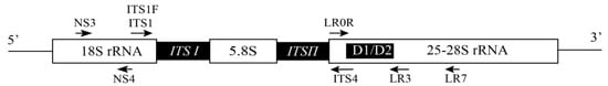
qPCR: quantitative PCR, ITS: internal transcribed spacer, GAPDH: glyceraldehydes-3-phosphate dehydrogenase, GS: glutamate synthetase, EF-1α: translation elongation factor 1-α, CAL: calmodulin, H3: histone subunit 3, ACT: actin, TUB2: ß-tubulin, RPB2: RNA polymerase subunit B2, COI: cytochrome oxidase subunit I, COII: cytochrome oxidase subunit II.

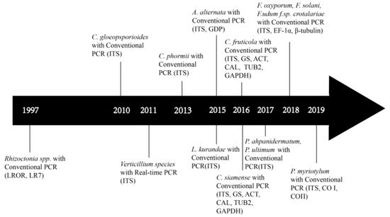
4. Development of Molecular Identification of Bast Fiber Fungal Pathogens

At present, most diagnosis of bast fiber diseases rely on disease symptoms and, when available, cultural characteristics of isolated fungal pathogens on artificial media. However, it is often difficult to identify the underlying pathogen based on those characters alone. For example, the disease symptoms of Verticillium wilt in hemp is very similar to Fusarium wilt and the pathogen species in both genera can invade a wide range of economical crops [37,38,39]. In addition, it is difficult to distinguish the species within most fungal genera based on morphological features alone. However, most of them are relatively easy to identify using molecular markers, as described below (Table 2; Table 3).
Table 3. Genes and PCR primers used for their amplification in fungal pathogens infecting bast fiber crops.

Target DNA

Primer Name and Sequence (5′-3′)

Size of PCR Product (bp)

Reference

18S

NS3

GCAAGTCTGGTGCCAGCAGCC

Not mentioned

[31]

NS4

CTTCCGTCAATTCCTTTAAG

28S

LR0R

GCAAGTCTGGTGCCAGCAGCC

Not mentioned

[31]

LR3

GCAAGTCTGGTGCCAGCAGCC

25S

LROR

ACCCGCTGAACTTAAGC

1431

[52]

LR7

TACTACCACCAAGATCT

ACT

ACT-512F

ATGTGCAAGGCCGGTTTCGC

300

[48]

ACT-783R

TACGAGTCCTTCTGGCCCAT

ß-tubulin

Vd-btub-1F

GCGACCTTAACCACCTCGTT

Not mentioned

[38]

Vd-btub-1R

CGCGGCTGGTCAGAGGA

VertBt-F

AACAACAGTCCGATGGATAATTC

Not mentioned

[38]

VertBt-R

GTACCGGGCTCGAGATCG

VITubF2

GCAAAACCCTACCGGGTTATG

143

[39]

VITubRl

AGATATCCATCGGACTGTTCGTA

VdTubF2

GGCCAGTGCGTAAGTTATTCT

82

[39]

VdTubR4

ATCTGGTTACCCTGTTCATCC

Bt2a

GGTAACCAAATCGGTGCTGCTTTC

Not mentioned

[26]

Bt2b

ACCCTCAGTGTAGTGACCCTTGGC

CAL

CL1

GARTWCAAGGAGGCCTTCTC

Not mentioned

[26]

CL2

TTTTTGCATCATGAGTTGGAC

CAL-228F

GAGTTCAAGGAGGCCTTCTCCC

Not mentioned

[50]

CAL-737R

CATCTTTCTGGCCATCATGG

EF-1α

EF-1

ATGGGTAAGGAGGACAAGAC

700

[34]

EF-2

GGAGGTACCAGTGATCATGTT

EF1-728F

CATCGAGAAGTTCGAGAAGG

Not mentioned

[50]

EF2

GGAGGTACCAGTGATCATGTT

EF1-728F

CATCGAGAAGTTCGAGAAGG

350

[48]

EF1-983R

TACTTGAAGGAACCCTTACC

Endochitinase

Vd-endoch-1F

CTCGGAGGTGCCATGTACTG

Not mentioned

[38]

Vd-endoch-1R

ACTGCCTGGCCCAGGTTC

GAPDH

Vd-G3PD-2F

CACGGCGTCTTCAAGGGT

Not mentioned

[38]

Vd-G3PD-1R

CAGTGGACTCGACGACGTAC

GDF1

GCCGTCAACGACCCCTTCATTGA

Not mentioned

[26]

GDR1

GGGTGGAGTCGTACTTGAGCATGT

gpd-1

CAACGGCTTCGGTCGCATTG

Not mentioned

[47]

gpd-2

GCCAAGCAGTTGGTTGTGC

GS

GSF1

ATGGCCGAGTACATCTGG

Not mentioned

[26]

GSR1

GAACCGTCGAAGTTCCAC

ITS

ITS1

TCCGTAGGTGAACCTGCGG

334-738

[24,30,35,36,37,38]

ITS4

TCCTCCGCTTATTGATATGC

Vd-ITS1-45-F

CCGGTCCATCAGTCTCTCTG

334

[37]

Vd-ITS2-379-R

ACTCCGATGCGAGCTGTAAC

ITS1-F

CTTGGTCATTTAGAGGAAGTAA

700

[34]

ITS4

TCCTCCGCTTATTGATATGC

VtF4

CCGGTGTTGGGGATCTACT

123

[39]

VtR2

GTAGGGGGTTTAGAGGCTG

ITS 4

TCCTCCGCTTATTGATATGC

Not mentioned

[26]

ITS 5

GGAAGTAAAAGTCGTAACAAGG

RPB2

bRPB2-6F

TGGGGYATGGTNTGYCCYGC

Not mentioned

[49]

bRPB2-7R

GAYTGRTTRTGRTCRGGGAAVGG

As early as 1997, a PCR-based method was used to help identify fungal pathogens of bast fiber crops. Specifically, McPartland et al. [52] amplified part of the 28S ribosomal RNA (rRNA) gene followed by EcoR I/Hind III digestion and electrophoresis to differentiate hemp fungal pathogens, and named two new species: Micropeltopsis cannabis sp. nov. and Orbilia luteola (Roum.) comb. nov. However, there were relatively few reports of fungal pathogens on bast fiber crops between 1998 and 2009, likely due to limited production of bast fiber crops and an emphasis on chemical fiber and other natural fibers. During this period, the acreage and production of bast fiber crops were low and there was limited research on these crops. Since 2009, with increasing production and research on bast fiber crops, there have been increasing reports on infectious diseases, including fungal diseases, on these crops [23]. This is especially true over the last five years when a large number of fungal pathogens were reported from bast fiber crops and many of these were identified based on molecular markers (Figure 1).
Figure 1. Development of molecular-based assays for the detection of fungal pathogens in bast fiber crops from 1997 until the present. For genus and species names, please see text and Table 2. Details of primers are shown in Table 3.
According to the National Center for Biotechnology Information (NCBI) PubMed, the most common literature on the molecular identification of fungal pathogens on bast fiber crops has been on hemp (including both industrial hemp and medicinal marijuana), accounting for ~45% of all published articles. This was then followed by flax and kenaf (at ~14% each), ramie (11%), and the rest being jute and sunn hemp. However, most of these reports were case reports.

5. Target DNA Selection and Molecular Assays of Fungal Pathogens on Bast Fiber Crops

Over the last three decades, several types of DNA-based methods have been developed and widely used to detect plant fungal pathogens. The invention of PCR technology using a thermostable polymerase by Kary Mullis gave birth to PCR in the early 1980s [4]. The invention of PCR has led to a diversity of PCR-based methods for fungal pathogen detections based on variations in DNA sequences within and among species (Figure 1, Table 2). Among these methods, qPCR is probably the most common molecular technology and it can be used for quantitative measurement of RNA and DNA, targeting both single nucleotide polymorphisms (SNPs) and copy number variations. qPCR allows not only the detection of whether a specific pathogen(s) is present in the sample, but also the quantification of pathogen levels in host tissues [5,6]. To improve the efficiency of conventional PCR, other methods have been coupled with PCR for plant fungal pathogen detection. For example, PCR in combination with enzyme-linked immunosorbent assay (ELISA) has been successfully applied to detect fungi, viruses, and bacteria, with high specificity [56]. Similarly, the highly specific IC-PCR approach can increase the sensitivity by 250 folds compared to conventional PCR amplification [7,8]. For absolute quantification without the need for references and standard curves, dd-PCR is the method of choice—this method is based on the combined technology of water–oil emulsion droplet and PCR [9]. In field conditions without ready access to laboratory equipment, LAMP can provide fast identifications of samples. LAMP uses six primers that are highly specific to target sites in a specific gene [10]. It can be carried out at a constant temperature in a short reaction time (<30 min). It is sensitive and cost-effective, potentially making it an ideal method for field detection of plant pathogens [57].
As shown in Table 2, PCR-based methods have been used as the main approach for detecting fungal pathogens in bast fiber crops. This pattern is similar to the detections of fungal pathogens in other crops in general. A number of DNA fragments and genes have been explored as potential targets for PCR-based detections, including the ribosomal RNA gene cluster, conserved housekeeping genes, and genes involved in the production of secondary metabolites, including mycotoxins [58,59,60]. Table 3 summarizes the genes and their primers that have been used for the detection and diagnostics of fungal pathogens on bast fiber crops. We would like to note that the molecular analyses reported so far for identifying fungal pathogens on bast fiber crops have been primarily using pure fungal strains, not those from diseased plant tissues. There is a large gap in applying these molecular methods in field conditions as a point-of-care test.
Among the DNA fragments that have been used for fungal pathogen detection, the most frequently used is the ribosomal RNA gene cluster. This gene cluster is composed of up to hundreds of repeating units with each unit containing the genes encoding the small (18S) ribosomal RNA subunit, the internal transcribed spacer (ITS) regions 1 and 2 that are separated by the 5.8S rRNA subunit, and the large (28S) ribosomal RNA subunit, with the intergenic spacer (IGS) region separating the adjacent units (Figure 2). The entire ITS fragment (which comprises ITS1, 5.8S rRNA, and ITS2) is typically 500–750 bp long and flanked by the 18S and 28S rRNA genes [61,62,63]. The ITS regions are present in all known fungi and have both highly conserved flanking regions located in the 5.8S, 18S, and 28S rRNA genes as well as the variable regions (located in the ITS1 and ITS2 regions). The conserved flanking regions allowed the development of highly conserved probes or primers to amplify most, if not all, fungi, while the variable regions allowed the development of species-specific markers [64,65]. Together, these features have contributed to ITS being the consensus fungal DNA barcode for the mycological community [64,65]. Furthermore, the ITS sequences obtained from the direct amplification and sequencing of environmental DNA samples have contributed to our increased understanding of fungal diversity from a variety of environments, including those from diseased plants and animals [65,66].
Figure 2. A schematic representation of the fungal ribosomal RNA gene cluster showing the locations of individual DNA fragments and the common primers used for PCR amplification.

This entry is adapted from the peer-reviewed paper 10.3390/pathogens9030223

This entry is offline, you can click here to edit this entry!
Academic Video Service