Nephrotoxic Effects of Paraoxon: History
Please note this is an old version of this entry, which may differ significantly from the current revision.
Subjects: Toxicology
Contributor:

Paraoxone as an organophosphorus compound has nephrotoxic properties for mammalian kidneys. In experiments on rats, even a single intoxication with paraoxone leads to the development of changes in the biochemical parameters of blood and urine, and causes structural changes in the renal tubules and ultrastructural changes in the glomerular basal membrane.

  • nephrotoxicity
  • rats
  • paraoxon
  • biomarkers
  • calbindin
  • chondroitin sulphate
  • creatinine clearance

1.Nephrotoxicity of organophosphate compounds

Damage to the kidneys and their impaired functions after a single contact with OPs, accompanied by the development of clinical manifestations of poisoning, was considered in the early 1990’s to be a rare manifestation of their toxicity  [1]. At that time, only three publications mentioning nephrotoxic effects after OPs poisoning in humans had been published in English-language literature [2][3][4]. Since the beginning of the 21st century, the nephrotoxicity [2] of OPs has been the subject of greater attention. In in vivo experiments with laboratory animals, nephrotoxicity has been established for many OPs compounds, including fenthion [5][6], diazinon [7], malathion [8][9], chlorpyrifos [10], dichlorfos [11], metamidophos [12], quinalfos [13], and methylparathion [14]. At the same time, an increasing number of publications has been devoted to the study of clinical cases of human poisoning with OPs compounds that were accompanied by nephrotoxic effects, such as acute kidney injury (AKI) and development of acute renal failure (ARF) [15][16][17].

1.1. Paraoxone nephrotoxicity

Paraoxon (O,O-diethyl O-(4-nitrophenyl) phosphate, POX) is a metabolic product of ethyl parathion, and is one of the most toxic pesticides for restricted use to control a broad spectrum of pests on alfalfa, barley, canola, corn, cotton, sorghum, soybeans, sunflowers, and wheat. POX as a potent cholinesterase inhibitor is responsible for the cholinergic crisis typical of parathion poisoning [18]. According to some authors, the histopathological manifestation of nephrotoxicity of POX in rats after a single sublethal dose poisoning is disorganization of proximal tubule epithelial cells with an increase in their diameter [19]. The results of in vitro studies showed that POX is able to induce apoptosis, which was detected in T-lymphocytic leukemia EL4 cells of mice. This stimulation occurs through a direct effect of POX on mitochondria, disrupting their transmembrane potential and causing release of cytochrome C into the cytosol with subsequent activation of caspase-9 [20].
The toxic effects of OPs are not limited to inhibition of cholinesterases. They are also capable of producing reactive oxygen species (ROS) that damage renal tubules and renal parenchyma, leading to hypovolemia and AKI. Oxidative stress is a major complication in the formation of renal pathology in OPs poisoning. POX in rats causes production of ROS and oxidative stress manifested by GSH depletion in kidney tissue [21]. In another study, it was found that the level of ROS production after POX poisoning is dose-dependent, and the induction of oxidative stress occurs in the following order: brain > liver > heart > kidney > spleen [22]. Administration of POX to rats at doses that cause the development of systemic toxicity also leads to vasoconstriction of the kidneys and a reduction in both efficacious renal blood flow (RBF) and glomerular filtration rate (GFR) [23].

2. Animal Models of Acute paraoxone Intoxication

The main biochemical characteristic of rodents is the presence of carboxylesterase in plasma, which is a target for OPs and can significantly distort their specific effects. To eliminate the effects of carboxylesterase in the first model (M1, POX2x group), POX was administered twice in doses of 110 g/kg and 130 g/kg subcutaneously, at 1 h intervals. In the second model (M2, CBPOX group), 1 h before POX poisoning at a dose of 130 g/kg subcutaneously, carboxylesterase activity was pre-inhibited by administration of the specific inhibitor cresylbenzodioxaphosphorin oxide (CBDP, 3.3 mg/kg intraperitoneally). In the third model (M3), POX was administered subcutaneously once at doses of LD16 (241 g/kg), LD50 (250 g/kg) and LD84 (259 g/kg).

2.1 Acute nephrotoxicity of paraoxone (POX) for rats

2.1.1. Plasma Biochemistry

In 1 and 2 Models of intoxication 3 h after paraoxon poisoning creatinine level increased by 35–40% in the CBPOX group (p < 0.05). Uric acid content in plasma was changed only in the POX2x group, with an increase of 20–40% by 1 and 7 days after poisoning. Since up to 80% of uric acid is excreted by the kidneys, these data provide further evidence of renal damage in acute OPs poisoning. Almost synchronous increase calcium and inorganic phosphate level and alkaline phosphatase activity was found in the CBPOX group after 3 h of poisoning.
After the poisoning with POX, endogenous creatinine clearance in rats in the M1/2 model decreased markedly within 24 h regardless of the mode of carboxylesterase inhibition. At the same time, 3 and 7 days after the poisoning, ECC in rats of all groups was not significantly different. This is evidence of a decrease in GFR in the early period after the poisoning, with subsequent recovery in the filtration level. A decrease in the effective RPF and GFR in rats after administration of POX at doses causing the development of systemic toxicity was demonstrated as early as in 1970 [23], and results of our studies agree with that publication. In humans, a decrease in ECC after OPs poisoning has also been registered, for example, with diazinon [24] and obidoxime [25].

2.1.2. Urinalysis

Microscopic examination of rat urine sediment in all models of acute paraoxon poisoning showed no evidence of acute kidney damage, including haematuria, erythrocyturia, pyuria, crystalluria and cylinduria as well as increased epithelial cell count. The only significant difference in urine biochemistry of poisoned rats was a marked glucosuria observed 24 h after the poisoning in both POX2x and CBPOX groups.

In 1 and 2 Models of intoxication at 24 h after poisoning, the CS level in the urine tended to increase: 11.3 ± 4.4 in the POX2x group; 10.1 ± 3.6 in the CBPOX group; and 5.7 ± 0.4 mg/mol creatinine in control animals. After 3 days, the CS content in the urine of poisoned rats was still elevated: 8.6 ± 1.8 in the POX2x group; 8.3 ± 0.9 (p < 0.05) in the CBPOX group; and 6.1 ± 0.6 mg/mol creatinine in control animals. At 7 days after poisoning, there was no significant difference in the content of chondroitin sulfate in the urine of poisoned and intact animals.
It should be noted that the role of CS both in OPs intoxication and in intoxication with other xenobiotics has been insufficiently studied. The level of CS excretion with urine also increases after poisoning at different doses of POX [26,27], as well as cyclophosphamide [28]. In this connection, at least for these two xenobiotics, a similar increase in CS excretion can be considered as a nonspecific marker of kidney (POX) and/or bladder (cyclophosphamide) damage.

2.1.4. Calbindin, KIM-1, TIMP-1 in Rat Urine

The level of these biomarkers of kidney injury in the urine of poisoned rats at 1 and 7 days after the poisoning showed no significant differences from that of control animals. However, 3 days after the poisoning, a significant increase in the concentration of calbindin was observed in the CBPOX group (Table 1).
Table 1. Calbindin, KIM-1, and TIMP-1 in daily urine of rats 3 days after exposure to POX.
  Control POX2x CBPOX
Calbindin 4101 ± 334 6419 ± 1201 7383 ± 990 *
KIM-1 21.8 ± 5.5 28.8 ± 6.0 24.1 ± 6.9
TIMP-1 32,755 ± 7830 13,078 ± 9771 19,174 ± 11,596

* p ≤ 0.05; Results are presented in ng/mol_creatinine.

Currently, calbindin is considered as an informative biomarker of damage of distal tubules and collecting tubes of the kidneys.  Changes in the morphometric characteristics of the tubule epithelium in 1–3 days after poisoning, and increased calbindin excretion, indicates tubule damage.

2.1.5. Histopathological Changes in the Kidneys

In 1 and 2 Models of intoxication within 24 h after the poisoning, there were no significant changes in the morphometry of the renal corpuscles, glomerulus, proximal, and distal convoluted tubules. Poisoning at a dose of LD16 does not lead to changes in the structure of kidney tissues and cells. After POX poisoning at LD50 and LD84 levels, microscopic examination of kidney sections revealed damage to tubule epihelial cells. By 1 and 3 days after the poisoning at a dose of LD84, the cell cytoplasm had become more homogeneous, and the granularity normally present in tubule epithelial cells had disappeared. It should be noted that, after 7 days of exposure to POX, the morphological characteristics of cytoplasm were completely restored. One day after the poisoning, there was an increase in the diameter of the tubule lumen in the LD84 group, and after 3 days of exposure to POX, their narrowing was observed (p < 0.05). The structure of the renal corpuscles 24 h after poisoning in the LD50 and LD84 groups showed a decrease in the ratio of the size of the renal corpuscles to the glomerular area (p < 0.05), indicating expansion of the Bowman′s capsule. Similar changes in the renal corpuscle are shown 3 days after exposure to POX, but were not observed at the 7 days’ checkpoint. The basal part of tubule epithelial cells clearly shows granularity. In the renal corpuscle, the cytoplasm of podocytes is weakly stained, and clear localization of nephrin at the edge of GBM is absent in all cases.

2.1.6. Ultrastructural Changes in Rat Kidneys after the Poisoning
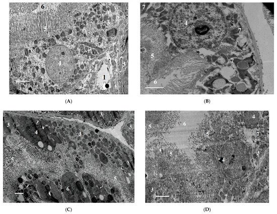
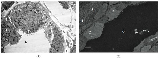
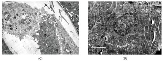
After POX exposure in dose LD84 were detected a decrease in the height of epithelial cells relative to the distal tubules 24 h after the poisoning (p < 0.05), and an increase in the proximal tubule epithelium 3 days after the poisoning (p < 0.05). In the epithelium of proximal tubules, the damages were expressed locally and were manifested as stretching of basal labyrinth, sloughing of microvilli on apical surface and presence of pycnotic nuclei in cells. Partial swelling and destruction of mitochondria, the presence of pyknotic nuclei, and the displacement of nuclei to apical cell surfaces were observed in the epithelium of distal tubules (Figure 1 and Figure 2).
Figure 1. (A)—control; (B)—LD50, 24 h after exposure to POX; (C)—LD84, 24 h after exposure to POX; (D)—LD84, 3 days after exposure to POX. (BD)—Vacuolization of cytoplasm, degeneration of mitochondria in epithelial cells of proximal tubules. (C)—Karyopiknosis of nuclei after acute poisoning with a dose at LD84. Increased height of proximal tubule epithelium 3 days after exposure to POX (D). 1—capillary lumen, 2—epithelial cell of the distal tubule, 3—epithelial cell of the distal tubule, 4—nucleus, 5—microvilli, 6—tubule lumen, 7—mitochondria. Double contrasting with lead nitrate and uranyl acetate. TEM, scale bar = 2 µm.
Figure 2. (A)—control; (B)—LD50, 24 h after exposure to POX; (C)—LD84, 24 h after exposure to POX; (D)—LD84, 3 days after exposure to POX. (B,C)—decrease in height of epithelial cells of distal convoluted renal tubules 24 h after exposure to POX (p < 0.05), and reduction in tubule lumen 3 days after exposure to POX (D) (p < 0.05); (C)—mitochondrial degradation. 1—capillary lumen, 2—epithelial cell of distal tubule, 3—epithelial cell of distal tubule, 4—nucleus, 5—microvessels, 6—tubule lumen, 7—mitochondria. Double contrasting with lead nitrate and uranyl acetate. TEM, scale bar = 2 µm.
After 7 days of exposure to POX at doses of LD50 and LD84, some signs of tubule epithelial cell destruction remained, in particular, the blocking of tubule lumen by cellular detritus and local destruction of apical cell surfaces. Thus, at this period after the poisoning, incomplete recovery of proximal tubule epithelium is observed. The structure of GFB shows no signs of significant damage after the poisoning with the doses studied. The urinary space contains a small amount of cellular detritus. Electron-dense deposits along basal membranes or in mesangium were not detected, and capillary lumen was free. Morphometric analysis of GBM 1 and 3 days after the poisoning revealed no statistically significant differences.  However, 7 days after the poisoning, a statistically significant (p < 0.05) increase in GBM thickness was observed: 0.212 ± 0.06 μm in the LD50 group; 0.209 ± 0.03 μm in the LD84 group; and 0.186 ± 0.04 μm in the control group.
By summarizing the results of the M3 model of exposure to POX, it should be noted that the LD16 dose had a minimal effect on the changes in the assessed parameters at the histological and ultrastructural level. In this regard, there is no reason to talk of a nephrotoxic effect of POX at this dose. However, as shown by the results of histopathological and EM studies, even a single ingestion of POX at doses of LD50 and LD84 leads to the development of morphological changes, especially those pronounced 1–3 days after the poisoning. As in the M1 and M2 models, at 7 days post-poisoning, most of these changes were compensated by the renal systems.

3. Conclusions

The nephrotoxic effects of POX in rats occur regardless of the mode of prior inhibition of carboxylesterase activity. The use of a conventional model of poisoning at a single dose of LD16 does not lead to the development of any abnormalities characterizing the nephrotoxicity of POX at this dose. At the same time, POX at the doses of LD50 and LD84 causes marked ultrastructural changes in the cells of tubules, which is a confirmation of its nephrotoxicity. Combined interpretation of the results with the data of biochemical study, biomarkers of renal damage and creatinine clearance suggests that, in the early period (1 to 3 days) after the poisoning, the changes registered are initially of afunctional nature, followed by development of changes in the structural elements of the nephron. Seven days after the poisoning, most of the revealed changes are normalized, but changes in GBM do not allow us to state that the consequences of poisoning are fully compensated.

This entry is adapted from the peer-reviewed paper 10.3390/ijms222413625

References

  1. Wedin, G.P. Nephrotoxicity of anticholinesterases. In Clinical and Experimental Toxicology of Organophosphates and Carbomates; Ballantyne, B., Marrs, T.C., Eds.; Britterworth-Heinemann Ltd.: Oxford, UK, 1992; pp. 195–202.
  2. Albright, R.K.; Kram, B.W.; White, R.P. Malathion exposure associated with acute renal failure. JAMA 1983, 250, 2469.
  3. Kushnir, A.; Finkelstein, Y.; Raikhlin, B.; Taitelman, U. Multihospital study of severe acute organophosphate insecticide poisoning. Vet. Hum. Toxicol. 1988, 30, 366.
  4. Wedin, G.P.; Pennente, C.M.; Sachdev, S.S. Renal involvement in organophosphatc poisoning. JAMA 1984, 252, 1408.
  5. Kaya, Y.; Bas, O.; Hanci, H.; Cankaya, S.; Nalbant, I.; Odaci, E.; Uydu, H.A.; Aslan, A. Acute renal involvement in organophosphate poisoning: Histological and immunochemical investigations. Ren. Fail. 2018, 40, 410–415.
  6. Kerem, M.; Bedirli, N.; Guerbuez, N.; Ekinci, O.; Bedirli, A.; Akkaya, T.; Sakrak, O.; Pasaoglu, H. Effects of Acute Fenthion Toxicity on Liver and Kidney Function and Histology in Rats. Turk. J. Med. Sci. 2007, 37, 281–288.
  7. Boroushaki, M.T.; Arshadi, D.; Jalili-Rasti, H. Protective effect of pomegranate seed oil against acute toxicity of diazinon in rat kidney. Iran. J. Pharm. Res. 2013, 12, 821–827.
  8. Possamai, F.P.; Fortunato, J.J.; Feier, G.; Agostinho, F.; de Quevedo, J.L.; Filho, D.W.; Pizzol, F.D. Oxidative stress after acute and sub-chronic malathion intoxication in Wistar rats. Environ. Toxicol. Pharmacol. 2007, 23, 198–204.
  9. Al-Attar, A.M. Physiological and Histopathological Investigations on the Effects of α-Lipoic Acid in Rats Exposed to Malathion. J. Biomed. Biotechnol. 2010, 2010, 203503.
  10. Ahmed, N.S.; Mohamed, A.S.; Abdel-Wahhab, M.A. Chlorpyrifos-induced oxidative stress and histological changes in retinas and kidney in rats: Protective role of ascorbic acid and alpha tocopherol. Pestic. Biochem. Physiol. 2010, 98, 33–38.
  11. Hou, Y.; Zeng, Y.; Li, S.; Qi, L.; Xu, W.; Wang, H.; Zhao, X.; Sun, C. Effect of quercetin against dichlorvos induced nephrotoxicity in rats. Exp. Toxicol. Pathol. 2014, 66, 211–218.
  12. Satar, S.; Satar, D.; Mete, U.O.; Suchard, J.R.; Topal, M.; Kaya, M. Ultrastructural Effects of Acute Organophosphate Poisoning on Rat Kidney. Ren. Fail. 2005, 27, 623–627.
  13. Eid, R.A. Apoptosis of rat renal cells by organophosphate pesticide, quinalphos: Ultrastructural study. Saudi J. Kidney Dis. Transpl. 2017, 28, 725–736.
  14. Fuentes-Delgado, V.H.; Martínez-Saldaña, M.C.; Rodríguez-Vázquez, M.L.; Reyes-Romero, M.A.; Reyes-Sánchez, J.L.; Jaramillo-Juárez, F. Renal damage induced by the pesticide methyl parathion in male Wistar rats. J. Toxicol. Env. Health Part A 2018, 81, 130–141.
  15. Lee, F.Y.; Chen, W.K.; Lin, C.L.; Lai, C.Y.; Wu, Y.S.; Lin, I.C.; Kao, C.H. Organophosphate Poisoning and Subsequent Acute Kidney Injury Risk: A Nationwide Population-based Cohort Study. Medicine 2015, 94, 2107.
  16. Zafar, R.; Munawar, K.; Nasrullah, A.; Haq, S.; Ghazanfar, H.; Sheikh, A.B.; Khan, A.Y. Renal failure due to organophosphate poisoning: A case report. Cureus 2017, 27, 523.
  17. Cavari, Y.; Landau, D.; Sofer, S.; Leibson, T.; Lazar, I. Organophosphate poisoning-induced acute renal failure. Pediatr. Emerg. Care 2013, 29, 646–647.
  18. Mohammadi, M.; Ghani, E.; Ghasemi, A.; Khoshbaten, A.; Asgari, A. Synaptosomal GABA uptake decreases in paraoxon-treated rat brain. Toxicology 2008, 244, 42–48.
  19. Sokolova, M.O.; Sobolev, V.E.; Reshetkina, D.A.; Nagibovich, O.A. Toxic effect of organophosphate compounds on the kidneys. Bull. Russ. Mil. Med. Acad. 2020, 22, 199–205. (In Russian)
  20. Saleh, A.M.; Vijayasarathy, C.; Masoud, L.; Kumar, L.; Shahin, A.; Kambal, A. Paraoxon induces apoptosis in EL4 cells via activation of mitochondrial pathways. Toxicol. Appl. Pharmacol. 2003, 190, 47–57.
  21. Abbasnezhad, M.; Jafari, M.; Asgari, A.R.; Hajihoseini, R.; Hajigholamali, M.; Salehi, M.; Salimian, M. The study regarding effect of paraoxon on oxidative stress. J. Maz. Univ. Med. Sci. 2009, 19, 16–26.
  22. Jafari, M.; Salehi, M.; Asgari, A.; Ahmadi, S.; Abbasnezhad, M.; Hajihoosani, R.; Hajigholamali, M. Effects of paraoxon on serum biochemical parameters and oxidative stress induction in various tissues of Wistar and Norway rats. Env. Toxicol. Pharmacol. 2012, 34, 876–887.
  23. Williams, R.I.; Pearson, J.E. Functional study of the renal effect of the anticholinesterase paraoxon. Arch. Int. Pharmacodyn. 1970, 184, 195–208.
  24. Abend, Y.; Goland, S.; Evron, E.; Sthoeger, Z.M.; Geltner, D. Acute renal failure complicating organophosphate intoxication. Ren. Fail. 1994, 16, 415–417.
  25. Bentur, Y.; Nutenko, I.; Tsipiniuk, A.; Raikhlin-Eisenkraft, B.; Taitelman, U. Pharmacokinetics of obidoxime in organophosphate poisoning associated with renal failure. J. Toxicol. Clin. Toxicol. 1993, 31, 315–322.
  26. Goncharov, N.V.; Terpilovskij, M.A.; Shmurak, V.I.; Belinskaya, D.A.; Avdonin, P.V. Rat (Rattus norvegicus) as a research object in the model of acute intoxication with organophosphorous compound. 1. Biochemical aspects. J. Evol. Biochem. Physiol. 2019, 55, 112–123.
  27. Sobolev, V.E.; Korf, E.A.; Goncharov, N.V. The Rat (Rattus norvegicus) as a Model Object for Acute Organophosphate Poisoning. 5. Morphofunctional Alterations in Kidneys. J. Evol. Biochem. Physiol. 2019, 55, 302–312.
  28. Sobolev, V.E.; Jenkins, R.O.; Goncharov, N.V. Sulfated glycosaminoglycans in bladder tissue and urine of rats after acute exposure to paraoxon and cyclophosphamide. Exp. Toxicol. Pathol. 2017, 69, 339–347.
More
This entry is offline, you can click here to edit this entry!
ScholarVision Creations