Cognitive Impairment in Heart Failure: History
Please note this is an old version of this entry, which may differ significantly from the current revision.
Contributor:

Cognitive impairment (CI) is common in heart failure (HF). Patients with HF demonstrate reduced global cognition as well as deficits in multiple cognitive domains compared to controls. Degree of CI may be related to HF severity. HF has also been associated with an increased risk of dementia. Anatomical brain changes have been observed in patients with HF, including grey matter atrophy and increased white matter lesions. Patients with HF and CI have poorer functional independence and self-care, more frequent rehospitalisations as well as increased mortality. Pathophysiological pathways linking HF and CI have been proposed, including cerebral hypoperfusion and impaired cerebrovascular autoregulation, systemic inflammation, proteotoxicity and thromboembolic disease.

  • heart failure
  • cognitive impairment
  • dementia
  • cerebral haemodynamics

1. Cognitive Changes in HF

Compared to healthy controls, patients with HF demonstrate reduced global cognition as well as deficits in multiple cognitive domains including executive function, psychomotor speed and verbal memory [9]. Sterling et al. found that the prevalence of CI among patients with incident HF (14.9%) was similar to controls without HF (13.4%) and was lower than reported in the general HF population. This suggests that CI may develop at some point after the onset for HF, rather than it being present prior to HF diagnosis or due to concomitant cardiovascular risk factors [21]. This is further supported by a study by Hammond et al. which reported a greater decline in Modified Mini Mental State test scores of 10.2 points over 5 years in patients with incident HF, compared to 5.8 points in controls [22].
HF has also been associated with an increased risk of dementia that may not be limited to vascular dementia [26,27]. Adelborg et al. found that patients with HF were 1.5 times more at risk of developing vascular dementia, and were also 1.3 times more likely to develop other dementias (defined as any dementia apart from vascular dementia or Alzheimer’s disease) over a 35-year follow-up period. However, they did not find a difference in the risk of Alzheimer’s disease between HF patients and controls [27]. In contrast, Qiu et al. reported an increased risk of both all-cause dementia and Alzheimer’s disease in patients with HF within a community-based cohort. Over a 9-year follow-up period, patients with HF were approximately 1.8 times more likely to develop incident all-cause dementia and 1.8 times more likely to develop Alzheimer’s disease [28]. Compared to the Adelborg study which identified incident dementia and Alzheimer’s disease from a psychiatric registry [27], Qiu et al. evaluated their study population on three separate follow-up sessions with a comprehensive clinical examination and cognitive test battery, with corroboration between two independent physicians. Therefore, potential misidentification of dementia and misclassification of dementia subtype in the Adelborg study may have contributed to these discrepancies in results [27]. In the general population, Jefferson et al. observed that a lower cardiac index (defined as cardiac output divided by body surface area measured in L/min/m2) among subjects of the Framingham Offspring Cohort was associated with higher all-cause dementia and Alzheimer’s risk [29].

1.1. HF Severity and Degree of CI

A dose–response relationship between HF and CI would further lend support to a connection between the two diseases. Patients with a more advanced New York Heart Association (NYHA) class demonstrated lower overall Z-scores compared to those with NYHA I or II disease and increased HF severity was associated with reduced memory, visuospatial ability, psychomotor speed and executive function [20]. Harkness et al. also found that the incidence of CI, defined as MoCA score < 26, was greater in HF patients with NYHA III or IV class (91%) compared to NYHA I or II class (52%) [23]. Hanon et al. reported more severe memory impairment, as evaluated by the delayed-recall Memory Impairment Screen (MIS-D), in patients with higher NYHA class [30] and Lee et al. found that NYHA class II or higher was independently associated with an increased likelihood of cognitive decline in patients with HF [31]. Similarly, another study reported poorer attention and memory in HF patients who scored higher on dyspnoea and fatigue rating scales [32]. Kindermann et al. observed poorer cognition in patients with decompensated HF compared to those with stable HF, and found that cognition improved after HF compensation [25]. In contrast, Huijts et al. found that although severe CI was present at baseline more often in HF patients with NYHA IV compared to NYHA II class, the prevalence of severe CI remained stable over 18 months in both groups. Moreover, baseline HF severity was not associated with cognitive decline [13]. These differing findings may be due to the authors’ use of the AMT to determine CI, which may be more susceptible to ceiling effects compared to other tools [33]. Myocardial stretch stimulates the release of pro B-type natriuretic peptide (proBNP), which is then rapidly cleaved into biologically active C-terminal BNP and inert N-terminal proBNP (NT-proBNP) [34]. BNP and NT-proBNP are both indicators of HF severity [35]. A connection between higher BNP levels and poorer attention and executive function was previously reported [36], in addition to reduced hippocampal volume in patients with higher BNP [37]. NT-proBNP has also been associated with an increased risk of dementia in an elderly community-dwelling population [38]. Overall, these studies suggest that HF severity may have an impact on the level of CI but the exact relationship remains to be elucidated.

1.2. The Impact of Ejection Fraction on CI

Left ventricular ejection fraction (EF), defined as a percentage of stroke volume over end-diastolic volume (SV/EDV ×100%), is the central measure of left ventricular systolic function. A lower left ventricular ejection fraction (EF), especially when <30%, has been associated with lower cognitive scores [39,40]. A study by Festa et al. showed that in patients 63 years old or older, EF < 30% was associated with poorer memory whereas memory was stable across all EF levels in younger patients [41]. It is unclear if this is due to poorer compensatory capacity, since age was not shown to affect dynamic cerebrovascular autoregulation in a healthy population [42]. Elderly patients do, however, appear to be more susceptible to watershed infarcts from cerebral hypoperfusion [43]. In contrast, a similar rate of cognitive decline was found in patients with HF with reduced (HFrEF) and preserved EF (HFpEF) [22] despite different patterns of cognitive deficits depending on predominance of systolic or diastolic dysfunction [36,44,45]. Concomitant severe systolic and diastolic dysfunction may worsen CI, especially in the form of poorer verbal fluency compared to those with systolic dysfunction alone [40].

1.3. Potential Confounders in the Association between HF and CI

HF and CI share several risk factors and studies which include a control group of patients with cardiovascular disease without HF may be useful to reduce the effects of potential confounding factors. Studies have shown that CI remains more common in patients with HF even when compared to these cardiac controls [2,24]. Vogels et al. reported that 25% of patients with HF had CI compared to 15% of those with cardiovascular disease without HF [2]. Another study found that the prevalence of abnormal performance on at least 3/7 tests in a neuropsychological battery was 57.9% and 43% in patients with severe and moderate HF, respectively, as compared to 34.3% in those with other cardiovascular diseases [24]. The degree of CI also appears to be greater in patients with HF, and HF and IHD patients were found to have a Cambridge Cognition Examination (CAMCOG) score of 2.8 and 1.8 less than healthy controls, respectively [16]. In contrast, a prospective study over 2 years found that while cognitive decline was greater in HF patients than in healthy controls, it was similar to those with coronary artery disease [46].

2. Proposed Aetiologies of CI in HF

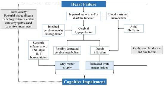
Several pathophysiological pathways have been proposed to contribute to the structural brain changes and CI among patients with HF. These are outlined in Figure 1.
Figure 1. Summary of potential pathophysiological pathways linking heart failure and cognitive impairment. Dotted line: the mechanisms by which systemic inflammation may contribute to brain changes and cognitive impairment in heart failure are not well described. Dashed line: proteotoxicity may be a shared disease pathology between specific cardiomyopathies and CI. IL, interleukin; TNF, tumour necrosis factor.

2.1. Cerebral Hypoperfusion and Impaired Autoregulation

Reduced cerebral blood flow (CBF) is one of the proposed mechanisms of brain injury and CI in HF [55,58]. Several studies have demonstrated lower CBF velocities on transcranial Doppler (TCD) in those with HF [59,60,61]. Furthermore, TCD measurements in patients with HF showed a decline over a 12-month period [62]. Lower CBF in HF patients impacted global cognitive function, attention and executive function [62,63], while reduced regional hippocampal CBF was associated with poorer performance on measures of delayed memory [64]. A low output state in HF may result in chronic cerebral hypoperfusion in patients with HF, making them more susceptible to watershed infarcts. Additionally, owing to the similar risk factors shared by patients with HF and cerebrovascular disease, patients with HF may also have a poorer collateral blood supply due to atheromatous stenosis of the cerebral arteries [43]. Some studies compared CBF measurements against structural neuroimaging or neuropsychological testing and evaluated the relationship between CBF and CI in HF [55,59]. Alosco et al. found that reduced CBF in patients with HF was associated with increased WMLs, which were in turn related to poorer MMSE scores [59]. Vogels et al. similarly described lower CBF in patients with HF, but did not find a correlation with brain changes on neuroimaging [55].
In the general population, Jefferson et al. reported higher MRI-assessed cardiac index to be positively related to total brain volume and information processing speed [58]. Similarly, a lower cardiac index was associated with increased dementia risk [29]. In a study of 4366 individuals from the United Kingdom Biobank, van Hout et al. found that individuals with subclinical reduced left ventricular EF had reduced total brain volume and GM volume, and also increased WMLs [65]. Interestingly, only WM and hippocampal volume loss were associated with CI, and both were not related to EF [65]. Arterial stiffness, microvascular damage, atherosclerosis and inflammation in HF may possibly confound the reported relationship between EF and CI. These pathophysiological mechanisms are adversely associated with both cardiac function and cognition, making it difficult to ascertain the true association between EF and CI [66,67]. However, Park et al. found that total brain volume and hippocampal volume remained associated with poorer left ventricular systolic function even after adjustment for cardiometabolic disease [37]. Left ventricular stroke volume and cardiac output have also been linked to CI [68]. The potential mechanisms by which cardiac dysfunction may influence brain atrophy are not well understood but may be related to decreased cerebral metabolism. Patients with HF with extensive hibernating myocardium had reduced cerebral metabolism in frontal and hippocampal areas in a study utilising 18F-flurodeoxyglucose positron emission tomography/computed tomography (18F-FDG PET/CT) imaging [69].
A reduced CBF is unlikely to be the sole explanation for cortical GM loss [16,54]. Brain areas such as the periventricular white matter, basal ganglia and hippocampus are susceptible to cerebral hypoperfusion due to their location at the junction of large-vessel arterial territories, or due to their irrigation by long-penetrating end arterioles. In contrast, the cortex has a rich dual blood supply and can better tolerate cerebral hypoperfusion [43,70]. The interplay of other cardiovascular risk factors may also contribute to cortical GM loss, since similar patterns of GM loss have been observed in HF and IHD patients [16]. Leeuwis et al. further argued that CBF may not be the main reason for CI in HF in light of their findings that while CBF was lower in patients with HF, this did not correspond with reduced cognitive function [71].
CBF has been shown to increase after heart transplantation [72] and heart transplantation has been associated with improved cognitive function [73,74]. Cognitive improvement has also been reported after left ventricular assist device (LVAD) placement [75,76]. However, these improvements were marginal and MoCA scores increased by approximately 1.6 following LVAD placement [76]. Schall et al., on the other hand, did not find a significant difference between pre- and post-operative cognitive scores after 7.7 months in their patients with dilated cardiomyopathy who underwent heart transplantation, despite greatly improved physical health [77]. A possible explanation is the shorter follow-up duration compared to other studies [73]. Several cognitive scores showed a non-statistically significant increase and a longer reassessment interval may have revealed further cognitive improvement [77]. Another reason may be the use of an extensive neuropsychological battery by Schall et al. compared to less rigorous screening measures such as the MoCA in other studies [74]. Patients with HF may also have diminished cerebrovascular autoregulation, with greater impairments in those with NYHA IV compared to NYHA II and III. Accordingly, cerebral oxygen saturations were found to be lower in patients with HF [78,79]. Previous studies have shown a blunted haemodynamic response and greater CBF reduction in patients with HF in response to upright posture [80,81]. More recently, Kharraziha et al. observed a more pronounced decrease in cerebral tissue oxygen saturations in response to head-up tilt in patients with HF [82]. While it is unclear how HF may lead to impaired autoregulation, it could result in increased susceptibility to low cardiac output states due to an inability to maintain CBF via vasodilatory mechanisms.
2.2. Systemic Inflammation

The systemic inflammatory state recognised in patients with HF may further contribute to CI in HF. Tumour necrosis factor (TNF)-alpha, interleukin (IL)-6 and cortisol are markers of inflammation which, together with high total plasma homocysteine (tHcy), have been associated with neuronal degeneration [83]. Increased secretion of cytokines was previously shown to correlate with decreased memory performance [84]. Patients with HF may demonstrate enhanced expression and release of inflammatory cytokines, with elevated levels of circulating cytokines proportionate to NYHA class and cardiac performance [85,86]. High tHcy was shown in a study by Almeida et al. to be independently associated with cerebral GM loss in HF [16].

2.3. Proteotoxicity

The possibility of proteotoxicity contributing to the development of both HF and CI has also been explored [87]. Misfolded proteins aggregate to form soluble oligomers, soluble aggregates and finally associate to form inclusion bodies. These aggregated proteins may induce cell death and this process is known as proteotoxicity. Misfolded proteins are associated with neurodegenerative diseases such as Alzheimer’s disease, Huntington’s disease and Parkinson’s disease [87,88]. Protein misfolding has also been implicated in certain cardiomyopathies. One such example is cardiac amyloidosis, where misfolded monocloncal immunoglobulin light chains or transthyretin results in the aggregation of amyloid fibrils. Extracellular deposition of these proteins in the myocardium results in myocardial distortion [89]. Upregulation of cytoskeletal, linkage and extracellular proteins have also been found in dilated cardiomyopathy [90]. Although some have suggested that protein misfolding may represent a shared pathophysiology between HF and neurodegenerative diseases [91], these specific cardiomyopathies are relatively uncommon causes of HF and hence proteotoxicity is unlikely to be a shared aetiological factor for HF and CI in the vast majority of patients with HF.

2.4. Thromboembolic Disease and Cerebral Infarction

There is considerable evidence in the literature linking atrial fibrillation (AF) and risk of CI and dementia [92]. Patients with HF and concomitant AF were shown to have worse global cognition and memory as well as reduced CBF velocities [93]. The association between AF and CI in those without clinical stroke suggests that occult embolic disease may contribute to cognitive decline in these patients [92]. While the impact of AF on CI is compelling [92], it cannot fully explain CI in HF as CI remains prevalent in patients with HF after controlling for AF [2]. In those with HF in sinus rhythm, downregulation of thrombomodulin, reduced myocardial contractility and resultant stasis of blood in HF may also lead to microemboli and occult cerebral infarction [94]. Hypercoagulability and increased risk of venous thromboembolism in HF further increase thromboembolic risk [95].

This entry is adapted from the peer-reviewed paper 10.3390/biology11020179

This entry is offline, you can click here to edit this entry!
ScholarVision Creations