Balancing Immune System to Treat Rheumatoid Arthritis: History
Please note this is an old version of this entry, which may differ significantly from the current revision.
Subjects: Immunology

Rheumatoid arthritis (RA) and other autoimmune inflammatory diseases are examples of imbalances within the immune system (disrupted homeostasis) that arise from the effects of an accumulation of environmental and habitual insults over a lifetime, combined with genetic predispositions. The Ligand Epitope Antigen Presentation System (LEAPS) therapies are capable of inhibiting ongoing disease progression in animal models. Whereas DMARDs ablate or inhibit specific proinflammatory cytokines or cells and JAK inhibitors (jakinibs) inhibit the receptor activation cascade for expression of proinflammatory cytokines, the LEAPS therapeutic vaccines specifically modulate the ongoing antigen-specific, disease-driving, proinflammatory T memory cell responses. This decreases disease presentation and changes the cytokine conversation to decrease the expression of inflammatory cytokines while increasing the expression of regulatory cytokines. 

  • immunotherapy
  • inflammatory
  • anti-inflammatory
  • cytokines
  • rheumatoid arthritis

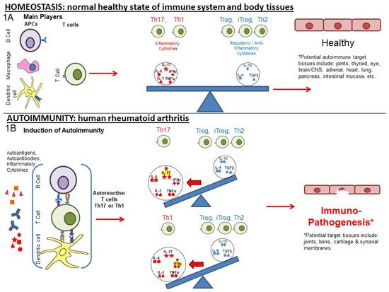
1. From Homeostasis to Autoimmunity

Rheumatoid arthritis (RA) and other autoimmune inflammatory diseases are examples of imbalances within the immune system (disrupted homeostasis) that arise from the effects of an accumulation of environmental and habitual insults over a lifetime, combined with genetic predispositions. The initial insults that lead to RA are thought to often originate in the lung and are connected with smoking. Lungs are constantly exposed to insults due to infections, injury, and inhalants, dust, silica, asbestos, and especially tobacco smoke [1,2,3]. In addition, bacterial infections in the oropharynx, leading to gingivitis or periodontal disease, are considered a possible key early factor (see reviews) [1,4,5,6,7]. The inflammatory responses that drive RA usually originate with autoimmune responses against normal, modified or immuno-mimetics of self-proteins that are often found within skeletal joints. Individuals may be genetically prone to or acquire an enhanced systemic inflammatory state due to infectious, metabolic, or other challenges, which are often related to the immunological maintenance of the gut microbiome. The accumulation of inflammatory insults over a lifetime generates such a state, which has been termed ”inflammaging” [8].

2. Post-Translational Modification and Its Role in Autoimmunity

Tobacco smoking and other challenges promote the activation of the enzyme peptidyl arginine deiminase (PADI, especially PADI-2 and PADI-4) in the lung and elsewhere. This enzyme converts arginine residues in proteins to citrulline in situ in a process called citrullination. Such post-translational modifications (PTM) can alter the immunogenicity of any protein containing that residue and create neo-epitopes, leading to the development of autoimmunity. Citrullinated vimentin, proteoglycan (PG), fibrin, and collagen are commonly found in the skeletal joints of RA patients [2]. Another PTM seen in RA involves lysine residues. Other in situ PTMs, such as on serine and threonine, alter the immunogenicity of other self-proteins and are being observed in a growing number of autoimmune diseases [1,2,3,9,10,11,12]. On an important note, PTM can affect cellular function and enhance the activation of neutrophils and macrophages, leading to the release of destructive enzymes from the recruited cells, such as matrix metalloproteinases (MMP, especially MMP-1, MMP-13), which are collagenases that can cause bone and cartilage destruction [13].

3. Role of Cytokines, Cells, and Their Interplay in Disrupting Immune Homeostasis

In the presence of systemic inflammation, potentially exacerbated by trauma, infection or other inflammatory trigger, autoimmune responses can be initiated against PTM proteins to alter the normal balance of immunity. Figure 1A represents the normal balance in immune pro-inflammatory and anti-inflammatory responses that promote immune homeostasis. The various actors include cells (T and B cells, macrophages, dendritic cells (DC), other blood cells) and pro-inflammatory and anti-inflammatory cytokines that affect the regulation of responses to self (auto)antigen.
Figure 1. Disruption of the homeostasis of the immune system promoting arthritis. (1A) Homeostasis: Antigen-presenting cells present peptides and cytokines to activate antigen-specific T cells influenced by environmental signals and cytokines. A balance of pro-inflammatory to humoral and regulatory responses promote immune homeostasis. Red symbols within dotted circles represent pro-inflammatory cytokines and blue symbols represent anti-inflammatory cytokines. (1B) Autoimmunity: Environmental factors (e.g., smoking), trauma (repetitive bone or cartilage injury), infections (microbial antigen mimicry), and genetic predisposition (e.g., MHC: HLA-DR4) can promote an inflammatory environment that promotes a self-sustaining disruption of immune balance that can result in rheumatoid arthritis (RA). Arthritogenic self-antigens are presented by DCs, macrophages, and B cells to T cells to generate auto-antibodies and self-reactive T cells, respectively, which promote inflammatory cytokine production that activates other cells and induces tissue remodeling and disruption.
As shown in Figure 1B, the disruption of immune homeostasis resulting in autoimmunity and RA can occur in an individual at risk of RA due to environmental, systemic, or genetic factors. The activation of inflammatory responses by exposure to the pathogen-associated molecular pattern molecules (PAMPs) of microbes or damage-associated molecular pattern molecules (DAMPs) released by tissue damage or trauma can activate processes that can initiate inflammatory responses to the PTM or other self-(auto)antigens that drive RA. These processes are driven by T helper (Th)1 and/or Th17 pro-inflammatory responses.
DCs process phagocytosed proteins, including citrullinated proteins if present, into peptides that then occupy major histocompatibility class (MHC I or MHC II) molecules and then activate the naive T cells that can recognize those peptide antigens. Depending on the cytokines produced by the DC, antigen-specific Th1 or Th17 pro-inflammatory T cells are activated [6,14]. These cell types are defined by different transcription factors (TFs) and the cytokines that they produce, setting up cytokine conversations.
Normally, the B cells and T cells that elicit autoimmune responses are either eliminated in the bone marrow and thymus, respectively (central tolerance), or controlled by regulatory responses (peripheral tolerance). Peripheral tolerance is mediated by regulatory T cells (Tregs) and induced T regs (iTregs), whose cytokine conversations involve IL-10 and/or tumor growth factor (TGF)-β. The iTregs are generated primarily from peripheral Th17 or other Th cells in the presence of high levels of IL-10 or TGF-β. Other cells, including macrophages and myeloid suppressor cells, can also produce these regulatory cytokines to control inappropriate or inflammatory responses. The tolerance that these cells impose can be overridden by infections and other challenges that disrupt the homeostatic balance and lead to the production of large amounts of acute phase cytokines (IL-1, IL-6, TNF-α), allowing responses to the microbial or modified antigens that mimic human antigens to initiate autoimmune responses [5,19,20].

4. Current and Older Therapeutic Approaches for RA

The current therapy for RA consists of either the treatment of symptomatology or the inhibition or ablation of the components of the inflammatory immune response (Table 1). Neither of these approaches address the imbalance of the antigen-specific immune response that is the root cause of the disease. Non-steroidal anti-inflammatory drugs (NSAIDs) are the first line of treatment used to alleviate pain and inflammation. Older ablative treatments with corticosteroid, methotrexate, and similar drugs can inhibit inflammation and are effective for many patients. The newer therapies, as shown in Table 1, are more selective in their inhibition of specific mediators of inflammation and can be organized into three main therapeutic approaches, as shown in Table 1 and Figure 3.
Table 1. Approaches to targeting inflammatory cytokines in RA and RA animal models with regard to targets, cytokines, and therapies.
Type Target ↓/↑ Modulation Regulated Immune Component, If Known [References] Generic and Product name, Regulatory Status Ref.
Therapeutic Vaccines Th1 IL-1, IL-17, IFN-γ, TNF-α [3,29,30] CEL-4000 (preclinical) [3,29,30]
Treg (FOXP3+), IL-4, IL-10, TGF-β [3,29,30]
Th17 TNF-α, IL-17, IL-6, MCP-1, IL-12p40 [27] CEL-2000 (preclinical) [27]
IL-12p70, IL-10 [27]
DMARDs TNF-α TNF-α [41] Adalimumab (Humira®) [41,42]
TNF-α TNF-α [43] Etanercept (Enbrel®) [43]
IL-1Ra IL-1 [44] Anakinra (Kineret®) [44]
IL-6R msR MCP-1 [42], IL-6 [45] Tocilizumab (Actemra®) [42,45]
IL-17 MCP-1 [42], IL-17A [46] Secukinumab (Cosentyx®) [42,46]
CD20 B cells as APCs: CD4+IFN-γ+, CD4+IL-17+ [47] Rituximab (Rituxan®) [47,48]
Anti-CD6 IL-17 [49], IFN-γ [49,50], IL-6, TNF-α [50] Itolizumab (Alzumab®) [49,50,51]
Agonistic Anti-CD137 IFN-γ [52,53], IDO [53] Utomilumab [52,53]
Anti-CTLA4 IL-17, IFN-γ [54] Abatacept (Orencia®) [54,55,56]
IL-35, IFN-β [54]
Anti-CD40 IL-6, RANKL [57], TNF-α, NF-κβ, IL-6, ICAM-1, VCAM-1, VEGF [58] Bi 655064 [57,58]
CD24 TNF-α, IL-6, MCP-1(CCL2), IL-1β [59] NF-κβ [60]   [59,61,62]
Jakinibs JAK3 > JAK1, JAK2 > TYK2 [63] Transcription: IL-2, IL-4, IL-7, IL-9, IL-15, IL-21, IL-6, IL-11, IL-13, IL-25, IL-27, IL-31, IFN-α, IFN-β, IL-10, IL-22, IFN-γ, > EPO, TPO, GH, G-CSF, GM-CSF, Leptin, IL-3, IL-5 > IL-12, IL-23, Type III IFNs [64]
in vitro: IL-6 by B cells, [65] IL-2, IL-4, IL-7, IL-15, IL-21, IL-6, and IFN-γ in CD4+ T cells. IL-17 in Th17 cells polarized via IL-23. IL-21 and IL-22 in Th17 [66], IFN-α, IL-6, IFN-γ, IL-2, IL-15, IL-4, GM-CSF [64] MCP-1 [42] IL-17 in CD4+T cells from AS, PSA, RA, and HC [67]
in vivo: IL-6 in human [68]
Tofacitinib (Xeljanz®)FDA approved (2012) [42,63,64,65,66,67,68,69,70,71]
in vitro: IL-2 in Th1. IL-17, IL-2 in Th17 cells (polarized via TGF-β1, IL-6) [66]
JAK3 > JAK1, TYK2, JAK2 [63] Transcription: IFN-α, IFN-β, IL-10, IL-22, IL-2, IL-4, IL-7, IL-9, IL-15, IL-2, IFN-γ > IL-6, IL-11, IL-13, IL-25, IL-27, IL-31, IL-12, IL-23, Type III IFNs, EPO, TPO, GH, G-CSF, GM-CSF, Leptin, IL-3, IL-5
in vitro: IL-4, IL-13, IFN-γ, TNF-α in PBMC after TCR stimulation, IL-4, IL-13, IFN-γ, TNF-α, IL-17A, GM-CSF in PBMC after IL-2 stimulation [72]
Peficitinib (Smyraf®) Japan Approved (2019) [63,72]
JAK2, JAK1 > TYK2 > JAK3 [63] Transcription: IL-6, IL-11, IL-13, IL-25, IL-27, IL-31, IFN-α, IFN-β, IL-10, IL-22, IFN-γ > IL-2, IL-4, IL-7, IL-9, IL-15, IL-21 > IL-12, IL-23, Type III IFNs, EPO, TPO, GH, G-CSF, GM-CSF, Leptin, IL-3, IL-5 [64]
in vitro: IL-6 in MoDCs, IFN-α secreted pDCs [65] MCP-1 [42] IL-17 in CD4+ T cells (AS, PSA, RA, and HC) [67]
Baricitnib (Olumiant®) FDA approved (2018) [42,63,64,65,67,69,70]
JAK2, JAK1 > TYK2 > JAK3 [63] Transcription: IFN-γ, EPO, TPO, GH, G-CSF, GM-CSF, Leptin, IL-3, IL-5 > IL-6, IL-11, IL-13, IL-25, IL-27, IL-31, IFN-α, IFN-β, IL-10, IL-22, IL-12, IL-23, Type III IFNs > IL-2, IL-4, IL-7, IL-9, IL-15, IL-21
in vitro: IL-10, IFN-γ, IL-6, TNF-α, IL-13 [73] IL-17 in CD4+ (AS, PSA, RA, and HC) [67]
in vivo: IFN-γ, IL-12p70, IL-6, G-CSF, IL-10, TNF-α [73]
Ruxolitinib (Jakafi®) FDA approved (2011) (myelofibrosis) [63,67,69,73,74]
in vitro: IL-2 [74]
JAK1 > JAK2 > TYK2 > JAK3 [63] Transcription: IL-6, IL-11, IL-13, IL-25, IL-27, IL-31 > IFN-α, IFN-β, IL-10, IL-22 > IFN-γ, > IL-2, IL-4, IL-7, IL-9, IL-15, IL-21 > EPO, TPO, GH, G-CSF, GM-CSF, Leptin, IL-3, IL-5, IL-12, IL-23, Type III IFNs [64]
in vitro: IL-2, IL-4, IFN-αB2, IFN-γ [71] IFN-α, IL-6, IFN-γ, IL-2, IL-15, IL-4 [64]
ex vivo: IL-6, GM-CSF [64]
in vivo: IFN-γ, IL-6, IL-1β, RANKL, MMP-3, MMP-13, IP10, XCL1, MCP-1, MIP-1b, MCP-3, MCP-5, M-CSF1, MDC, SCF, KC/GRO, IL-1α [71] SAA, IL-6, IL-1β, GM-CSF, TNF-RI, Resistin, TNF-α, MMP-3, YKL40, VEGF, MMP-1, IL-12, IL-2, IFN-γ, IL-13, IL-5, IL-21, IL-23, IL-17A, IL-7, IL-10, CXCL10, CXCL13, MCP-1, VCAM-1, MIP-1a [75]
Filgotinib (Jyseleca®) EMA & Japan approved (2020) [63,64,69,71,75]
JAK1 > JAK2 > JAK3 > TYK2 [63] Transcription: IL-6, IL-11, IL-13, IL-25, IL-27, IL-31, IFN-α, IFN-β, IL-10, IL-22, IFN-γ EPO, TPO, GH, G-CSF, GM-CSF, Leptin, IL-3, IL-5 IL-2, IL-4, IL-7, IL-9, IL-15, IL-21 > IL-12, IL-23, Type III IFNs [64]
in vitro: IFN-α, IL-6, IFN-γ, IL-2, IL-4, IL-15, G-CSF [64]
Upadacitinib (Rinvoq®) FDA approved (2019) [63,64,70]
JAK2 > JAK1 > TYK2 > JAK3 [76] See main text
in vitro: VCAM-1, IL-6 [74]
Fedratinib (Inrebic®) (2019) (myelofibrosis) [63,74,77,78,79]
in vitroIL-2 [74]
Footnotes: Heat map colors: Red: proinflammatory; Blue: anti-inflammatory; Gray: either possibility. For abbreviations used above, see abbreviation section before the author contribution section For jakinibs: extrapolated expectations are based on the inhibited JAK/STAT pathways indicated by experimental data. Canonical JAK signaling pathways: JAK1/JAK3: IL-2, IL-4, IL-7, IL-9, IL-15, IL-21; JAK1/JAK2: IFN-γ; JAK1/TYK2: IFN-α, IFN-β, IL-10, IL-22; JAK1/JAK2/TYK2: IL-6, IL-11, IL-13, IL-25, IL-27, IL-31; JAK2/TYK2: IL-12, IL-23, Type III IFNs; JAK2/JAK2: EPO, TPO, GH, G-CSF, GM-CSF, Leptin, IL-3, IL-5 [68,80,81,82].

5. Grouping of the Therapeutic Approaches

The three groups of therapies shown in Table 1 are presented with the immunological target of the treatment, the cytokine(s) that are affected and whether their amount is increased or decreased and examples of specific drugs for that type of treatment. The information in Table 1 is presented as a “heat map” to assist in revealing trends and patterns. Highlighted in red are the five pro-inflammatory cytokines (IL-1(α or β), IL-6, IL-17, IFN-γ, and TNF-α), in Blue are two anti-inflammatory cytokines (IL-4 and IL-10) and in gray are IL-2, since it seems to play a key regulatory role, and IL-12p70, which promotes IFN-γ responses, which can be pro-inflammatory or anti-inflammatory.
The Group I therapies, i.e., LEAPS therapeutic vaccines, focus on the cytokine-secreting, antigen-specific T cells, but not individual cytokines per se. These treatments promote a modification of the cytokine conversation that affects both anti-inflammatory cytokines and pro-inflammatory cytokines, focusing upstream on the source of the response, the memory T cell.
Group II therapies focus on ablations of individual cytokine action by neutralizing the cytokine or blocking its receptor. Neutralizing monoclonal antibodies or receptor antagonists prevent the action of individual acute phase cytokines, TNF-α, IL-1, IL-6 or mediators of Th17 responses, such as IL-23 or IL-17. Alternatively, antibodies to surface differentiation antigens (CDs) are used to eliminate or inhibit specific cells and their functions. Antibody to CD20 lessens the number of B cells to reduce these RA-promoting, antigen-presenting cells. These therapeutic products, monoclonal antibodies, soluble receptor antagonists, agonists, or modified soluble receptors are collectively referred to as Disease-Modifying Antirheumatic Drugs (DMARDs).
The first developed and therefore oldest of the group (II) cytokine-focused therapies are the biological response modifiers (BRM), such as neutralizing monoclonal antibodies, solubilized receptors (sR) and modified soluble receptors (msR) (e.g., for IL-2R: MR-IL-2). As monoclonal antibodies, they are highly specific for the antigenic epitopes present on the target molecules [83,84] and act extracellularly. However, it should be noted that some protein subunits are shared between several cytokines, such as IL-12p40 for both IL-12 and IL-23 cytokines, so that multiple cytokines and their consequences may be affected [85,86]. Similarly, monoclonal antibodies directed towards cell surface markers (e.g., CD20) (other than cytokine receptors), may affect several different types of cells expressing that marker [48,84]. The Group II agents are often administered by injection as an intravenous (IV) bolus or infusion over time because of their much larger sizes (e.g., about 150 kDa for monoclonal antibodies).
Group III therapies focus on JAK inhibitors (abbreviated to jakinhib-1, -2 or -3, etc.) which target the JAK/STAT transduction of receptor signals, which activates the transcription and production of one or more cytokines, including not only the pro-inflammatory cytokines, but also potentially therapeutic anti-inflammatory cytokines, notably IL-2, IL-4, IL-10, and TGF-β [64,66,68,82,86,87,88,89].

6. Comparisons of LEAPS, Monoablative, and Jakinib Therapies

LEAPS peptide therapeutic vaccines are designed to have an immunomodulatory effect on the T cells driving the disease, as illustrated in Figure 3A,B. In so doing, the LEAPS peptides affect the entire cytokine conversation, increasing the expression of some and decreasing other cytokines to return immunobalance, rather than acting on a single cytokine [3,27,29,30,31].

The monoablative therapies (shown in Figure 3C,D) use a neutralizing monoclonal antibody or receptor-antagonist to specifically block the action of one of the disease-associated cytokines after secretion and not its synthesis. The targets for these therapies include IL-1β, IL-6, IL-17A, IL-23, IL-12, and TNF-α and, under certain circumstances, IFN-γ. These monoablation therapies only indirectly affect other pro-inflammatory cytokines and do not upregulate anti-inflammatory cytokines to rebalance the cytokine conversation.

The third group (III) of therapies (Table 1, Figure 3E–G) are the jakinibs, which act by inhibiting specific receptor-associated JAK/STAT tyrosine kinases, ultimately inhibiting the synthesis and secretion of multiple cytokines (multi-ablative therapy) that are activated by the specific JAK cascade. The jakinibs are small-molecule (~300Da) inhibitors acting on the Janus kinases JAK1, JAK2, JAK3 or TYK2 and have the major advantage of being taken orally [63]. The JAK enzymes most often work in pairs as homo- or heterocomplexes activating STAT molecules to create transcriptional activators and promote the expression of groups of cytokines and other genes. JAK activation or inhibition also influences the expression of different cell surface receptors, including CD4, CD80 and CD86 on T, B and other cells and their associated immune responses [65]. The expression of some JAK enzymes is more restricted to certain cell types than others, such as JAK3 for immune system cells such as B, T, and NK cells. This is a therapeutic advantage.

As can be seen in Table 1, there are at least five different jakinibs approved for RA treatment in the USA, Europe, or Japan and several others are under investigation, each unique in terms of the molecule’s binding preference for its particular cognate JAK or JAK-associated molecule. Different manifestations of treatment occur depending on the relative selectivity of binding and whether it is reversible or irreversible. The representative jakinibs are shown as different-colored (red, blue and orange) hexagonal shapes in Figure 3 for the three examples of jakinibs that downregulate inflammatory and anti-inflammatory cytokines. Jakinib A (Figure 3E red hexagon) has activity which is JAK 3>1 and downregulates the expression of IL-6, IL-17, IFN-γ, TNF-α, IL-4, and IL-10. Jakinib B (Figure 3F blue hexagon) is a JAK 2>1 inhibitor downregulating IL-6, IL-17, IFN-γ, TNF-α, and IL-10. Jakinib C (Figure 3G orange hexagon) is a JAK 1>2 inhibitor downregulating IL-1, IL-6, IL-17, IFN-γ, TNF-α, IL-4, and IL-10. It should be noted that a jakinib specific for only JAK2 cannot be used since the inhibition of JAK2/STAT is associated with lethality early in life [63,77].

Although TNF-α, IL-1, and IL-17 are major targets for monoablation therapy, they are not directly affected by the jakinibs. However, they may be indirectly affected by the inhibition of expression of other cytokines that are supposedly not involved in the JAK signaling pathway; for example, IL-17 is affected indirectly by several of these jakinibs [66,67,77]. Similarly, an indirect effect of jakinibs may also promote the expression of some anti-inflammatory cytokines. The combination therapy targeting several cytokines, as possibly seen for the JAK inhibitors, may be effective, although this is still being debated and new clinical studies will be needed [2,99,111,112].

This entry is adapted from the peer-reviewed paper 10.3390/biomedicines10010044

This entry is offline, you can click here to edit this entry!