You're using an outdated browser. Please upgrade to a modern browser for the best experience.
Parasite Druggable Targets of Toxoplasmosis: History
Please note this is an old version of this entry, which may differ significantly from the current revision.
Subjects: Allergy
Contributor: Hiba El Hajj

Toxoplasmosis is a prevalent disease affecting a wide range of hosts including approximately one-third of the human population. It is caused by the sporozoan parasite Toxoplasma gondii (T. gondii), which instigates a range of symptoms, manifesting as acute and chronic forms and varying from ocular to deleterious congenital or neuro-toxoplasmosis. Toxoplasmosis may cause serious health problems in fetuses, newborns, and immunocompromised patients. Recently, associations between toxoplasmosis and various neuropathies and different types of cancer were documented. In the veterinary sector, toxoplasmosis results in recurring abortions, leading to significant economic losses. Treatment of toxoplasmosis remains intricate and encompasses general antiparasitic and antibacterial drugs. The efficacy of these drugs is hindered by intolerance, side effects, and emergence of parasite resistance. Furthermore, all currently used drugs in the clinic target acute toxoplasmosis, with no or little effect on the chronic form.

  • acute toxoplasmosis
  • chronic toxoplasmosis
  • parasite therapeutic targets
  • neuropathies
  • antiparasitic drugs
  • immunomodulatory drugs

1. Introduction

Toxoplasma gondii (T. gondii) is an obligate intracellular protozoan parasite infecting a broad range of animals, including one-third of the world’s human population [1]. Because of its high prevalence in the United States, the Centers for Disease Control and Prevention classified toxoplasmosis among the neglected parasitic infections that require a public health action control [2]. The pathogenesis of T. gondii varies among patients. The acute form of the disease develops a few days after the infection and is asymptomatic in more than 80% of immunocompetent individuals [1,3]. The remaining patients may exhibit general flu-like symptoms, fever, myalgia, and cervical posterior adenopathy, among other symptoms [1]. In certain regions, e.g., French Guiana and Latin America, severe symptoms including fatal pneumonitis, myocarditis, meningoencephalitis, and polymyositis were noted in some immunocompetent patients who contracted atypical strains of T. gondii [4].
Congenital toxoplasmosis occurs in sero-negative pregnant women acquiring T. gondii as primary infection. The parasite crosses the blood–placenta barrier and reaches the fetus [5]. The transmission rate and severity of congenital toxoplasmosis depend on the gestational trimester at which the infection is acquired [6,7]. Transmission rates of 25% are estimated when the infection occurs during the first trimester, while 54 and 65% transmission rates are estimated when infection occurs during the second or third trimesters, respectively [7,8]. Infection of the fetus during the first trimester often leads to abortion. However, in cases of stillbirth, the baby suffers from severe aberrations of the brain (hydrocephalus, intracranial calcifications, deafness, mental retardation, seizures) and the eyes (retinochoroiditis that may lead to blindness (reviewed in [9])). Infection of the fetus during the second or third trimester is less likely to cause abortion; however, retinochoroiditis and learning difficulties may occur after birth [10]. Retinochoroiditis is the most common symptom of ocular toxoplasmosis and predominantly results from an acquired congenital toxoplasmosis. It presents with posterior uveitis, vitritis, focal necrotizing granulomatous retinitis, and reactive granulomatous choroiditis [11]. It is worth noting that the acquisition of toxoplasmosis during pregnancy varies according to regions [12,13], and atypical T. gondii genotypes were identified and led to re-infection of previously sero-positive pregnant women [14], resulting in a more severe congenital disease [15].
Following an acute infection, T. gondii targets the brain and skeletal muscles, where it persists as latent tissue cysts responsible for chronic toxoplasmosis (reviewed in [16]). The switch from the acute to the chronic form is triggered by the host immune response, among other factors (reviewed in [17,18]). Although the direct symptoms of chronic toxoplasmosis are not fully characterized in healthy individuals, chronic toxoplasmosis was regarded as clinically asymptomatic [19]. Yet, the brain immune response triggers inflammation, which disrupts neuronal connectivity and associates with ventricular dilatation [20,21,22]. In addition, chronic toxoplasmosis correlates with several neuropathies [19], behavioral disorders, and cancers [23]. Nonetheless, direct molecular proofs remain to be elucidated [24,25] (Daher et al., in press).
Immunosuppression triggers the reactivation of chronic toxoplasmosis, leading to serious complications and potential fatality [1,26,27,28]. Reactivation was mostly documented in HIV patients or patients treated with immunosuppressive therapies prior to solid organ or hematopoietic stem cell transplantation [18,29,30,31,32,33,34,35]. Indeed, among solid organ transplanted patients, orthotopic heart transplant recipients presented with the highest risk of reactivation of toxoplasmosis, owing to the high propensity of T. gondii cysts in striated muscles [36]. Chemotherapy administration, in particular, rituximab, also triggered the reactivation of toxoplasmosis [37,38,39,40]. HIV patients with reactivated toxoplasmosis manifest predominantly with neurological symptoms including toxoplasmic encephalitis, encephalopathy, meningoencephalitis, headache, seizures, and poor coordination, while transplanted patients exhibit a more disseminated status [4,41].

2. Emerging Therapeutic Targets in Toxoplasma gondii Infections

Despite all prophylactic approaches to prevent the infection with T. gondii, an available human vaccine is still out of reach. While the search for a vaccine has been highly pursued, currently used drugs target only the acute form of the disease. Chronic toxoplasmosis, which represents the more prevalent form and associates with dreadful clinical outcomes, reaching fatality in immunocompromised patients, remains an unmet medical need. An ideal drug against toxoplasmosis should affect multiple stages of the parasite life cycle (i.e., tachyzoites responsible for acute toxoplasmosis and bradyzoites responsible for chronic toxoplasmosis). Furthermore, these drugs should (1) target the parasite biology, (2) exhibit low toxicity and tolerable side effects, (3) have high bioavailability, and (4) cross the blood–brain barrier and reach the brain, where the propensity for neuronal cysts is high [81]. A number of preclinical studies were conducted in vitro and prolonged mice survival in vivo (reviewed in [82]). A comprehensive summary of tested drugs and compounds over a decade extending from 2006 to 2016 reported 80 clinically available drugs and a large number of new compounds with more than 40 mechanisms of action. Several target-based drug screens were also identified. These include different kinases, mitochondrial electron transport chain, fatty acid synthesis, DNA synthesis, and replication, among several others [59]. In the following sections, we will provide a comprehensive overview of the different parasite targets and their corresponding emerging drugs (Figure 1, Table 2).
Figure 1. Schematic representation of known Toxoplasma gondii therapeutic targets.
Table 2. Summary of Toxoplasma gondii therapeutic targets and their corresponding drugs.

2.1. Targeting the Apicoplast

In apicomplexan parasites, the apicoplast is a nonphotosynthetic organelle formed following endosymbiosis of a green algae. This organelle assumes several functions including the biosynthesis of fatty acids, lipoic acid, and isoprenoids, among other metabolites [83]. The absence of apicoplasts in mammalian cells made them an excellent therapeutic target, and several attempts to target their parasitic functions have been tested (Figure 1, Table 2). These therapeutic targets span several apicoplast enzymes involved in fatty acid synthesis and metabolism. These include the fatty acid synthase II (FASII) [84], the acetyl CoA carboxylase (ACC) that catalyzes the formation of Malonyl-CoA, and the β-ketoacyl ACP synthase III (FabH) [85,86,87]. Clodinafop, thiolactomycin, and triclosan proved efficient in targeting these enzymes and blocking the parasite fatty acid synthesis [85,86].
Another apicoplast therapeutic drug target is the isoprenoid synthesis pathway. Isoprenoid is a precursor of ubiquinone and sterols, playing an important role in cell signaling. 1-deoxy-D-xylulose-5-phosphate (DOXP) was identified in the apicoplast and plays a role in the biosynthesis of isoprenoid [88]. Two key enzymes featured in the DOXP pathway, the DXP reducto-isomerase and DXP synthase, were identified in apicomplexan parasites including T. gondii and are absent in humans, posturing these enzymes as attractive therapeutic drug targets [88]. Fosmidomycin antibiotic proved efficient against these two enzymes; however, resistance problems, high concentrations, and low bioavailability hindered its activity [89,90].
Because of its prokaryotic nature, the apicoplast’s DNA is circular, and its unwinding and supercoiling during replication is controlled and mediated by DNA topo-isomerases including the DNA gyrase [91,92,93]. This enzyme is absent in humans and was the target of several quinolone and fuoroquinolone antibiotics. These include ciprofloxacin, trovafloxacin, ofloxacin and temafloxacin, which exhibited in vitro and in vivo efficacy against T. gondii infections [94,95]. In addition to DNA replication, the prokaryotic nature of the apicoplast also confers a mechanism of protein synthesis independent of that occurring in the nucleus of the parasite. Indeed, the apicoplast encodes proteins and RNA indispensable to its ribosomes, hence its specific proteins [96]. Clindamycin, spiramycin, and azithromycin, known to affect prokaryotic protein synthesis, exhibited toxoplasmicidal activities in vitro and in vivo [97].

2.2. Targeting the Invasion Complex

2.2.1. Microneme Organelles

Micronemes are small rod-shaped organelles of the apical complex of T. gondii. They play a chief role in attachment, gliding, motility, and egress during the invasion process required for parasite survival [98,99,100]. Owing to their uniqueness in apicomplexan parasites, their absence in mammalian cells, and their pivotal role in invasion, micronemes and especially their calcium-mediated secretion have been at the core of parasite targeting. In that sense, T. gondii calcium-dependent protein kinase 1 (TgCDPK1), a parasite cytosolic serine/threonine-protein kinase regulating the calcium-dependent pathway, is essential for micronemal protein secretion. The inhibition of this enzyme impairs host cell invasion capacity [101,102,103]. Several bumped kinase inhibitors (BKIs) selectively inhibited TgCDPK1 [104,105] (Figure 1, Table 2). BKI-1294, a pyrazolo-pyrimidine-based compound, resulted in high inhibition of invasion in vitro and high efficiency against acute toxoplasmosis in vivo when given orally [105]. This same inhibitor also proved efficient against congenital toxoplasmosis in a murine model [106]. The promising efficacy of BKI-1294 was hindered by its cardiac toxicity, halting its clinical development [107,108,109]. Consequently, BKI-1294 was chemically modified by maintaining its TgCDPK1 selectivity and efficacy and reducing its cardiac toxicity. Compound 32 was thus developed and proved efficacious in vitro and in vivo, in particular, through reducing brain cysts [108]. Other pyrazolo-pyrimidine inhibitors of TgCDPK1 were also tested. Another compound (called compound 24) exhibited in vitro nanomolar and submicromolar activity on TgCDPK1 and inhibited parasite proliferation [110]. This BKI analog showed an excellent oral bioavailability, reduced acute and chronic toxoplasmosis in mice, and, more importantly, delayed reactivation of the chronic disease following immunosuppression [110]. Recently, BKI-1748, a 5-aminopyrazole-4-carboxamide compound inhibited proliferation of Neospora caninum and Toxoplasma gondii in vitro. The safety of this compound was tested in zebrafish with no embryonic impairment up to 10 μM, and in pregnant mice, with no pregnancy interference at a dose of 20 mg/kg/day. The efficacy of BKI-1748 was assessed in standardized pregnant mouse models infected with tachyzoites or oocysts of T. gondii and resulted in increased pup survival and profound inhibition of vertical transmission [111]. SP230, an imidazo[1,2-b]pyridazine salt targeting TgCDPK1, proved efficient against murine acute toxoplasmosis in mice. Importantly, administration of SP230 yielded significant efficacy against congenital toxoplasmosis. SP230 resulted in the reduction of parasite burden in 97% of fetuses [112].

2.2.2. Rhoptry Organelles

Rhoptries are organelles of the apical complex discharging their content during parasite invasion. Their protein content plays several roles including contributing to the formation of moving junction and the parasitophorous vacuole membrane (PVM). Furthermore, other functions were unraveled, and some rhoptry proteins play roles as virulence factors, others manipulate host signaling, while others play a role in immune evasion [113,114,115].
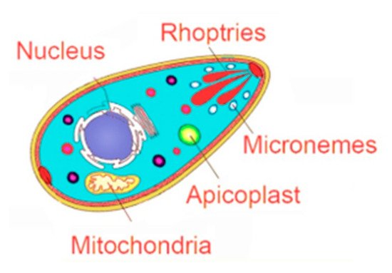
ROP2 family contains a group of proteins, with some members sharing more than 70% identity, while other members are structurally more divergent [116]. While the members of this family evolved with all the elements to be active kinases, some members (ROP2, ROP4, ROP7, ROP5) lost some key motifs or residues in the kinase activity domain over time to acquire other functions [116,117]. For instance, ROP2 contributes [118], but is not the only factor [119], to the recruitment of the host mitochondria around the PVM. ROP5 exhibits an inverted topology in the PVM as compared to other members of the family [120], and protein forms a complex with ROP17 and ROP18 (which retained their kinase activity), hence controlling the virulence in mice [121,122]. In that sense, ROP5 and ROP18 allele combinations are tightly related to T. gondii virulence [122,123,124,125], and ROP5 teams up with ROP18 and complements its activity to inhibit the accumulation of the IFN-γ-induced immunity-related GTPases (IRGs) in vivo, hence contributing to the pathogenesis and immune evasion [126]. Owing to the role of ROP5 and ROP18 in virulence, attempts to use this complex as a vaccine strategy were promising in mice [127]. In addition, recombinant ROP5 and ROP18 were evaluated for their diagnostic potential in human toxoplasmosis [128]. ROP16 and ROP18 were also proven as virulence factors through targeting the host cell nucleus and exhibiting their kinase activity to phosphorylate key proteins involved in cell cycle and different signaling pathways [129]. ROP18 is expressed in genotypes I/II demonstrating their role in controlling the virulence of the parasite [130], and transfection of the virulent ROP18 allele into a nonpathogenic type III strain confers virulence and enhances mortality in vivo [131]. Through its kinase activity, ROP18 phosphorylates GTPases, promoting macrophage survival and virulence [132] and ensuring an immune evasion strategy for virulent strains [133]. ROP16, on the other hand, is expressed in genotypes I/III and also plays a key role in the virulence of the parasite [130]. ROP16 phosphorylates STAT3 and STAT6 [134], hence downregulating IL-12, which plays a chief role in mounting an immune response against T. gondii infection [130]. ROP16 also suppresses T cell activity, hence ensuring immune cell evasion [135]. Moreover, direct phosphorylation of STAT3 by ROP16 mimics the IL-10 activity and downregulates IFN-γ, hence enhancing the virulence of T. gondii [134]. Recently, ROP16-mediated activation of STAT6 proved important for type III T. gondii survival through suppression of host cell reactive oxygen species production [136]. Moreover, ROP16 kinase activity silences the cyclin B1 gene promoter, hijacking the function of the host cell epigenetic machinery [137]. The role of ROP proteins in the virulence of the parasite makes them excellent drug target candidates to combat toxoplasmosis. A high-throughput screen to identify small molecule inhibitors of ROP18 identified several inhibitors belonging to oxindoles, 6-azaquinazolines, and pyrazolopyridines chemical scaffolds. Treatment of IFN-γ-activated cells with one of these inhibitors enhanced immunity-related GTPase recruitment to wild type parasites [138]. Thiazolidinone derivatives inhibited T. gondii in vitro, and in silico analysis demonstrated that the best binding affinity of these derivatives was observed in the active site of kinase proteins with a possible effect of one derivative in the active site of ROP18 [139] (Figure 1, Table 2).

2.3. Targeting the Parasite Mitochondrial Electron Transport Pathway

In apicomplexan parasites, the mitochondrial electron transport chain is of central importance for energy production [140]. This complex, present in the mitochondrial electron transport chain, was targeted by several mitochondrial inhibitors, hindering cell respiration and leading to parasite death (Figure 1, Table 2). Atovaquone, clinically used in the treatment and prophylaxis of toxoplasmosis, is an inhibitor of the hydroquinone oxidation site of the bc1 complex [48]. Emerging resistance of the parasite limited its use [141]. Different quinolone derivatives including the endochin-like quinolones (ELQs), which target the hydroquinone reduction site of bc1, have been developed. ELQ-271 and ELQ-316 inhibited parasite growth at nanomolar concentrations in vitro and reduced the number of brain cysts in murine models [142,143,144]. Another compound, ELQ-400, alleviated the burden of acute toxoplasmosis in mice and demonstrated 100% cure rates upon infection of mice with a type I lethal strain [143,145].
Naphthoquinones bind to the hydroquinone oxidation site of the bc1 complex. Seven naphthoquinones exhibited an anti-T. gondii inhibitory effect in vitro. Three out of seven (para-hydroxynaphthoquinones) were able to enhance survival of mice following infection with a virulent T. gondii strain (Ferreira et al., 2002) (Figure 1, Table 2).

2.4. Targeting the Interconversion between Tachyzoites and Bradyzoites

Histone acetylase (HAT) and histone deacetylase (HDAC) enzymes controlling histone acetylation regulate and control the parasite gene expression during the back and forth interconversion between acute and chronic toxoplasmosis. Targeting these enzymes is a plausible therapeutic scenario. The cyclopeptide FR235222, a TgHDAC3 inhibitor, induced in vitro conversion to bradyzoites and inhibited parasite growth [146]. To decrease the toxicity of FR235222, W363 and W399 derivatives were generated and exhibited equivalent IC50 to the mother compound in vitro [147].
Rolipram, a phosphodiestrase-4 (PDE4) inhibitor interfered with the interconversion from tachyzoites to bradyzoites through immunomodulatory activities and significantly reduced the cyst burden in the brains of chronically infected mice [148]. Guanabenz, an FDA-approved antihypertensive drug, interferes with translational control in tachyzoite and bradyzoite stages through inhibition of dephosphorylation of T. gondii eukaryotic initiation factor 2 (TgeIF2). This inhibitor protected mice against acute toxoplasmosis and reduced the brain cyst numbers in chronically infected mice [81].
T. gondii mitogen-activated protein kinase (MAPK) regulates parasite proliferation, response to stress, and stage differentiation. Pyridinylimidazole inhibited TgMAPK1, caused morphological changes, and reduced the virulence of T. gondii [149,150,151]. In conclusion, targeting the interconversion between tachyzoites and bradyzoites can be a promising therapeutic approach.
Heat shock proteins (HSPs) promote host cell invasion, parasite growth, survival, as well as stage conversion from tachyzoite to bradyzoite, hence from the acute to the chronic form of infection [152,153]. HSP60 and 70 are important in the development and survival of T. gondii. While HSP60 is responsible for stage-specific induction of the respiratory pathway, HSP70 plays a role in stage differentiation and virulence [154]. HSP70 protects the parasite from the host immune system. Treatment of mice with quercetin and oligonucleotide reduced HSP70 expression in a virulent T. gondii strain [155]. The 3D structures for T. gondii Hsp60 and Hsp70 were performed by homology modeling, and a virtual screening of 1560 compounds from the NCI Diversity Set III was analyzed and demonstrated that the major exhibited interactions were hydrogen bonding and hydrophobic interactions in binding to HSP60 and HSP70, providing guidelines for the development of inhibitors for these parasitic heat shock proteins [156].

This entry is adapted from the peer-reviewed paper 10.3390/microorganisms9122531

This entry is offline, you can click here to edit this entry!
Academic Video Service