Ammonia nitrogen is the major oxygen-consuming pollutant in aquatic environments. Exposure to ammonia nitrogen in the aquatic environment can lead to bioaccumulation in fish, and the ammonia nitrogen concentration is the main determinant of accumulation. In most aquatic environments, fish are at the top of the food chain and are most vulnerable to the toxic effects of high levels of ammonia nitrogen exposure. In fish exposed to toxicants, ammonia-induced toxicity is mainly caused by bioaccumulation in certain tissues. Ammonia nitrogen absorbed in the fish enters the circulatory system and affects hematological properties. Ammonia nitrogen also breaks balance in antioxidant capacity and causes oxidative damage. In addition, ammonia nitrogen affects the immune response and causes neurotoxicity because of the physical and chemical toxicity.
- ammonia nitrogen
- fish
- oxidative stress
- neurotoxicity
- immune response
1. Introduction

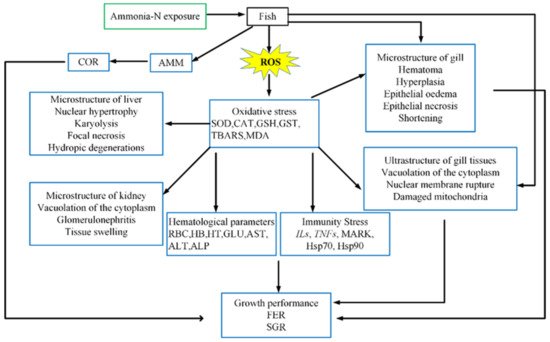
2. Bioaccumulation
3. Hematological Parameters
| Exposure Route | Exposure Type | Fish Specie | Ammonia Concentration |
Exposure Time | Response Concentration |
Response * | Reference |
|---|---|---|---|---|---|---|---|
| RBC (Billion/mL) | |||||||
| Sea water | Waterborne exposure |
Takifugu rubripes | 5, 50, 100, 150 mg/L | 24, 48, 96 h | 100, 150 mg/L | − | Gao et al. [40] 2021 |
| Freshwater | Waterborne exposure |
Cyprinus carpio | 106mg/L | 24 h | 106 mg/L | − | Hoseini et al. [43] 2019 |
| Megalobrama amblycephala | 5, 10, 15, 20 mg/L | 9 weeks | 20 mg/L | − | Zhang et al. [11] 2019 | ||
| Ht (%) | |||||||
| Sea water | Waterborne exposure |
Takifugu rubripes | 5, 50, 100, 150 mg/L | 24, 48, 96 h | 50, 100, 150 mg/L | − | Gao et al. [40] 2021 |
| Piaractus mesopotamicus |
1, 2, 3 mg/L | 96 h | 2, 3 mg/L | + | Edison et al. [51] 2015 | ||
| Freshwater | Waterborne exposure |
Cyprinus carpio | 106 mg/L | 24 h | 10 6mg/L | − | Hoseini et al. [43] 2019 |
| Megalobrama amblycephala | 5, 10, 15, 20 mg/L | 9 weeks | 20 mg/L | − | Zhang et al. [11] 2019 | ||
| Hb (g/L) | |||||||
| Sea water | Waterborne exposure |
Takifugu rubripes | 5, 50, 100, 150 mg/L | 24, 48, 96 h | 50, 100, 150 mg/L | − | Gao et al. [40] 2021 |
| Piaractus mesopotamicus |
1, 2, 3 mg/L | 96 h | 2, 3 mg/L | − | Edison et al. [51] 2015 | ||
| Freshwater | Waterborne exposure |
Cyprinus carpio | 106 mg/L | 24 h | 106 mg/L | − | Hoseini et al. [43] 2019 |
| Megalobrama amblycephala | 5, 10, 15, 20 mg/L | 9 weeks | 20 mg/L | − | Zhang et al. [11] 2019 | ||
| Glucose (mg/dL) | |||||||
| Sea water | Waterborne exposure |
Takifugu rubripes | 5, 50, 100, 150 mg/L | 24, 48, 96 h | 50, 100, 150 mg/L | + | Gao et al. [40] 2021 |
| Litopenaeus vannamei | 0.32, 0.44, 0.60 mg/L | 6 h, 12 h, 1 day, 2 days | 0.32, 0.44, 0.60 mg/L | + | Cui et al. [52] 2017 | ||
| Piaractus mesopotamicus |
1, 2, 3 mg/L | 96 h | 2, 3 mg/L | + | Edison et al. [51] 2015 | ||
| Freshwater | Waterborne exposure |
Pelteobagrus fulvidraco | 100 mg/L | 24, 48, 72 h | 100 mg/L | + | Zhao et al. [53] 2021 |
| Megalobrama amblycephala | 5, 10, 15, 20 mg/L | 9 weeks | 20 mg/L | − | Zhang et al. [11] 2019 | ||
| Cyprinus carpio | 0.5 mg/L | 24 h | 0.5 mg/L | + | Mirghaed et al. [54] 2019 | ||
| Total protein (g/dL) | |||||||
| Sea water | Waterborne exposure |
Takifugu rubripes | 5, 50, 100, 150 mg/L | 24, 48, 96 h | 50, 100, 150 mg/L | − | Gao et al. [40] 2021 |
| Epinephelus fuscoguttatus ♀ × E. lanceolatus ♂ | 1, 2, 4, 8 mg/L | 1week, 2 weeks | 8 mg/L | − | Kim et al. [48] 2020 | ||
| Freshwater | Waterborne exposure |
Pelteobagrus fulvidraco | 100 mg/L | 24, 48, 72 h | 100 mg/L | + | Zhao et al. [53] 2021 |
| Inject | Ctenopharynodon idellus | 9 μL | 96 h | 9 μL | × | Xing et al. [55] 2016 | |
| AST (U/L) | |||||||
| Freshwater | Waterborne exposure |
Pelteobagrus fulvidraco | 100 mg/L | 24, 48, 72 h | 100 mg/L | + | Zhao et al. [53] 2021 |
| Megalobrama amblycephala | 5, 10, 15, 20 mg/L | 9 weeks | 20 mg/L | + | Zhang et al. [11] 2019 | ||
| Cyprinus carpio | 106 mg/L | 24 h | 106 mg/L | + | Hoseini et al. [43] 2019 | ||
| ALT (U/L) | |||||||
| Sea water | Waterborne exposure |
Takifugu rubripes | 5, 50, 100, 150 mg/L | 24, 48, 96 h | 50, 100, 150 mg/L | + | Gao et al. [40] 2021 |
| Freshwater | Waterborne exposure |
Pelteobagrus fulvidraco | 100 mg/L | 24, 48, 72 h | 100 mg/L | + | Zhao et al. [53] 2021 |
| Megalobrama amblycephala | 5, 10, 15, 20 mg/L | 9 weeks | 20 mg/L | × | Zhang et al. [11] 2019 | ||
| Cyprinus carpio | 106 mg/L | 24 h | 106 mg/L | + | Hoseini et al. [43] 2019 | ||
| ALP (U/L) | |||||||
| Freshwater | Waterborne exposure |
Pelteobagrus fulvidraco | 100 mg/L | 24, 48, 72 h | 100 mg/L | × | Zhao et al. [53] 2021 |
| Megalobrama amblycephala | 5, 10, 15, 20 mg/L | 9 weeks | 20 mg/L | × | Zhang et al. [11] 2019 | ||
| Cyprinus carpio | 106 mg/L | 24 h | 106 mg/L | + | Hoseini et al. [43] 2019 | ||
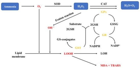
4. Oxidative Stress
| Exposure Route | Exposure Type |
Fish Specie | Ammonia Concentration |
Exposure Periods | Response Concentration |
Target Organs | Response * | Reference |
|---|---|---|---|---|---|---|---|---|
| SOD (Superoxide dismutase) | ||||||||
| Sea water | Waterborne exposure |
Dicentrarchus labrax | 20 mg/L | 12, 48, 84, 180 h | 20 mg/L | Blood | × | Sinha et al. [74] 2015 |
| Epinephelus fuscoguttatus ♀ × E. lanceolatus ♂ | 1, 2, 4, 8 mg/L | 1week, 2 wk | 4, 8 mg/L | Liver, Gill | + | Kim et al. [48] 2020 | ||
| Scophthalmus maximus | 5, 20, 40 mg/L | 24, 48, 96 h | 20, 40 mg/L | Liver | + | Jia et al. [75] 2020 | ||
| Chlamys farreri | 20 mg/L | 1, 12, 24 d | 20 mg/L | Blood | + | Wang et al. [76] 2012 | ||
| Takifugu rubripes | 5, 50, 100, 150 mg/L | 24 h | 50, 100, 150 mg/L | Gill | + | Gao et al. [40] 2021 | ||
| 48, 96 h | 50, 100, 150 mg/L | Gill | − | |||||
| Freshwater | Waterborne exposure |
Carassius auratus | 10, 50 mg/L | 30 d | 10, 50 mg/L | Liver | − | Qi et al. [9] 2017 |
| Megalobrama amblycephala | 5, 10, 15, 20 mg/L | 9 weeks | 20 mg/L | Liver | − | Zhang et al. [11] 2019 | ||
| Cyprinus carpio | 106 mg/L | 24 h | 106 mg/L | Blood | × | Hoseini et al. [43] 2019 | ||
| Oreochromis niloticus | 5, 10 mg/L | 70 days | 5, 10 mg/L | Liver, Muscle | + | Hegazi et al. [77] 2010 | ||
| CAT (Catalase) | ||||||||
| Sea water | Waterborne exposure |
Dicentrarchus labrax | 20 mg/L | 12, 48, 84, 180 h | 20 mg/L | Blood | + | Sinha et al. [74] 2015 |
| Scophthalmus maximus | 5, 20, 40 mg/L | 24, 48, 96 h | 20, 40 mg/L | Liver | + | Jia et al. [75] 2020 | ||
| Takifugu rubripes | 5, 50, 100, 150 mg/L | 24 h | 50, 100, 150 mg/L | Gill | + | Gao et al. [40] 2021 | ||
| 48, 96 h | 50, 100, 150 mg/L | Gill | − | |||||
| Freshwater | Waterborne exposure |
Carassius auratus | 10, 50 mg/L | 30 days | 10, 50 mg/L | Liver | × | Qi et al. [9] 2017 |
| Megalobrama amblycephala | 5, 10, 15, 20 mg/L | 9 weeks | 20 mg/L | Liver | − | Zhang et al. [11] 2019 | ||
| Cyprinus carpio | 106 mg/L | 24 h | 106 mg/L | Blood | − | Hoseini et al. [43] 2019 | ||
| Corbicula fluminea | 10, 25 mg/L | 24, 48 h | 10 mg/L | Digestive gland | + | Zhang et al. [78] 2020 | ||
| 10, 25 mg/L | 24, 48 h | 10 mg/L | Gill | × | ||||
| 10, 25 mg/L | 24, 48 h | 25 mg/L | Digestive gland | − | ||||
| 10, 25 mg/L | 24, 48 h | 25 mg/L | Gill | + | ||||
| GST (Glutathione-S-transferase) | ||||||||
| Sea water | Waterborne exposure |
Dicentrarchus labrax | 20 mg/L | 12, 48, 84, 180 h | 20 mg/L | Blood | + | Sinha et al. [74] 2015 |
| Takifugu rubripes | 5, 50, 100, 150 mg/L | 24 h | 50, 100, 150 mg/L | Gill | + | Gao et al. [40] 2021 | ||
| 48, 96 h | 50, 100, 150 mg/L | Gill | − | |||||
| Epinephelus fuscoguttatus♀ × E. lanceolatus ♂ | 1, 2, 4, 8 mg/L | 1 week | 4, 8 mg/L | Liver, Gill | + | Kim et al. [48] 2020 | ||
| Epinephelus fuscoguttatus♀ × E. lanceolatus ♂ | 1, 2, 4, 8 mg/L | 2 weeks | 4, 8 mg/L | Liver, Gill | − | |||
| Freshwater | Waterborne exposure |
Carassius auratus | 10, 50 mg/L | 30 days | 10, 50 mg/L | Liver | × | Qi et al. [9] 2017 |
| Paralichthys orbignyanus | 5, 10 mg/L | 70 days | 5, 10 mg/L | Liver, Muscle | + | Hoseini et al. [43] 2019 | ||
| Cyprinus carpio L. | 10, 20, 30 mg/L | 6, 24, 48 h | 30 mg/L | Liver | + | Li et al. [10] 2019 | ||
| 10, 20, 30 mg/L | 6, 24, 48 h | 10, 20, 30 mg/L | Gill | + | ||||
This entry is adapted from the peer-reviewed paper 10.3390/ani11113304
References
- Chang, Z.W.; Chiang, P.C.; Cheng, W.; Chang, C.C. Impact of ammonia exposure on coagulation in white shrimp, Litopenaeus vannamei. Ecotoxicol. Environ. Saf. 2015, 118, 98–102.
- Romano, N.; Zeng, C.S. Toxic effects of ammonia, nitrite, and nitrate to decapod crustaceans: A review on factors Influencing their toxicity, physiological consequences, and coping mechanisms. Rev. Fish. Sci. 2013, 21, 1–21.
- Randall, D.J.; Tsui, T.K.N. Ammonia toxicity in fish. Mar. Pollut. Bull. 2002, 45, 17–23.
- Eddy, F.B. Ammonia in estuaries and effects on fish. J. Fish Biol. 2005, 67, 1495–1513.
- Armstrong, D.A.; Chippendale, D.; Knight, A.W.; Colt, J.E. Interaction of Ionized and un-Ionized ammonia on short-term survival and growth of prawn larvae, Macrobrachium rosenbergii. Biol. Bull. 1978, 154, 15–31.
- Wang, L.S.; Xu, Q.Y.; Wang, C.A.; Li, J.N.; Chen, D.; Zhao, Z.G.; Luo, L.; Du, X. Effects of dietary α-ketoglutarate supplementation on the antioxidant defense system and HSP70 and HSP90 gene expression of hybrid sturgeon Acipenser schrenckii ♀ × Acipenser baerii ♂exposed to ammonia-N stress. Aquacult. Res. 2017, 48, 2266–2277.
- Benli, A.C.K.; Koksal, G.; Ozkul, A. Sublethal ammonia exposure of Nile tilapia (Oreochromis niloticus L.): Effects on gill, liver and kidney histology. Chemosphere 2008, 72, 1355–1358.
- Cong, M.; Wu, H.F.; Cao, T.F.; Ji, C.L.; Lv, J.S. Effects of ammonia nitrogen on gill mitochondria in clam Ruditapes philippinarum. Environ. Toxicol. Pharmacol. 2019, 65, 46–52.
- Qi, X.Z.; Xue, M.Y.; Yang, S.B.; Zha, J.W.; Wang, G.X.; Ling, F. Ammonia exposure alters the expression of immune-related and antioxidant enzymes-related genes and the gut microbial community of crucian carp (Carassius auratus). Fish Shellfish Immunol. 2017, 70, 485–492.
- Li, L.H.; Qi, H.X. Effect of acute ammonia exposure on the glutathione redox system in FFRC strain common carp (Cyprinus carpio L.). Environ. Sci. Pollut. Res. 2019, 26, 27023–27031.
- Zhang, W.; Xia, S.; Zhu, J.; Miao, L.; Ren, M.; Lin, Y.; Ge, X.; Sun, S. Growth performance, physiological response and histology changes of juvenile blunt snout bream, Megalobrama amblycephala exposed to chronic ammonia. Aquaculture 2019, 506, 424–436.
- Yan, X.; Chen, Y.; Dong, X.; Tan, B.; Liu, H.; Zhang, S.; Chi, S.; Yang, Q.; Liu, H.; Yang, Y. Ammonia toxicity induces oxidative stress, inflammatory response and apoptosis in hybrid grouper (♀ Epinephelus fuscoguttatus × ♂ E. lanceolatus). Front. Mar. Sci. 2021, 8, 667432.
- Liew, H.J.; Sinha, A.K.; Nawata, C.M.; Blust, R.; Wood, C.M.; de Boeck, G. Differential responses in ammonia excretion, sodium fluxes and gill permeability explain different sensitivities to acute high environmental ammonia in three freshwater teleosts. Aquat. Toxicol. 2013, 126, 63–76.
- Nakada, T.; Westhoff, C.M.; Kato, A.; Hirose, S. Ammonia secretion from fish gill depends on a set of Rh glycoproteins. FASEB J. 2007, 21, 1067–1074.
- Dosdat, A.; Person-Le Ruyet, J.; Coves, D.; Dutto, G.; Gasset, E.; Le Roux, A.; Lemarie, G. Effect of chronic exposure to ammonia on growth, food utilisation and metabolism of the European sea bass (Dicentrarchus labrax). Aquat. Living Resour. 2003, 16, 509–520.
- Lemarie, G.; Dosdat, A.; Coves, D.; Dutto, G.; Gasset, E.; Person-Le Ruyet, J. Effect of chronic ammonia exposure on growth of European seabass (Dicentrarchus labrax) juveniles. Aquaculture 2004, 229, 479–491.
- Wilkie, M.P.; Pamenter, M.E.; Duquette, S.; Dhiyebi, H.; Sangha, N.; Skelton, G.; Smith, M.D.; Buck, L.T. The relationship between NMDA receptor function and the high ammonia tolerance of anoxia-tolerant goldfish. J. Exp. Biol. 2011, 214, 4107–4120.
- Gao, N.; Zhu, L.; Guo, Z.; Yi, M.; Zhang, L. Effects of chronic ammonia exposure on ammonia metabolism and excretion in marine medaka Oryzias melastigma. Fish Shellfish. Immunol. 2017, 65, 226–234.
- Smart, G.R. Investigation of the toxic mechanisms of ammonia to fish—gas exchange in rainbow trout (Salmo gairdneri) exposed to acutely lethal concentrations. J. Fish Biol. 1978, 12, 93–104.
- Wee, N.L.J.; Tng, Y.Y.M.; Cheng, H.T.; Lee, S.M.L.; Chew, S.F.; Ip, Y.K. Ammonia toxicity and tolerance in the brain of the African sharptooth catfish, Clarias gariepinus. Aquat. Toxicol. 2007, 82, 204–213.
- Jayakumar, A.R.; Norenberg, M.D. The Na-K-Cl Co-transporter in astrocyte swelling. Metab. Brain Dis. 2010, 25, 31–38.
- Marquez, J.; Cardona, C.; Campos-Sandoval, J.A.; Penalver, A.; Tosina, M.; Mates, J.M.; Martin-Rufian, M. Mammalian glutaminase isozymes in brain. Metab. Brain Dis. 2013, 28, 133–137.
- Rao, V.L.R. Nitric oxide in hepatic encephalopathy and hyperammonemia. Neurochem. Int. 2002, 41, 161–170.
- Rodrigo, R.; Erceg, S.; Felipo, V. Neurons exposed to ammonia reproduce the differential alteration in nitric oxide modulation of guanylate cyclase in the cerebellum and cortex of patients with liver cirrhosis. Neurobiol. Dis. 2005, 19, 150–161.
- Rodrigo, R.; Cauli, O.; Boix, J.; ElMlili, N.; Agusti, A.; Felipo, V. Role of NMDA receptors in acute liver failure and ammonia toxicity: Therapeutical implications. Neurochem. Int. 2009, 55, 113–118.
- Braissant, O.; McLin, V.A.; Cudalbu, C. Ammonia toxicity to the brain. J. Inherit. Metab. Dis. 2013, 36, 595–612.
- Chew, S.F.; Gan, J.; Ip, Y.K. Nitrogen metabolism and excretion in the swamp eel, Monopterus albus, during 6 or 40 days of estivation in mud. Physiol. Biochem. Zool. 2005, 78, 620–629.
- Van der Linden, A.; Verhoye, M.; Nilsson, G.E. Does anoxia induce cell swelling in carp brains? In vivo MRI measurements in crucian carp and common carp. J. Neurophysiol. 2001, 85, 125–133.
- Ip, Y.K.; Leong, M.W.F.; Sim, M.Y.; Goh, G.S.; Wong, W.P.; Chew, S.E. Chronic and acute ammonia toxicity in mudskippers, Periophthalmodon schlosseri and Boleophthalmus boddaerti: Brain ammonia and glutamine contents, and effects of methionine sulfoximine and MK801. J. Exp. Biol. 2005, 208, 1993–2004.
- Hegazi, M.M.; Hasanein, S.S. Effects of chronic exposure to ammonia concentrations on brain monoamines and ATPases of Nile tilapia (Oreochromis niloticus). Comp. Biochem. Physiol. C Toxicol. Pharmacol. 2010, 151, 420–425.
- Cheng, C.H.; Yang, F.F.; Ling, R.Z.; Liao, S.A.; Miao, Y.T.; Ye, C.X.; Wang, A.L. Effects of ammonia exposure on apoptosis, oxidative stress and immune response in pufferfish (Takifugu obscurus). Aquat. Toxicol. 2015, 164, 61–71.
- Zhang, M.Z.; Li, M.; Wang, R.X.; Qian, Y.X. Effects of acute ammonia toxicity on oxidative stress, immune response and apoptosis of juvenile yellow catfish Pelteobagrus fulvidraco and the mitigation of exogenous taurine. Fish Shellfish. Immunol. 2018, 79, 313–320.
- Fazio, F.; Saoca, C.; Sanfilippo, M.; Capillo, G.; Spano, N.; Piccione, G. Response of vanadium bioaccumulation in tissues of Mugil cephalus (Linnaeus 1758). Sci. Total Environ. 2019, 689, 774–780.
- Fazio, F.; Saoca, C.; Ferranteili, V.; Cammilleri, G.; Capillo, G.; Piccione, G. Relationship between arsenic accumulation in tissues and hematological parameters in mullet caught in Faro Lake: A preliminary study. Environ. Sci. Pollut. Res. 2019, 26, 8821–8827.
- Kim, J.H.; Kang, Y.J.; Kim, K.I.; Kim, S.K.; Kim, J.H. Toxic effects of nitrogenous compounds (ammonia, nitrite, and nitrate) on acute toxicity and antioxidant responses of juvenile olive flounder, Paralichthys olivaceus. Environ. Toxicol. Pharmacol. 2019, 67, 73–78.
- Kim, J.H.; Kang, J.C. Effects of sub-chronic exposure to lead (Pb) and ascorbic acid in juvenile rockfish: Antioxidant responses, MT gene expression, and neurotransmitters. Chemosphere 2017, 171, 520–527.
- Kim, J.H.; Kang, J.C. The chromium accumulation and its physiological effects in juvenile rockfish, Sebastes schlegelii, exposed to different levels of dietary chromium (Cr6+) concentrations. Environ. Toxicol. Pharmacol. 2016, 41, 152–158.
- Yang, W.; Xiang, F.; Sun, H.; Chen, Y.; Minter, E.; Yang, Z. Changes in the selected hematological parameters and gill Na+/K+ ATPase activity of juvenile crucian carp Carassius auratus during elevated ammonia exposure and the post-exposure recovery. Biochem. Syst. Ecol. 2010, 38, 557–562.
- Elbialy, Z.I.; Salah, A.S.; Elsheshtawy, A.; Rizk, M.; Abualreesh, M.H.; Abdel-Daim, M.M.; Salem, S.M.R.; Askary, A.E.; Assar, D.H. Exploring the multimodal role of yucca schidigera extract in protection against chronic ammonia exposure targeting: Growth, metabolic, stress and inflammatory responses in Nile iilapia (Oreochromis niloticus L.). Animals 2021, 11, 2072.
- Gao, X.Q.; Fei, F.; Huang, B.; Meng, X.S.; Zhang, T.; Zhao, K.F.; Chen, H.B.; Xing, R.; Liu, B.L. Alterations in hematological and biochemical parameters, oxidative stress, and immune response in Takifugu rubripes under acute ammonia exposure. Comp. Biochem. Physiol. C Toxicol. Pharm. 2021, 243, 108978.
- Kavitha, C.; Ramesh, M.; Kumaran, S.S.; Lakshmi, S.A. Toxicity of Moringa oleifera seed extract on some hematological and biochemical profiles in a freshwater fish, Cyprinus carpio. Exp. Toxicol. Pathol. 2012, 64, 681–687.
- Praveena, M.; Sandeep, V.; Kavitha, N.; Jayantha Rao, K. Impact of tannery effluent, chromium on hematological parameters in a fresh water fish, Labeo Rohita (Hamilton). Res. J. Anim. Vet. Fish. Sci. 2013, 1, 1–5.
- Hoseini, S.M.; Yousefi, M.; Hoseinifar, S.H.; van doan, H. Antioxidant, enzymatic and hematological responses of common carp (Cyprinus carpio) fed with myrcene- or menthol-supplemented diets and exposed to ambient ammonia. Aquaculture 2019, 506, 246–255.
- Zeitoun, M.M.; El-Azrak, K.E.-D.M.; Zaki, M.A.; Nemat-Allah, B.R.; Mehana, E.-S.E. Effects of ammonia toxicity on growth performance, cortisol, glucose and hematological response of Nile Tilapia (Oreochromis niloticus). Aceh J. Anim. Sci. 2016, 1, 21–28.
- Hoseini, S.M.; Tarkhani, R. Effect of short-term treatment with potassium permanganate on stress markers and blood biochemistry in goldfish Carassius auratus. Aquacult. Res. 2013, 44, 869–875.
- David, M.; Mushigeri, S.B.; Shivakumar, R.; Philip, G.H. Response of Cyprinus carpio (Linn) to sublethal concentration of cypermethrin: Alterations in protein metabolic profiles. Chemosphere 2004, 56, 347–352.
- Asthana, S.; Fatma, F. Effect of ammonia inhalation on serum protein of Albino rat. Natl. Acad. Sci. Lett. India 2008, 31, 117–119.
- Kim, J.H.; Cho, J.H.; Kim, S.R.; Hur, Y.B. Toxic effects of waterborne ammonia exposure on hematological parameters, oxidative stress and stress indicators of juvenile hybrid grouper, Epinephelus lanceolatus ♂× Epinephelus fuscoguttatus ♀. Environ. Toxicol. Pharmacol. 2020, 80, 103453.
- Das, P.C.; Ayyappan, S.; Jena, J.K.; Das, B.K. Acute toxicity of ammonia and its sub-lethal effects on selected haematological and enzymatic parameters of mrigal, Cirrhinus mrigala (Hamilton). Aquacult. Res. 2004, 35, 134–143.
- Iheanacho, S.C.; Odo, G.E. Neurotoxicity, oxidative stress biomarkers and haematological responses in African catfish (Clarias gariepinus) exposed to polyvinyl chloride microparticles. Comp. Biochem. Physiol. C Toxicol. Pharmacol. 2020, 232, 10.
- Barbieri, E.; Bondioli, A.C.V. Acute toxicity of ammonia in Pacu fish (Piaractus mesopotamicus, Holmberg, 1887) at different temperatures levels. Aquac. Res. 2015, 46, 565–571.
- Cui, Y.; Ren, X.; Li, J.; Zhai, Q.; Feng, Y.; Xu, Y.; Ma, L. Effects of ammonia-N stress on metabolic and immune function via the neuroendocrine system in Litopenaeus vannamei. Fish Shellfish. Immunol. 2017, 64, 270–275.
- Zhao, H.; Peng, K.; Wang, G.; Mo, W.; Huang, Y.; Cao, J. Metabolic changes, antioxidant status, immune response and resistance to ammonia stress in juvenile yellow catfish (Pelteobagrus fulvidraco) fed diet supplemented with sodium butyrate. Aquaculture 2021, 536, 736441.
- Mirghaed, A.T.; Fayaz, S.; Hoseini, S.M. Effects of dietary 1,8-cineole supplementation on serum stress and antioxidant markers of common carp (Cyprinus carpio) acutely exposed to ambient ammonia. Aquaculture 2019, 509, 8–15.
- Xing, X.D.; Li, M.; Yuan, L.X.; Song, M.Z.; Ren, Q.Y.; Shi, G.; Meng, F.X.; Wang, R.X. The protective effects of taurine on acute ammonia toxicity in grass carp Ctenopharynodon idellus. Fish Shellfish Immunol. 2016, 56, 517–522.
- Roda, J.F.B.; Lauer, M.M.; Risso, W.E.; Martinez, C.B.D. Microplastics and copper effects on the neotropical teleost Prochilodus lineatus: Is there any interaction? Comp. Biochem. Physiol. A-Mol. Integr. Physiol. 2020, 242, 11.
- Kim, J.H.; Kang, J.C. Changes in hematological parameters, plasma cortisol, and acetylcholinesterase of juvenile rockfish, Sebastes schlegelii supplemented with the dietary ascorbic acid. Aquacult. Rep. 2016, 4, 80–85.
- Habte-Tsion, H.M.; Liu, B.; Ge, X.P.; Xie, J.; Xu, P.; Ren, M.C.; Zhou, Q.L.; Pan, L.K.; Chen, R.L. Effects of dietary protein level on growth performance, muscle composition, blood composition, and digestive enzyme activity of wuchang bream (Megalobrama amblycephala) fry. Isr. J. Aquacult. Bamidgeh. 2013, 65, 9.
- Peyghan, R.; Takamy, G.A. Histopathological, serum enzyme, cholesterol and urea changes in experimental acute toxicity of ammonia in common carp Cyprinus carpio and use of natural zeolite for prevention. Aquac. Int. 2002, 10, 317–325.
- Murthy, C.R.K.; Rao, K.V.R.; Bai, G.; Norenberg, M.D. Ammonia-induced production of free radicals in primary cultures of rat astrocytes. J. Neurosci. Res. 2001, 66, 282–288.
- Halliwell, B. Antioxidant defence mechanisms: From the beginning to the end (of the beginning). Free. Radic. Res. 1999, 31, 261–272.
- Suzuki, H.; Yanaka, A.; Shibahara, T.; Matsui, H.; Nakahara, A.; Tanaka, N.; Muto, H.; Momoi, T.; Uchiyama, Y. Ammonia-induced apoptosis is accelerated at higher pH in gastric surface mucous cells. Am. J. Physiol. Gastroint. Liver Physiol. 2002, 283, G986–G995.
- Madeira, D.; Narciso, L.; Cabral, H.N.; Vinagre, C.; Diniz, M.S. Influence of temperature in thermal and oxidative stress responses in estuarine fish. Comp. Biochem. Physiol. A-Mol. Integr. Physiol. 2013, 166, 237–243.
- Rama, S.; Manjabhat, S.N. Protective effect of shrimp carotenoids against ammonia stress in common carp, Cyprinus carpio. Ecotoxicol. Environ. Saf. 2014, 107, 207–213.
- Livingstone, D.R. Contaminant-stimulated reactive oxygen species production and oxidative damage in aquatic organisms. Mar. Pollut. Bull. 2001, 42, 656–666.
- Kim, J.H.; Yu, Y.B.; Choi, J.H. Toxic effects on bioaccumulation, hematological parameters, oxidative stress, immune responses and neurotoxicity in fish exposed to microplastics: A review. J. Hazard Mater. 2021, 413, 125423.
- Xu, K.H.; Zhang, Y.D.; Huang, Y.M.; Wang, J. Toxicological effects of microplastics and phenanthrene to zebrafish (Danio rerio). Sci. Total Environ. 2021, 757, 8.
- Kim, J.H.; Kang, J.C. Oxidative stress, neurotoxicity, and non-specific immune responses in juvenile red sea bream, Pagrus major, exposed to different waterborne selenium concentrations. Chemosphere 2015, 135, 46–52.
- Gurer-Orhan, H.; Sabir, H.U.; Ozgunes, H. Correlation between clinical indicators of lead poisoning and oxidative stress parameters in controls and lead-exposed workers. Toxicology 2004, 195, 147–154.
- Kim, J.H.; Kang, J.C. Oxidative stress, neurotoxicity, and metallothionein (MT) gene expression in juvenile rock fish Sebastes schlegelii under the different levels of dietary chromium (Cr6+) exposure. Ecotoxicol. Environ. Saf. 2016, 125, 78–84.
- Chen, D.C.; Ning, F.Y.; Zhang, J.Y.; Tang, Y.; Teng, X.H. NF-kappa B pathway took part in the development of apoptosis mediated by miR-15a and oxidative stress via mitochondrial pathway in ammonia-treated chicken splenic lymphocytes. Sci. Total Environ. 2020, 729, 9.
- Han, Q.; Zhang, J.Y.; Sun, Q.; Xu, Y.M.; Teng, X.H. Oxidative stress and mitochondrial dysfunction involved in ammonia-induced nephrocyte necroptosis in chickens. Ecotoxicol Environ. Saf. 2020, 203, 9.
- Liu, C.; Wu, F.C.; Que, H.Y.; Zhang, G.F. Relationships of growth and mortality to enzymatic activity, and the relative mRNA expression of cultured scallops Patinopecten yessoensis in the Yellow Sea, China. J. Oceanol. Limnol. 2019, 37, 1409–1422.
- Sinha, A.K.; AbdElgawad, H.; Zinta, G.; Dasan, A.F.; Rasoloniriana, R.; Asard, H.; Blust, R.; de Boeck, G. Nutritional status as the key modulator of antioxidant responses induced by high environmental ammonia and salinity stress in European sea bass (Dicentrarchus labrax). PLoS ONE 2015, 10, 29.
- Jia, R.; Liu, B.L.; Han, C.; Huang, B.; Lei, J.L. Effects of ammonia exposure on stress and immune response in juvenile turbot (Scophthalmus maximus). Aquac. Res. 2017, 48, 3149–3162.
- Wang, X.Q.; Wang, L.L.; Yao, C.; Qiu, L.M.; Zhang, H.; Zhi, Z.; Song, L.S. Alternation of immune parameters and cellular energy allocation of Chlamys farreri under ammonia-N exposure and Vibrio anguillarum challenge. Fish Shellfish. Immunol. 2012, 32, 741–749.
- Hegazi, M.M.; Attia, Z.I.; Ashour, O.A. Oxidative stress and antioxidant enzymes in liver and white muscle of Nile tilapia juveniles in chronic ammonia exposure. Aquat. Toxicol. 2010, 99, 118–125.
- Zhang, T.; Yan, Z.; Zheng, X.; Wang, S.; Fan, J.; Liu, Z. Effects of acute ammonia toxicity on oxidative stress, DNA damage and apoptosis in digestive gland and gill of Asian clam (Corbicula fluminea). Fish Shellfish. Immunol. 2020, 99, 514–525.
- Hang, W.W.; Du, M.R.; Fang, J.G.; Gao, Y.P.; Mao, Y.Z.; Chen, Q.L.; Lin, F.; Jiang, Z.J. Response of Yesso scallop Patinopecten yessoensis to acute temperature challenge: Physiological and biochemical parameters. J. Oceanol. Limnol. 2019, 37, 321–329.
- Kim, S.H.; Kim, J.H.; Park, M.A.; Hwang, S.D.; Kang, J.C. The toxic effects of ammonia exposure on antioxidant and immune responses in Rockfish, Sebastes schlegelii during thermal stress. Environ. Toxicol. Pharmacol. 2015, 40, 954–959.
- Sinha, A.K.; AbdElgawad, H.; Giblen, T.; Zinta, G.; de Rop, M.; Asard, H.; Blust, R.; de Boeck, G. Anti-oxidative defences are modulated differentially in three freshwater teleosts in response to ammonia-induced oxidative stress. PLoS ONE 2014, 9, 19.
- Lin, Y.C.; Chen, J.C. Acute toxicity of ammonia on Litopenaeus vannamei Boone juveniles at different salinity levels. J. Exp. Mar. Biol. Ecol. 2001, 259, 109–119.
- Wang, J.; Li, J.J.; Xu, N.; Li, J.; Li, Z.H.; Chen, Y.F.; Yang, Z. Responses of Takifugu obscurus fertilized eggs and larvae to increased ammonia exposure. Environ. Sci. Pollut. Res. 2015, 22, 15976–15984.
- Li, M.; Yu, N.; Qin, J.G.; Li, E.C.; Du, Z.Y.; Chen, L.Q. Effects of ammonia stress, dietary linseed oil and Edwardsiella ictaluri challenge on juvenile darkbarbel catfish Pelteobagrus vachelli. Fish Shellfish. Immunol. 2014, 38, 158–165.
- Sun, H.J.; Lu, K.; Minter, E.J.A.; Chen, Y.F.; Yang, Z.; Montagnes, D.J.S. Combined effects of ammonia and microcystin on survival, growth, antioxidant responses, and lipid peroxidation of bighead carp Hypophthalmythys nobilis larvae. J. Hazard. Mater. 2012, 221, 213–219.
- Li, C.H.; Ni, D.J.; Song, L.S.; Zhao, J.; Zhang, H.; Li, L. Molecular cloning and characterization of a catalase gene from Zhikong scallop Chlamys farreri. Fish Shellfish. Immunol. 2008, 24, 26–34.
- Xue, S.Q.; Lin, J.W.; Han, Y.; Han, Y. Ammonia stress-induced apoptosis by p53-BAX/BCL-2 signal pathway in hepatopancreas of common carp (Cyprinus carpio). Aquac. Int. 2021, 29, 1895–1907.
- Luo, Y.; Su, Y.; Lin, R.Z.; Shi, H.H.; Wang, X.R. 2-Chlorophenol induced ROS generation in fish Carassius auratus based on the EPR method. Chemosphere 2006, 65, 1064–1073.
- Maltez, L.C.; Stringhetta, G.R.; Enamorado, A.D.; Okamoto, M.H.; Romano, L.A.; Monserrat, J.M.; Sampaio, L.A.; Garcia, L. Ammonia exposure and subsequent recovery trigger oxidative stress responses in juveniles of Brazilian flounder Paralichthys orbignyanus. Fish Physiol. Biochem. 2017, 43, 1747–1759.
- Ponton, D.E.; Caron, A.; Hare, L.; Campbell, P.G.C. Hepatic oxidative stress and metal subcellular partitioning are affected by selenium exposure in wild yellow perch (Perca flavescens). Environ. Pollut. 2016, 214, 608–617.
- Li, Z.H.; Zlabek, V.; Velisek, J.; Grabic, R.; Machova, J.; Randak, T. Modulation of antioxidant defence system in brain of rainbow trout (Oncorhynchus mykiss) after chronic carbamazepine treatment. Comp. Biochem. Physiol. C Toxicol. Pharmacol. 2010, 151, 137–141.
- Antonio-Garcia, M.T.; Masso-Gonzalez, E.L. Toxic effects of perinatal lead exposure on the brain of rats: Involvement of oxidative stress and the beneficial role of antioxidants. Food Chem. Toxicol. 2008, 46, 2089–2095.
- Li, M.; Gong, S.; Li, Q.; Yuan, L.; Meng, F.; Wang, R. Ammonia toxicity induces glutamine accumulation, oxidative stress and immunosuppression in juvenile yellow catfish Pelteobagrus fulvidraco. Comp. Biochem. Physiol. C Toxicol. Pharmacol. 2016, 183–184, 1–6.
- Almroth, B.C.; Sturve, J.; Berglund, A.; Forlin, L. Oxidative damage in eelpout (Zoarces viviparus), measured as protein carbonyls and TBARS, as biomarkers. Aquat. Toxicol. 2005, 73, 171–180.
- Buege, J.A.; Aust, S.D. Microsomal lipid peroxidation. Methods Enzymol. 1978, 52, 302–310.
- Barhoumi, B.; Clerandeau, C.; Gourves, P.Y.; Le Menach, K.; El Megdiche, Y.; Peluhet, L.; Budzinski, H.; Baudrimont, M.; Driss, M.R.; Cachot, J. Pollution biomonitoring in the Bizerte lagoon (Tunisia), using combined chemical and biomarker analyses in grass goby, Zosterisessor ophiocephalus (Teleostei, Gobiidae). Mar. Environ. Res. 2014, 101, 184–195.
- Zhang, Z.W.; Liu, Q.; Cai, J.Z.; Yang, J.; Shen, Q.; Xu, S.W. Chlorpyrifos exposure in common carp (Cyprinus carpio L.) leads to oxidative stress and immune responses. Fish Shellfish. Immunol. 2017, 67, 604–611.
- Al-Ghanim, K.A.; Ahmad, Z.; Al-Balawi, H.F.A.; Al-Misned, F.; Maboob, S.; Suliman, E.M. Effects of a low-radiotoxicity uranium salt (uranyl acetate) on biochemical and hematological parameters of the catfish, Clarias gariepinus. Chin. J. Oceanol. Limnol. 2016, 34, 109–117.