TP53 and Testicular Germ Cell Tumors: History
Please note this is an old version of this entry, which may differ significantly from the current revision.
Subjects: Cell Biology
Contributor:

Germ cell tumors (GCTs) are the most common solid malignancies in young men. Despite the high frequency of these cancers within this defined age group, the discovery of the exceptional sensitivity of these tumors to the platinum DNA crosslinking compound cisplatin has led to the survival of most patients, with the current five-year survival rate exceeding 95%.

  • human malignant germ cell tumors
  • mediastinal germ cell tumors
  • testicular germ cell tumors
  • TP53

1. Introduction

As GCTs are derived from embryonic germ cells, closely resembling embryonic stem cells, their hypersensitivity to DNA-damaging agents is often traced back to their early embryonic phenotype [1][2][3]; for instance, similarly to embryonic stem cells, GCTs often display a low/inefficient DNA damage response and, as opposed to most solid malignancies, GCTs that are naïve to systemic treatment rarely harbor TP53 mutations, irrespective of histology [4][5]. Moreover, the wild-type TP53 status of GCTs, combined with a pluripotent phenotype, high levels of PUMA and NOXA, and, often, low expression levels of CDKN1A (P21), result in a cellular disbalance and a favor towards apoptosis over DNA repair [6][7][8][9][10]. Furthermore, a physiological antagonist of P53, mouse double minute 2 homologue (MDM2), has been illustrated to be especially important in P53 regulation in GCTs, as it has been shown to hamper the apoptotic response via binding to P53 and can be a putative important clinical target [3][11][12][13][14]. It has already been shown that the inhibition of MDM2 and disruption of the MDM2–P53 interaction can potentiate apoptosis and sensitize GCT cells to cisplatin [11][12]. On the other hand, no correlation has been identified between the levels of MDM2 and the treatment response [5]. Furthermore, the existence of many MDM2 binding partners, and the reported synergy between MDM2 antagonists and (targeted) therapy, both in GCTs and other cancers, make this an interesting and relevant target as well [11][12][15][16]. Histologically and clinically, GCTs can be divided into two main subtypes, referring partly to their pluripotent potential, namely, seminomas and non-seminomas [1][2]. While patients presenting with seminomas have an excellent prognosis, patients harboring non-seminomas have a mixed prognosis, based on tumor histology (e.g., embryonal carcinoma (EC), yolk sac tumor (YST), choriocarcinoma (CC), or teratoma (TE)), therapy naivety or chemotherapeutic resistance, and anatomical location, mainly focusing on extra-cranial GCTs of the mediastinum versus the testis [1][2][4][9][17]. Apart from tumor histology and origin, the P53 pathway and deregulation thereof has been studied in light of GCT treatment resistance [3][4][5][8][9][11][12][14][18]. Even though P53′s have many implications in resistance, no clear-cut result has been obtained that displays their role in clinical resistance, especially related to informative in vitro models [5][18]. In this entry, we focused on the latter (i.e., mediastinal GCTs vs. testicular GCTs) and developed a novel approach to shed light on the difference in treatment resistance between testicular and mediastinal GCTs. This is an important issue, as it is currently unclear whether mediastinal GCTs are more resistant to treatment because of their TP53 mutations, or whether these mutations simply occur more in these tumors as these tumors harbor different intrinsic resistance mechanisms.

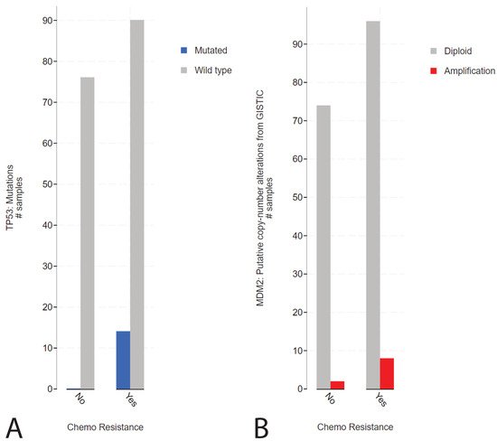
2. Presence of TP53 Mutations in Refractory Cisplatin-Resistant GCTs with a Specificity towards Mediastinal Localization

To elucidate the function of P53 in (resistant) GCTs, we initially used the cBioPortal online tool. We investigated the MSKCC data set on refractory GCTs previously reported by Bagrodia and colleagues in 2016 [4]. The rationale for investigating this data set was based on the abundant presence of detailed clinical data, including anatomical location, treatment, number of chemotherapy cycles, patient survival and outcome, and tumor histology. Supplemental Figure S1A illustrates the presence of TP53 (and MDM2) alterations in cisplatin-sensitive and -resistant GCTs. Strikingly, while GCTs rarely harbor TP53 mutations, in line with their embryonic phenotype [3], alterations in TP53 are detected in cisplatin-resistant patients. Furthermore, we observe that MDM2 amplifications become increasingly abundant in patients with cisplatin-resistant GCTs. Note that, as expected, alterations regarding TP53 are often missense mutations or deep deletions. When comparing the disease-free survival of patients with alterations in the TP53 gene to patients with wild-type TP53 (unaltered group), we observed a highly significant (logrank test p-value of 1.991 × 10⁻⁶) decrease in disease-free survival in patients harboring TP53 alterations (Supplemental Figure S1B). As previously reported, there could be a bias in this analysis, associated with the type of genetic aberration in relation to the anatomic location of GCTs [4]. The tumors of patients harboring TP53 mutations often localize to the mediastinum, whereas the tumors of patients harboring MDM2 amplifications primarily localize to the testis (Supplemental Figure S1C,D). Interestingly, TP53 or MDM2 aberrations occur significantly more frequently in patients with chemotherapy-resistant tumors (Figure 1A,B).
Figure 1. cBioPortal analysis of the tumor resistance in TP53- or MDM2-altered patients in the MSKCC, J Clin Oncol 2016 data set. (A) Bar graph displaying the number of patients with sensitive or resistant cisplatin, patients harboring wild-type (grey) or mutated (blue) TP53 are plotted. (B) Bar graph displaying the number of patients with sensitive or resistant cisplatin, patients harboring wild-type (grey) or amplified (red) MDM2 are plotted [4][19][20].

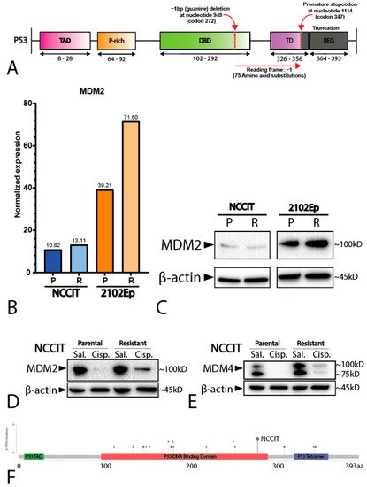
2. Mediastinal GCT Cell Line NCCIT Harbors Low Levels of MDM2 and Mutant TP53 whereas Testicular GCT Cell Line 2102Ep Harbors Wild-Type TP53 and High Levels of MDM2

To study the difference between mediastinal and testicular GCTs, we used the well-established and -characterized NCCIT and 2102Ep GCT (EC) cell lines. While 2102Ep originates from the testis, NCCIT originates from the mediastinum, with a similar differentiation state [21]. Furthermore, similarly to most GCTs, 2120Ep has a wild-type TP53 status, whereas NCCIT carries a hemizygous one-base-pair deletion at nucleotide 949 (codon 272), resulting in a frameshift and a premature STOP codon at codon 347 (Figure 2A). This observation is in line with the finding of TP53 mutations in mediastinal GCTs (see above).
Figure 2. Characterization of the cell lines NCCIT and 2102Ep. (A) Schematic overview of the hemizygous mutation present in the NCCIT cell line. (B) Bar graph displaying the normalized expression (RNA-seq) of MDM2 in the NCCIT and 2102Ep parental and resistant cell lines. (C) Western blot showing the protein levels of MDM2 in the NCCIT and 2102Ep parental and resistant cell lines. (D,E) Western blot displaying the MDM2 (D) and MDM4 (E) protein levels after treatment with sublethal cisplatin doses (1 µM) or saline vehicle control. (F) Mutational position of TP53 mutations in patients in the MSKCC, J Clin Oncol 2016 data set. The mutation found in NCCIT is highlighted with a blue dot.
Additionally, we employed matched isogenic clones of both NCCIT and 2102Ep that have acquired a cisplatin resistance phenotype through long-term sublethal exposure to cisplatin (see Materials and Methods section for details). RNA sequencing (RNA-seq) analysis showed that both the parental and resistant NCCIT cell lines had lower normalized MDM2 expression than both the 2102Ep cell lines, with 2102Ep resistance displaying the highest levels of MDM2 (Figure 2B), supported by Western blotting showing that the resistant 2102Ep subclone had higher levels of MDM2 (Figure 2C). In contrast, the expression levels of MDM4 were similar between all the cell lines (Supplemental Figure S2). Principal component analysis of the matched parental and resistant cell lines showed no major differences and demonstrated close similarities between the matched subclones (data not shown). To determine whether NCCIT had an active DNA damage response and possible P53 pathway activation, despite a low MDM2 level, we treated NCCIT cells with sublethal (1 µM) levels of cisplatin for 24 h prior to protein analysis via Western blotting. Both the NCCIT parental and resistant cell lines showed a clear decrease in MDM2 and MDM4 after exposure to cisplatin, an effect that was not visible in the saline vehicle control condition (Figure 2D,E). This indicates a functional DNA-damage sensing pathway upstream of MDM2 and MDM4, and, therefore, suggests an intact regulation of P53 downstream of MDM2 and MDM4, despite the suggested null status of TP53 as described in the literature [8][11][18][22].

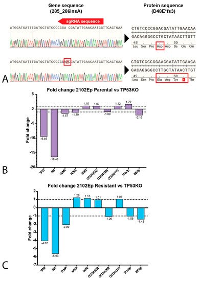
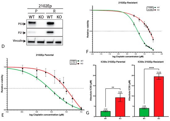
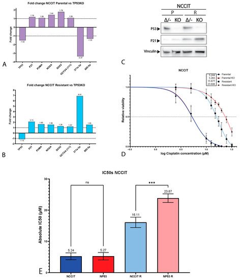
3. P53 Is Involved in Cisplatin Resistance in Both Wild-Type (Testicular) and Mutant (Mediastinal) GCT Cell Lines

It is largely accepted that the chemotherapeutic hypersensitivity of GCTs is partly due to their wild-type TP53 status [3][9][23][24][25]. However, despite its TP53 mutant status, the NCCIT cell line is considered to be inherently sensitive to cisplatin. Thus, we further compared the mutational status of the NCCIT cell line to the mutations found in refractory GCT patients (Figure 2F). When comparing the intrinsic TP53 mutation in the NCCIT cell line to the TP53 mutations present in refractory GCT patients, we observed that most mutations found in patients disrupt the DNA-binding domain of TP53, a well-known mutational hotspot [4][26]. In contrast, the intrinsic TP53 mutation of NCCIT appears to largely spare the DNA-binding domain and is, therefore, more C-terminally located than most mutations found in refractory patients, suggesting the possibility for residual protein activity. Furthermore, the enrichment of mutations in TP53 in refractory patients, together with a bias towards mediastinal anatomical localization (and, hence, a more resistant phenotype), suggests that TP53 mutations could add additively to inherent cisplatin resistance mechanisms [4][9][17]. Based on these observations, we decided to further test the involvement of TP53 in cisplatin resistance in the approach described. Therefore, we generated isogenic CRISPR/Cas9-mediated TP53 knock-out clones of both 2102Ep and NCCIT, as well as their resistant counterparts. Sanger sequencing, after mono-clonal picking and expansion, revealed mono-clonal sequence traces and a one-base-pair insertion at amino acid 48, resulting in a premature STOP codon at amino acid 51 (Figure 3A). We were able to obtain clones harboring this mutation for all the investigated cell lines. No major copy number changes between the original and TP53 knock-out NCCIT and 2102Ep subclones were identified based on Infinium Global Screening Array-24 v3.0 BeadChipGSA (GSA) profiling (Supplemental Figure S3). Gene expression analysis using RT-qPCR indicated a clear reduction in both TP53 and CDKN1A (P21) expression in both 2102Ep parental TP53 knock-out lines (~9.46 and ~16.45, respectively) and 2102Ep-resistant TP53 knock-out lines (~4.07 and 5.63, respectively), which was also confirmed by Western blot (Figure 3B–D). No differences were observed in P53 target gene expression (PUMA/NOXA) or differentiation marker expression (SOX2, OCT3/4, miR371a-3p, or miR885-5p); the latter indicates that the loss of TP53 expression had no effect on the differentiation status. The miR371a-3p expression levels were checked because of its many implications in GCTs (mostly as a biomarker and marker of pluripotency in these tumors), together with the implications of P53 pathway regulation [2][15][27][28][29][30][31][32][33]. Strikingly, after treating 2102Ep parental and resistant cells, as well as their isogenic TP53 knock-out clones, with cisplatin, we identified a clear significant (parental p = 0.0049, resistant p ≤ 0.0001) shift in cisplatin resistance when comparing the TP53 knock-out clone to its wild-type counterpart, with the 2102Ep-resistant TP53 knock-out clone demonstrating the highest cisplatin resistance (Figure 3E–G). When we performed this approach with the NCCIT cell line, we obtained clones with the same one-base-pair insertion mutation (A) found in the 2102Ep cell lines (Figure 3A). However, we found no strong reduction in either TP53 or CDKN1A expression, P53 target gene expression, or differentiation marker expression (Figure 4A,B). Interestingly, however, we did observe a reduction in miR371a-3p expression (3.37-fold) in the parental TP53 knock-out clone compared to its parental counterpart, while we observed an increase in miR371a-3p expression (6.91-fold) in the NCCIT-resistant TP53 knock-out clone compared to its NCCIT-resistant counterpart (Figure 4A,B). Western blotting confirmed that the TP53 knock-out lines had lost P53 protein expression; however, strikingly, the levels of P21 were increased in the NCCIT-resistant TP53 knock-out line compared to its NCCIT-resistant counterpart (and both other lines; Figure 4C). Moreover, TP53 knock-out in the NCCIT clones resulted in no shift in cisplatin resistance in the NCCIT parental clone, and a major significant (p = 0.0005) shift in cisplatin resistance in the NCCIT-resistant TP53 knock-out clone compared to its NCCIT-resistant counterpart (Figure 4D,E).
Figure 3. Characterization of 2102Ep TP53 knock-out cell lines. (A) SnapGene genome sequence alignments of the CRISPR/Cas9 target site of the TP53 gene. The knock-out cell line (bottom sequence) shows a one-base-pair insertion (A) at amino acid 49, resulting in a premature STOP codon at amino acid 51. (B,C) Bar graphs showing the fold change in expression between 2102Ep parental cell line and its isogenic TP53 knock-out clone (B) or 2102Ep-resistant cell line and its isogenic TP53 knock-out clone (C). (D) Western blots showing the protein levels of P53, P21 and vinculin (as loading control) in 2102Ep parental and resistant cell lines and their isogenic TP53 knock-out clones. (E,F) S-curves showing the viability of the parental (E) and resistant (F) 2102Ep cell lines and their corresponding knock-out when treated with cisplatin for 72 h. Graphs represent three biological replicates with three technical replicates each. (G) Bar plots displaying IC50 values of all 2102Ep cell lines. Both cell line pairs show significant differences in IC50 values after knock-out (parental p = 0.0049, resistant p ≤ 0.0001, unpaired Student’s t-test). Mean ± SD: 2102Ep parental 2.62 ± 0.33, 2102Ep parental TP53 KO 9.28 ± 2.02, 2102Ep resistant 4.06 ± 0.32, and 2102Ep resistant TP53 KO 19.50 ± 1.36. Graphs represent three biological replicates with three technical replicates each. ** p ≤ 0.01, **** p ≤ 0.0001.
Figure 4. Characterization of NCCIT TP53 knock-out cell lines. (A,B) Bar graphs showing the fold change in expression between NCCIT parental cell line and its isogenic TP53 knock-out clone (A) or NCCIT-resistant cell line and its isogenic TP53 knock-out clone (B). (C) Western blots showing the protein levels of P53, P21 and vinculin (as loading control) in NCCIT parental and resistant cell lines and their isogenic TP53 knock-out clones. (D) S-curves showing the viability of the NCCIT cell lines (parental and resistant and TP53 knock-out lines) when treated with cisplatin for 72 h. Graphs represent three biological replicates with three techinical replicates each. (E) Bar plots displaying IC50 values of all NCCIT cell lines. The NCCIT-resistant cell line shows a significant difference in IC50 values after knock-out (p = 0.0005, one-way ANOVA, Tukey’s multiple comparisons post hoc test). Mean ± SD: NCCIT parental 5.24 ± 1.09, NCCIT parental TP53 KO 5.27 ± 1.18, NCCIT resistant 16.11 ± 1.67, and NCCIT resistant TP53 KO 23.87 ± 1.38. ns = p > 0.05, *** p ≤ 0.001.

This entry is adapted from the peer-reviewed paper 10.3390/ijms222111774

References

  1. Oosterhuis, J.W.; Looijenga, L. Testicular Germ-Cell Tumours in a Broader Perspective. Nat. Rev. Cancer 2005, 5, 210–222.
  2. Oosterhuis, J.W.; Looijenga, L.H.J. Human Germ Cell Tumours from a Developmental Perspective. Nat. Rev. Cancer 2019, 19, 522–537.
  3. Timmerman, D.; Remmers, T.; Hillenius, S.; Looijenga, L. Mechanisms of TP53 Pathway Inactivation in Embryonic and Somatic Cells—Relevance for Understanding (Germ Cell) Tumorigenesis. Int. J. Mol. Sci. 2021, 22, 5377.
  4. Bagrodia, A.; Lee, B.; Lee, W.; Cha, E.K.; Sfakianos, J.P.; Iyer, G.; Pietzak, E.J.; Gao, S.P.; Zabor, E.C.; Ostrovnaya, I.; et al. Genetic Determinants of Cisplatin Resistance in Patients with Advanced Germ Cell Tumors. J. Clin. Oncol. 2016, 34, 4000–4007.
  5. Kersemaekers, A.-M.F.; Mayer, F.; Molier, M.; Van Weeren, P.C.; Oosterhuis, J.W.; Bokemeyer, C.; Looijenga, L.H. Role of P53 and MDM2 in Treatment Response of Human Germ Cell Tumors. J. Clin. Oncol. 2002, 20, 1551–1561.
  6. Ulbright, T.M.; Orazi, A.; De Riese, W.; De Riese, C.; Messemer, E.J.; Foster, R.S.; Donohue, J.P.; Eble, J.N. The Correlation of P53 Protein Expression with Proliferative Activity and Occult Metastases in Clinical Stage I Non-Seminomatous Germ Cell Tumors of the Testis. Mod. Pathol. 1994, 7, 64–68.
  7. Filion, T.M.; Qiao, M.; Ghule, P.N.; Mandeville, M.; van Wijnen, A.J.; Stein, J.L.; Lian, J.B.; Altieri, D.C.; Stein, G.S. Survival Responses of Human Embryonic Stem Cells to DNA Damage. J. Cell. Physiol. 2009, 220, 586–592.
  8. Gutekunst, M.; Oren, M.; Weilbacher, A.; Dengler, M.A.; Markwardt, C.; Thomale, J.; Aulitzky, W.E.; Van Der Kuip, H. p53 Hypersensitivity is the Predominant Mechanism of the Unique Responsiveness of Testicular Germ Cell Tumor (TGCT) Cells to Cisplatin. PLoS ONE 2011, 6, e19198.
  9. Jacobsen, C.; Honecker, F. Cisplatin Resistance in Germ Cell Tumours: Models and Mechanisms. Andrology 2015, 3, 111–121.
  10. Bloom, J.C.; Loehr, A.R.; Schimenti, J.C.; Weiss, R.S. Germline Genome Protection: Implications for Gamete Quality and Germ Cell Tumorigenesis. Andrology 2019, 7, 516–526.
  11. Bauer, S.; Mühlenberg, T.; Leahy, M.; Hoiczyk, M.; Gauler, T.; Schuler, M.; Looijenga, L. Therapeutic Potential of Mdm2 Inhibition in Malignant Germ Cell Tumours. Eur. Urol. 2010, 57, 679–687.
  12. Koster, R.; Timmer-Bosscha, H.; Bischoff, R.; Gietema, J.; De Jong, S. Disruption of the MDM2–p53 Interaction Strongly Potentiates p53-Dependent Apoptosis in Cisplatin-Resistant Human Testicular Carcinoma Cells via the Fas/FasL Pathway. Cell Death Dis. 2011, 2, e148.
  13. Gutekunst, M.; Mueller, T.; Weilbacher, A.; Dengler, M.A.; Bedke, J.; Kruck, S.; Oren, M.; Aulitzky, W.E.; Van Der Kuip, H. Cisplatin Hypersensitivity of Testicular Germ Cell Tumors Is Determined by High Constitutive Noxa Levels Mediated by Oct-4. Cancer Res. 2013, 73, 1460–1469.
  14. Lobo, J.; Jerónimo, C.; Henrique, R. Cisplatin Resistance in Testicular Germ Cell Tumors: Current Challenges from Various Perspectives. Cancers 2020, 12, 1601.
  15. Mego, M.; Van Agthoven, T.; Gronesova, P.; Chovanec, M.; Miskovska, V.; Mardiak, J.; Looijenga, L.H.J. Clinical Utility of Plasma miR-371a-3p in Germ Cell Tumors. J. Cell. Mol. Med. 2019, 23, 1128–1136.
  16. Fåhraeus, R.; Olivares-Illana, V. MDM2’s Social Network. Oncogene 2013, 33, 4365–4376.
  17. Zhou, Z.-T.; Wang, J.-W.; Yang, L.; Wang, J.; Zhang, W. Primary Germ Cell Tumor in the Mediastinum-Report of 47 Cases. Zhonghua Zhong Liu Za Zhi 2006, 28, 863–866.
  18. Burger, H.; Nooter, K.; Boersma, A.W.; Kortland, C.J.; Stoter, G. Lack of Correlation Between Cisplatin-Induced Apoptosis, p53 Status and Expression of Bcl-2 Family Proteins in Testicular Germ Cell Tumour Cell Lines. Int. J. Cancer 1997, 73, 592–599.
  19. Cerami, E.; Gao, J.; Dogrusoz, U.; Gross, B.E.; Sumer, S.O.; Aksoy, B.A.; Jacobsen, A.; Byrne, C.J.; Heuer, M.L.; Larsson, E.; et al. The cBio Cancer Genomics Portal: An Open Platform for Exploring Multidimensional Cancer Genomics Data: Figure 1. Cancer Discov. 2012, 2, 401–404.
  20. Gao, J.; Aksoy, B.A.; Dogrusoz, U.; Dresdner, G.; Gross, B.; Sumer, S.O.; Sun, Y.; Jacobsen, A.; Sinha, R.; Larsson, E.; et al. Integrative analysis of complex cancer genomics and clinical profiles using the cBioPortal. Sci. Signal. 2013, 6, l1.
  21. de Jong, J.; Stoop, H.; Gillis, A.J.M.; Hersmus, R.; van Gurp, R.J.H.L.M.; van de Geijn, G.-J.M.; van Drunen, E.; Beverloo, H.B.; Schneider, D.T.; Sherlock, J.K.; et al. Further Characterization of the First Seminoma Cell Line TCam-2. Genes Chromosom. Cancer 2008, 47, 185–196.
  22. Li, J.; Kurokawa, M. Regulation of MDM2 Stability After DNA Damage. J. Cell. Physiol. 2015, 230, 2318–2327.
  23. Spierings, D.; De Vries, E.E.G.; Vellenga, E.; De Jong, S. The Attractive Achilles Heel of Germ Cell Tumours: An Inherent Sensitivity to Apoptosis-Inducing Stimuli. J. Pathol. 2003, 200, 137–148.
  24. Kerley-Hamilton, J.S.; Pike, A.M.; Li, N.; DiRenzo, J.; Spinella, M.J. A p53-Dominant Transcriptional Response to Cisplatin in Testicular Germ Cell Tumor-Derived Human Embyronal Carcinoma. Oncogene 2005, 24, 6090–6100.
  25. Romano, F.J.; Rossetti, S.; Conteduca, V.; Schepisi, G.; Cavaliere, C.; Di Franco, R.; La Mantia, E.; Castaldo, L.; Nocerino, F.; Ametrano, G.; et al. Role of DNA Repair Machinery and P53 in the Testicular Germ Cell Cancer: A Review. Oncotarget 2016, 7, 85641–85649.
  26. Baugh, E.H.; Ke, H.; Levine, A.J.; Bonneau, A.R.; Chan, C.S. Why are there Hotspot Mutations in the TP53 Gene in Human Cancers? Cell Death Differ. 2018, 25, 154–160.
  27. Voorhoeve, P.M.; le Sage, C.; Schrier, M.; Gillis, A.J.; Stoop, H.; Nagel, R.; Liu, Y.-P.; van Duijse, J.; Drost, J.; Griekspoor, A.; et al. A Genetic Screen Implicates miRNA-372 and miRNA-373 as Oncogenes in Testicular Germ Cell Tumors. Cell 2006, 124, 1169–1181.
  28. Lobo, J.; Gillis, A.J.; Berg, A.V.D.; Dorssers, L.C.J.; Belge, G.; Dieckmann, K.-P.; Roest, H.P.; Van Der Laan, L.J.W.; Gietema, J.; Hamilton, R.J.; et al. Identification and Validation Model for Informative Liquid Biopsy-Based microRNA Biomarkers: Insights from Germ Cell Tumor In Vitro, In Vivo and Patient-Derived Data. Cells 2019, 8, 1637.
  29. Almstrup, K.; Lobo, J.; Mørup, N.; Belge, G.; Meyts, E.R.-D.; Looijenga, L.H.J.; Dieckmann, K.-P. Application of miRNAs in the Diagnosis and Monitoring of Testicular Germ Cell Tumours. Nat. Rev. Urol. 2020, 17, 201–213.
  30. Murray, M.J.; Bell, E.; Raby, K.L.; Rijlaarsdam, M.; Gillis, A.J.M.; Looijenga, L.; Brown, H.; Destenaves, B.; Nicholson, J.C.; Coleman, N.S. A pipeline to Quantify Serum and Cerebrospinal Fluid Micrornas for Diagnosis and Detection of Relapse in Paediatric Malignant Germ-Cell Tumours. Br. J. Cancer 2016, 114, 151–162.
  31. Murray, M.J.; Halsall, D.J.; Hook, C.E.; Williams, D.M.; Nicholson, J.C.; Coleman, N.; Sweet, W.; Duh, Y.-J.; Greenfield, L.; Tarco, E.; et al. Identification of MicroRNAs from the miR-371∼373 and miR-302 Clusters as Potential Serum Biomarkers of Malignant Germ Cell Tumors. Am. J. Clin. Pathol. 2011, 135, 119–125.
  32. Syring, I.; Bartels, J.; Holdenrieder, S.; Kristiansen, G.; Müller, S.C.; Ellinger, J. Circulating Serum miRNA (miR-367-3p, miR-371a-3p, miR-372-3p and miR-373-3p) as Biomarkers in Patients with Testicular Germ Cell Cancer. J. Urol. 2015, 193, 331–337.
  33. Leão, R.; Albersen, M.; Looijenga, L.H.; Tandstad, T.; Kollmannsberger, C.; Murray, M.J.; Culine, S.; Coleman, N.; Belge, G.; Hamilton, R.J.; et al. Circulating MicroRNAs, the Next-Generation Serum Biomarkers in Testicular Germ Cell Tumours: A Systematic Review. Eur. Urol. 2021, 80, 456–466.
More
This entry is offline, you can click here to edit this entry!