The vulvar area is a common site of contact dermatitis due to the thin skin, easily traversable by irritant and allergic substances. The nonkeratinized vulvar vestibule is likely to be more permeable than the keratinized portions of the vulva and thus more susceptible to exogenous topical agents. The vulva is an area of occlusion due to both its intrinsic anatomical structure and the frequent use of occlusive napkins or underwear, which increase penetration or absorption of both irritants and allergens. Furthermore, women at different ages, due to urine and feces as children and to vaginal mucosal atrophy and the increase in the vulvar pH in menopause, may have an altered barrier function and, in incontinent elderly subjects, the use of diapers may contribute to increased susceptibility to irritants and allergens.
1. Irritant Contact Dermatitis
Irritant contact dermatitis (ICD) is the result of a direct damage to the skin by various chemical or physical stimuli. The initiating event is the disruption of the epidermal barrier (i.e., the stratum corneum), with consequent increased skin permeability. This results in an inflammatory cutaneous reaction, caused by proinflammatory mediators released from keratinocytes and by the activation of innate immunity [
2,
3]. Risk factors for vulvar ICD are multifactorial and include the type of irritant, the length of exposure, the presence of previous dermatoses, and the host’s susceptibility. Women with an atopic diathesis (particularly atopic dermatitis) are more susceptible to ICD as a result of the impaired barrier function of their skin. Furthermore, vulvar skin shows an increased susceptibility to some irritants (maleic acid and benzalkonium chloride) [
1]. ICD is more common than allergic contact dermatitis, but the exact prevalence is unknown. The most common vulvar irritants are reported in
Table 1.
Table 1. List of the most common irritants responsible for vulvar ICD.
| Vulvar Irritants |
|
| Strong Irritants |
Weak Irritants |
| Imiquimod |
Friction/rubbing |
| Trichloroacetic acid |
Urine/feces/vaginal secretions/sweat/semen/saliva |
| Podophyllin |
Sanitary napkins |
| 5-fluorouracil |
Soaps/detergents |
| |
Antiseptic or scented wipes |
| Sodium hypochlorite |
Deodorants |
Irritant contact dermatitis from strong irritants (caustics, topical medicaments) can have a rapid onset within minutes or hours of being exposed and presents with erythema, patches, papules, vesicles, bullae, and scaling. In chronic diseases (mostly due to weak cumulative allergens such as detergents or friction), lichenification and fissuring are more typical features. The main symptoms of ICD are burning, stinging, and, less frequently, itching or pain. In most cases the dermatitis is localized to the site of contact. In particular, when due to napkins ICD is located on the convex areas of the vulva, sparing the folds.
Avoiding use of the offending agents and providing patients education, together with the prescription of potent topical steroids to reduce inflammation, are crucial to the control of symptoms and signs.
2. Allergic Contact Dermatitis
Allergic contact dermatitis (ACD) is the consequence of a T-lymphocytes mediated immune reaction to small, molecular weight chemicals (haptens) that penetrate the skin and activate innate immunity and then the adaptive immunity [
3]. During the sensitization phase, naive T cells are activated in a process that involves Langerhans cells and dermal dendritic cells; in the elicitation phase, T cells migrate into the skin and induce skin damage through the release of proinflammatory cytokines and by killing hapten-loaded keratinocytes.
The sensitization phase of ACD results in the expansion of skin-homing hapten-specific T cells that, upon subsequent hapten challenge, migrate into the skin and induce the skin damage through the release of proinflammatory cytokines and by killing hapten-loaded keratinocytes. Vulvar ACD may occur as a primary disorder or may complicate an underlying vulvar dermatosis. The risk of ACD increases in the case of pre-existing ICD and with the use of multiple topical treatments.
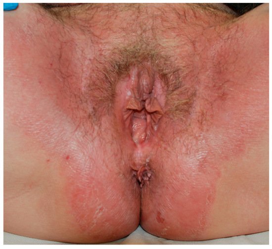
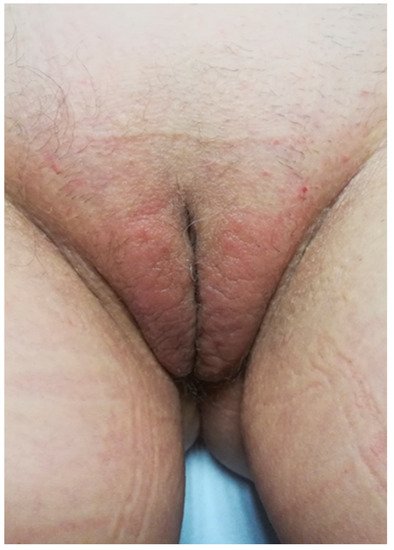
Vulvar ACD may develop as an acute eczema where the allergen was applied. In that case an itching vesicular or exudative eczema develops on previously healthy skin. However, the onset is often a complication of previous different cutaneous dermatoses (lichen sclerosus, psoriasis, atopic dermatitis, etc.) presenting as a local aggravation or exacerbation of symptoms and signs (Figure 1). The diagnosis may not be easy because of the confounding clinical aspects related to the preexisting dermatosis. In this case, history and the clinical aspect are very important in differentiating a lichen flare-up with an allergic dermatitis. The appearance of acute inflammatory lesions as erythema, edema, and vesiculation suggests contact sensitization. Furthermore, a poor response to an appropriate topical corticosteroid therapy could be indicative of contact sensitization to these molecules. The prolonged contact with the allergen can cause lichenification. (Figure 2).
Figure 1. A case of psoriasis complicated by allergic contact dermatitis due to topical medications.
Figure 2. Lichenification following persistent allergic contact dermatitis.
ACD can often severely affect quality of life for women who already suffer from a debilitating vulvar disease.
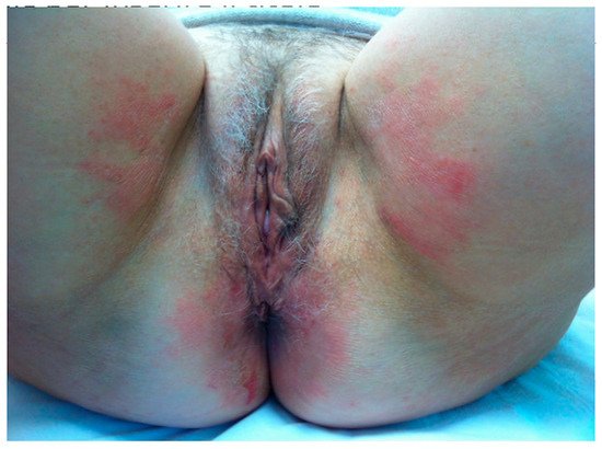
Sometimes the area of involvement spreads over the borders of the vulva not only due to the spread of inflammation but also because of the modality of contact with the allergens [
4]. (
Figure 3).
Figure 3. A case of allergic contact dermatitis in which the area of involvement spreads over the borders of the vulva.
Distant localizations may also develop due to inadvertent hand transfer or rubbing of the adjacent areas (e.g., thighs). The contamination of clothing or napkins may lead to persistent dermatitis. Furthermore, a rapid spread to distant sites (auto-eczematization) may also result from the absorption and diffusion of allergens inducing a sort of id-dermatitis.
Clinically, vulvar irritation and allergic dermatitis can be difficult to distinguish, and diagnosis is made on the basis of history, clinical investigation, and patch testing.
A particular situation in vulvar ACD may be represented by the so-called “Connubial dermatitis”, a dermatitis that occurs because of contact with substances transferred to the patients’ skin by her partner. The diagnosis of connubial dermatitis should be considered in cases of probable allergic contact eczema when patch test results are apparently inconsistent with the patient’s clinical history. In these situations, it may be necessary to extend medical investigation to the patient’s partner as well, in order to clarify the source of allergic contacts when no obvious exposures can be found.
Vulvar ACD is frequently linked to direct contact with cosmetics and detergents or medicaments. Less frequently textiles or dyes are described. Sometimes unsuspected allergens such as nail varnish (ectopic contact dermatitis) can be the cause.
A review of the studies concerning vulvar ACD is reported in
Table 2 [
5,
6,
7,
8,
9,
10,
11,
12,
13,
14,
15,
16,
17,
18,
19].
Table 2. Review of the studies concerning vulvar ACD.
| Authors |
|
Pathologies |
N° Patients |
%
Positiveness |
%
Relevant p.t. |
Allergens |
| Doherty et al. |
1990 |
Vulvar itching |
50 |
78 |
- |
nickel, fragrances, neomycin, local anesthetics |
| Marren et al. |
1992 |
Vulvar dermatoses |
135 |
47 |
29 |
nickel, fragrances, preservatives, ethylenediamine, topical medicaments |
| Brenan et al. |
1996 |
Chronic vulvar symptoms |
700 |
42 |
- |
nickel, fragrances, ethylenediamine |
| Goldsmith et al. |
1997 |
Anogenital dermatoses |
201 |
39 |
28 |
antibiotics, local anesthetics, fragrances, corticosteroids |
| Lewis et al. |
1997 |
Vulvar symptoms |
121 |
58.7 |
49 |
local anesthetics, fragrances, neomycin |
| Lucke et al. |
1998 |
Vulvar dermatoses |
55 |
65 |
- |
nickel, fragrances, medicaments, dyes |
| Bauer et al. |
2000 |
Anogenital symptoms |
351 |
47 |
34.8 |
nickel, fragrances, local anesthetics |
| Crone et al. |
2000 |
Vulvar dermatoses |
38 |
47 |
28 |
fragrances, preservatives, medicaments |
| Virgili et al. |
2003 |
Vulvar lichen simplex chronicus |
61 |
47.5 |
26 |
nickel, preservatives, fragrances, medicaments |
| Nardelli et al. |
2004 |
Vulvar symptoms |
92 |
38 |
16 |
medicaments |
| Utas et al. |
2008 |
Vulvar itching |
50 |
52 |
16 |
preservatives, fragrances, medicaments |
| Haverhoek et al. |
2008 |
Vulvar pruritus |
43 |
81.4 |
44 |
preservatives, fragrances, medicaments |
| Warshsoaw et al. |
2008 |
Anogenital dermatoses |
570 |
44.1 |
27 |
medicaments, corticosteroids |
| Vermaat et al. |
2008 |
Anogenital dermatoses |
53 |
66 |
20 |
fragrances, spices |
| O’Gorman et al. |
2013 |
Vulvar itching |
90 |
69 |
39 |
preservatives, fragrances, medicaments |
| Al-Niaimi at al. |
2014 |
Vulvar symptoms |
282 |
54 |
49 |
nickel, fragrances, neomycin |
| Trivedi et al. |
2018 |
Vulvar itching |
- |
64 |
54 |
preservatives, fragrances |
It is not surprising that a high level of sensitization (39–78%) is found testing patients affected by different vulvar disorders (vulvar symptoms, vulvar dermatoses, or anogenital symptoms). In selected conditions as well, like lichen simplex chronicus [
20], similar percentages can be found. The reported incidence of clinically relevant patch test results for patients presenting with vulvar complaints are likewise high, ranging from 16% to 54% [
5,
6,
7,
8,
9,
10,
11,
12,
13,
14,
15,
16,
17,
18,
19].
Fragrances, preservatives, and topical medicaments (especially corticosteroids, neomycin, and topical anesthetics) are the relevant allergens usually found. Although some authors consider nickel a relevant positivity for the disease, in the majority of cases it is considered a non-relevant allergen that simply reflects the high level of sensitization in the general population [
9,
15,
20].
This entry is adapted from the peer-reviewed paper 10.3390/allergies1040019


