Lipid Oxidation in Food Systems: History
Please note this is an old version of this entry, which may differ significantly from the current revision.
Contributor:

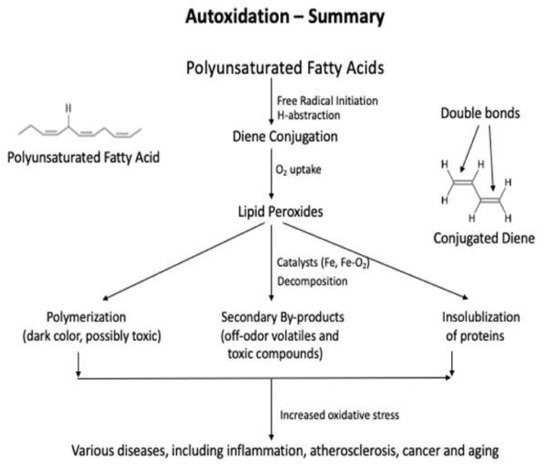
Lipids are significant nutrients for humans and help many functional and regulatory activities in the human body, such as signal transduction, myelination, and synaptic plasticity. Lipids are also involved in the structural developments of the human body . In food, lipid content and fatty acid composition are the two critical congenital parameters to the susceptibility of food to oxidative changes. Lipid content and the fatty acid composition of fat of farm animals varies significantly depending on animal species and the diet. Lipid oxidation causes quality deterioration in food. Depending upon the reaction mechanisms and factors involved, lipid oxidation can be divided into autoxidation, photo-oxidation, and enzyme-catalyzed oxidation. Autoxidation is the most common process of lipid oxidation in foods and is divided into initiation, propagation, and termination stages. 

  • lipid oxidation
  • primary oxidation products
  • secondary oxidation products
  • antioxidant capacity

1. Introduction

Autoxidation is the most common process of lipid oxidation in foods and is divided into initiation, propagation, and termination stages. During the initiation step, free radicals abstract the liable hydrogen atoms from the methylene group of polyunsaturated fatty acids. Then, the fatty acid rearrangement (diene conjugation) stabilizes the fatty acid radical [1][2][3][4]. In the presence of oxygen, the conjugated dienes become the peroxyl (lipid) radical with high reactivity. The total bis-allylic carbon determines the rate of peroxyl radical formation in a fatty acid molecule. Reactive oxygen species (ROS) are the most common reactive compounds that abstract a free hydrogen atom from the lipid molecules [1][4]. During the propagation, the conjugated diene becomes a highly reactive lipid radical (LOO) in the presence of O2 and abstracts a hydrogen atom from an adjacent polyunsaturated fatty acid. Once this propagation process is started, it will continue until the termination step, where the unstable peroxyl radicals become stable non-radical [3]. The summary of the lipid oxidation is shown in Figure 1 [4].
Figure 1. A schematic diagram of lipid oxidation pathway and the types of products produced.

2. Methods Used to Detect the Primary Oxidation Products

The three most used methods for the primary oxidation products in foods are iodometric, ferric thiocyanate, and diene conjugation methods, and they are the direct methods that determine the amount of hydroperoxides formed by oxidation in foods [5][6]. The peroxide value (PV) analysis is performed using titration or spectrophotometer, and the diene conjugation method determines all the conjugated fatty acids, including lipid peroxides, in the food products after extracting lipids from them. All three methods are used to determine the early stage of lipid oxidation, and their sensitivities are lower than those of the measurements of the secondary products. The values of the primary products can indicate the potentials of lipid oxidation at an early stage when the products are exposed to catalysts or further processed and, thus, may disagree with the sensory quality of the food.

2.1. Methods Used to Detect the Primary Oxidation Products

The three most used methods for the primary oxidation products in foods are iodometric, ferric thiocyanate, and diene conjugation methods, and they are the direct methods that determine the amount of hydroperoxides formed by oxidation in foods [5][6]. The peroxide value (PV) analysis is performed using titration or spectrophotometer, and the diene conjugation method determines all the conjugated fatty acids, including lipid peroxides, in the food products after extracting lipids from them. All three methods are used to determine the early stage of lipid oxidation, and their sensitivities are lower than those of the measurements of the secondary products. The values of the primary products can indicate the potentials of lipid oxidation at an early stage when the products are exposed to catalysts or further processed and, thus, may disagree with the sensory quality of the food.

2.1.1. Peroxide Values: Iodometric and Ferric Thiocyanate Assays

The iodometric assay uses the 2H+ that proceeds, according to the following equations:
ROOH + 2H+ + 2I → ROH + H2O + I2,
I + I2  I3.
The liberated iodine (I2) is titrated with iodide Equation (1), or the reaction product of I + I2, triiodide anion (I3) is measured using a spectrophotometer at 353 nm Equation (2). This method requires the extraction of lipids from the sample using Folch’s solution (chloroform: methanol = 2:1). A precaution is needed to minimize oxygen in the reaction solution because molecular oxygen interferes with the reaction and increases the background, lowering the method’s sensitivity. Thus, significant efforts should be directed to minimize oxygen in the reaction solution [7]. Under anaerobic conditions, iodometric assay displays high sensitivity and exact stoichiometry and can be carried out with minimum apparatus. Another precaution needed is eliminating any substances that promote the decomposition of hydroperoxides (e.g., transition metal ions) or that react with iodine (e.g., acetone).
The ferric thiocyanate assay also uses lipid extraction before analysis. The principle is the oxidation of ferrous iron (Fe2+) to ferric iron (Fe3+) by one-electron reduction of hydroperoxides (LOOH), followed by the homolytic cleavage of LOOH, to produce lipid alkoxyl radical (LO), which is highly reactive, and further react with ferrous ion, solvent molecules, and LOOH Equations (3) and (4) [8].
LOOH + Fe2+ ---→ LO + OH + Fe3+
LO + Fe2+ + H+ --→ LOH + Fe3+

The ferric irons (Fe3+) formed Equations (3) and (4) make complexes with thiocyanate, and the absorbance at 500 nm determines the amount of hydroperoxide present in the sample [9]. The final amount of Fe2+ oxidized depends on the nature of the solvent and the amount of LOOH present. However, the amount of ferrous iron oxidized depends on the amount of LOOH present in the sample when the solvent conditions are the same. The ferric thiocyanate method is simpler than the iodometric method, and the ferrous iron has lower sensitivity to oxygen than the iodide [10]. This method also has been successfully used to determine lipid oxidation levels in insect-based foods [11][12]. The method is easy, rapid, and sensitive and is responsive to mono- and polyunsaturated fatty acids (PUFA) hydroperoxides.

2.1.2. Conjugated Diene Analysis

Autoxidation is a chain reaction in polyunsaturated fatty acids. When a hydrogen atom is abstracted from PUFA by reactive oxygen (e.g., hydroxyl radical), a double bond neighboring the oxygen-deprived carbon moves to the other double bond and forms conjugated dienes, which stabilizes the molecule [4]. In the presence of oxygen, the PUFA peroxyl radical propagates the free radical to a new PUFA, and it becomes a hydroperoxide with a conjugated form of PUFA. Thus, the analysis of conjugated diene represents the degree of oxidation at an early stage. The conjugated dienes absorb at 233 nm and, thus, can be assayed by recording the increase in the absorbance of extracted lipids at the wavelength [13]. This method is simple and determines all the conjugated forms of dienes in lipids during the early stage of lipid oxidation. The results of diene conjugation agree well with those of the peroxide and iodine values [14]. However, the diene conjugation peak is distinct at 233 nm, stands only in fully peroxidized lipids, and the peak becomes obscure in partially peroxidized lipids due to the nonperoxidized lipid and the extracted contaminants [6].

2.2. Direct Methods to Determine the Secondary Oxidation Products

Even though determining the oxidation level using the primary compounds is simple, large quantities of secondary (degraded) products are produced during the oxidation. Many methods are practised based on the secondary degraded products, but this review discusses the most practised methods for plant and animal-based food products.

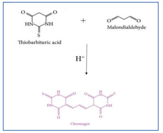
2.2.1. Thiobarbituric Acid Reactive Substances (TBARS) Method

The thiobarbituric reactive substances (TBARS) assay is one of the most popular methods to determine lipid oxidation in meat and fish products. Malonaldehyde (MA) is an aldehyde produced during the breakdown of unsaturated fatty acids [15][16][17]. One molecule of MA reacts with two molecules of thiobarbituric acid when heated in an acidic solution to form a unique Schiff base compound that gives a pink color, and the color can be measured using a visible (at 532–535 nm) or fluorescent (excitation 515 nm, emission 553 nm) spectrophotometers [16][17].

Absorption Spectrometry

The chromogen formation from the reaction of thiobarbituric acid (TBA) and MA is shown in Figure 2 [18]. The reaction rate depends on the concentration of TBA, pH and the temperature used [19]. The MA can be either extracted by distillation or with an acid solution before reacting with TBA [20]. TBA is added to a sample homogenate to react with MA in the sample [6][21] directly. The distillation method is commonly used for raw, cured, and cooked meat products but is a time-consuming method. In this method, the MA is extracted with trichloroacetic acid (TCA) at a controlled rate, and the distillate is collected, reacted with TBA, and the absorbance is measured using a spectrophotometer [22]. The acid extraction is used mainly for raw and cooked meat and fish samples, which use 7.5% TCA, followed by filtration and reaction with TBA, in a boiling water bath for 30–40 min [17]. This method has also been used to determine the lipid oxidation in edible insect-based foods [11][12]. In the direct reaction method, two volumes of TBA/TCA solution (15% TCA/20 mM TBA) are added to one sample homogenate volume and are reacted at boiling temperature for 15 min. The color formed in the supernatant is collected after centrifugation, and the absorbance is read using a spectrophotometer at 532 nm. The quantification of MA in meat is performed using a standard curve and is expressed as mg of MDA/kg food [23][24]. TBARS method is easy, simple, low cost, and reproducible, and the values are well correlated with the sensory properties of the final product. However, TBA can react with other oxidized compounds in the food product and produce false-positive results [19][16][25]. The nitrite used in the cured meat products can react with MA under acidic conditions and lower value in cured meat. Therefore, the addition of sulfanilamide for both distillation and extraction methods are needed [15][16]. Production of yellow or orange chromogen is another drawback in this test and is mainly due to the reaction of sugars (sucrose, glucose, and fructose), water-soluble proteins, peptides, and free amino acids, such as Lys and Arg, in meat and meat products with the TBA. Besides the carbohydrates, phenylpropanoid-type pigments can also show absorbance at 532 nm, which result in overestimation in the oxidation [19]. The MDA can also be converted to other organic compounds with storage in fish and fish-based products, leading to a low MDA value [26]. Therefore, the TBARS method is unsuitable for determining the oxidation level in high carbohydrate-containing food materials and egg yolk.
Figure 2. Formation of chromophore during the TBARS assay.

Fluorometric Spectrometry

Fluorometric measurement of TBARS started from the observation of Bernheim et al. [27], who found that the oxidation of unsaturated fatty acids produced 3 carbon atoms, presumably MA, which reacted with TBA and produced a red pigment [27]. However, many constituents of food components, such as amino acids, proteins, and carbohydrates, can react with TBA [28][29]. Yagi modified the fluorometric TBARS method to determine the peroxide level in the serum or plasma using a fluorometer. He collected the water-soluble TBARS in the serum or plasma samples by precipitating proteins and lipid oxidation products under acidic conditions. Then, the precipitant was collected, suspended in distilled water, and reacted with TBA at 95 °C for 1 h. The reaction products were extracted using n-butanol, and the fluorescence intensity was measured at 553 nm with excitation at 515 nm [30]. Yagi’s fluorometric method had greater sensitivity than the spectrophotometric method but was not appropriate for meat. So, Jo and Ahn [15] further modified the method of Yagi for use in meat. They directly added TBA to the meat homogenates under acidic conditions and then reacted them at 90 °C for 15 min, extracted the reacted lipid oxidation products using the n-butanol and pyridine mixture, and then measured the intensity of fluorescence at 550 nm with 520 nm excitation. The fluorometric methods showed high sensitivity with little interference by the meat components [15]. In comparison, the fluorometric methods of Yagi and Jo and Ahn are more sensitive than the conventional spectrophotometric methods and are suitable for samples with low TBARS, such as blood serum and fresh meat samples [15][30].

2.2.2. Chromatographic Methods

Because the conventional TBARS methods have insufficient specificity and sensitivity, alternative analytical approaches have been developed. The chromatographic methods are developed to determine the primary and secondary oxidation products present in the food. Kakuda et al. [31] developed an HPLC method to assess the amount of MA in aqueous distillates of chicken meat. They found that HPLC was more sensitive than spectrophotometric methods in assessing lipid oxidation products in aqueous distillates of chicken. Hydroperoxides and malonaldehyde, the primary and secondary oxidation products in foods, can also be extracted under acidic conditions first, and then the oxidation products are determined using an HPLC [32]. The method is simple, and the sensitivity is high compared with other methods but is time-consuming. Product identification and quantification can be easily made if the HPLC unit is coupled with a mass spectrometer [19][16][32][33]. Recently, a comprehensive two-dimensional liquid chromatography (LC × LC) was developed to determine lipid oxidation products using a light-scattering detector (ELSD). This method can determine both polar and non-polar lipid oxidation products. However, sample preparation for the HPLC method is complicated [33], analysis time per sample is long, and the equipment cost is high. Therefore, the industrial applications of this method are limited.
Gas chromatography equipped with mass spectrometry is another method used to determine the lipid oxidation products in foods. Specific marker compounds, such as hexanal and total aldehydes, were the most successfully used volatile compounds to determine the degree of oxidation in various foods, including meat, milk, dried, and plant-based foods, using a GC-MS [23]. In addition, the typical end products of lipid oxidation, such as aldehydes, ketones, hydrocarbons, and alcohols, that reacted with TBA are used to determine the degree of oxidation using a GC-MS [24]. Ahn et al. [34] showed that the amounts of propanal, pentanal, hexanal, 1-pentanol, and total volatiles were correlated highly (p < 0.01) with the TBARS values of cooked meat, but hexanal and total volatiles represented the degree of lipid oxidation better than any individual volatile compounds in cooked meat. GC method is also a sensitive method, but equipment cost is high, and analysis is time-consuming [19][34][35]. Thus, conventional spectrophotometric TBARS methods are still preferred over chromatographic methods because of their simplicity.

2.3. Indirect Methods Used to Detect the Secondary Oxidation Products

2.3.1. Fluorometric Method

The fluorometric method is an indirect method widely used to determine the level of lipid oxidation in raw fish, meat-based products, and animal by-products, such as blood [36][26]. It can also be used on plant oils rich in polyunsaturated fatty acids (PUFA) [37][38]. In addition, the fluorometric method has been used to determine lipid oxidation in low-moisture foods, such as cereals [39]. In the fluorometric method, MA is converted to tetramethoxy-propane, which eliminates the false fluorescence readings by vitamin A, nicotinamide adenine dinucleotide (NADH), proteins, and amino acids. Thus, this method is more suitable for protein-rich foods with more ionic and covalent bonds, such as fish [40]. The most common method includes lipid extraction using a chloroform-methanol-water solvent. The collected organic layer is mixed with sodium sulfate and filtered. Then the mixture is dried with nitrogen gas at a temperature not exceeding 35 °C. The MA equivalent in lipid extracts is measured by reacting with TBA. BHT is added to the extraction solvent, and the secondary reaction products of lipid oxidation are measured by fluorescence excitation (360 nm) and emission (440 nm) of the aqueous and organic layers that contain chloroform and methanol (2:1) [37][38]. The fluorescence produced is reliable and sensitive to detecting lipid and water-soluble hydroperoxides in meat and fish samples [41]. The sensitivity of this method is higher than the absorption spectrophotometric methods (TBARS method) and is reproducible, simple, and needs a tiny amount of sample to detect the level of oxidation in meat and meat-based products.
Lipofuscin is commonly used as a fluorescent marker, but some other markers, such as 1,4-dihydropyridines and 1-palmitoyl-2-((2-(4-(6-phenyltrans-1,3,5-hexatrienyl)-phenyl)-ethyl)-carbonyl)-sn-glycero-3-phosphocholine (DPH-PC), diphenyl-1-pyrenylphosphine (DPPP), are also used as a new type of fluorescent compounds in food. These markers are dissolved in a chloroform/ methanol mixture, and the solvents are removed using nitrogen. The dried sample is mixed with phosphate buffer, and the reaction progress is observed at 430 nm (excitation at 354 nm) at 37 °C using a fluorimeter [41]. With these markers, image processing is introduced as a novel method to determine the level of oxidation in foods [40][41].

2.3.2. Sensory Analysis

Sensory evaluation evokes, measures, analyzes, and interprets human responses to the properties of food materials. A trained or untrained panelist can determine the sensory attributes, such as smell, flavor, and taste of the food product [42]. Sensory evaluation is considered a critical method since it directly interprets oxidation levels in a food product [43][44][45]. In a practical situation, this evaluation is performed by field panels, consumer panels, free choice profiling panels, quantitative description analysis panels, and expert panels, especially with low-moisture foods, such as cereals [39][42][46]. Sensory evaluation is cost-effective and simple and can be applied to all plant- and animal-based foods in liquid, semi-solid, and solid forms. However, the sensory analysis shows high regional variations and low repeatability because of the personal preference and experience, the cultural backgrounds, the age and sex of panelists, and the time of the evaluation performed. Therefore, a sensory, along with the chemical and instrumental, analysis is recommended. As discussed above, many methods are available to determine the degree of lipid oxidation in food, but one method cannot fit all food products. Thus, selecting a proper and effective method for a specific food is essential.

2.4. Methods Used to Detect the Primary and Secondary Oxidation Products

p-Anisidine test (p-AV) and total oxidation index (TOTOX) are two common methods used in determining lipid oxidation in food products, especially edible oils. Both p-anisidine and TOTOX tests determine both the primary and secondary oxidation products in oil [47]. However, the anisidine test uses aldehydes, such as 2-alkenals and 2,4-alkadineals, as the main oxidation markers, and the level of anisidine produced is measured using the UV-spectrophotometer at 350 nm [47][48].
TOTOX indicates the overall oxidation states, and it can give a good explanation of the final quality of the edible oil. It is calculated using the following equation:
TOTOX= p-Anisidine value + 2 Peroxide value.
Both p-AV and TOTOX methods are simple, require less technical knowledge, and are commonly used to determine the quality of the EPA/DHA-containing oils and finished products [48][49]. However, p-AV is not suitable for determining the oxidation in omega-3-rich oils with intense color or specific flavorings added to the final oil [49]. Table 1 shows the summary of lipid oxidation methods with their principles, applications, advantage, and disadvantages of each method.
Table 1. Summary of the common methods used in lipid oxidation.
Lipid Oxidation Analysis Method Principle of the Method Possible Applications Advantages of the Method Disadvantages of the Method References
Peroxide value (PV): Iodometric and ferric thiocyanate Oxidation of iodide by hydroperoxides or by oxidation of Fe2+ to Fe3+.
Use a spectrophotometer to obtain the final reading.
Plant oils and liquid food products, edible insects. Simple and cheap.
Direct readings.
Under anaerobic conditions, the sensitivity is high.
Depend on the titration skills of the person.
Only applicable to liquid-based products.
[7][8][9][10][11][12]
Conjugated diene analysis Isomeric hydroperoxides will make conjugated dienes with the removal of oxygen and determine 1,4-dienes produced.
Measured at 233 nm.
Suitable for PUFA-containing foods. Gives actual values of LDL oxidation during the early stages of oxidation.
Simple and cheap.
Depend on the composition and size of the lipoproteins.
Small, conjugated dienes are difficult to detect.
[4][6][13][14]
TBARS assay Detect the production of chromogen due to the reaction of MA and TBA.
Read absorption at 532 nm. HPLC, GC-MS, and fluorometer are also used.
Meat and meat-based products.
Fish and fish-based products.
Can be used in cured meat products, edible insects.
Simple and
fast detection.
Easy to detect
and low cost.
Reproducible and
correlate well with sensory attributes.
MA and TBA can react with other organic compounds present in food. Absorption spectrophotometer is not suitable for detection at low levels. [19][22][15][16][17][18][20][21][23][24][25][26]
Chromatography methods By using the HPLC or GC. Determine the specific compounds produced. All types of raw and processed foods. Oxidative stress-related diseases. Sensitive and accurate. Identification and quantification can be made. The cost of the equipment is high. The complexity of the method and the fact that it is time-consuming. [19][16][23][24][32][33][34][35]
Fluorometric method Use different fluorescent porphyrins to interact with MDA produced during oxidation. Animal-based products. Can be used to determine the changes in human serum/plasma. Low moisture foods. Fast and accurate.
Non-destructive
Sensitive. Image produced can be further used. Can be used to detect unstable oxidized compounds.
High cost of the equipment used.
Complexity of the method.
[36][26][37][38][39][40][41]
Sensory analysis Use trained or untrained human panelist to determine the level of oxidation through sensory attributes, such as odor, taste, and acceptability. All animals and plant-based foods, cereals. Gives the overall quality of the food. Direct interpretation.
Can be used for liquid, semi-solid, and solid foods.
Depends on the individual participants and time variations.
Depends on the region.
Reproducibility difficult. Ethical clearance is needed.
[42][43][44][45][46]
p-Anisidine test Determine the level of anisidine produced from the secondary aldehydes produced. Oil and oil-based products. Simple, less technical knowledge needed. Problems in omega-3-rich oils that contain intense colors or containing specific flavorings. [47][48][49]
Total oxidation index (TOTOX) Determine the total oxidizied products. Oil and oil-based products. Simple calculation. Similar problem associated with p-anisidine test. [47][48][49]

This entry is adapted from the peer-reviewed paper 10.3390/antiox10101587

References

  1. Min, B.; Ahn, D.U. Mechanism of lipid peroxidation in meat and meat products—A review. Food Sci. Biotechnol. 2005, 14, 152–163.
  2. Guéraud, F.; Atalay, M.; Bresgen, N.; Cipak, A.; Eckl, P.M.; Huc, L.; Jouanin, I.; Siems, W.; Uchida, K. Chemistry and biochemistry of lipid peroxidation products. Free Rad. Res. 2010, 44, 1098–1124.
  3. Ayala, A.; Munoz, M.F.; Argüelles, S. Lipid peroxidation: Production, metabolism, and signaling mechanisms of malondialdehyde and 4-hydroxy-2-nominal. Oxid. Med. Cell Longev. 2014, 360438.
  4. Huang, X.; Ahn, D.U. Lipid oxidation and its implications to meat quality and human health. Food Sci. Biotechnol. 2019, 28, 1275–1285.
  5. Barthel, G.; Grosch, W. Peroxide value determination—Comparison of some methods. JAOCS 1974, 51, 540–544.
  6. Buege, J.A.; Aust, S.D. Microsomal lipid peroxidation. Method Enzymol. 1978, 52, 302–310.
  7. Pryor, W.A.; Castle, L. Chemical methods for the detection of lipid hydroperoxides. Method Enzymol. 1984, 5, 293–299.
  8. Labeque, R.; Marnett, L.J. 10-Hydroperoxy-8,12-octadecadienoic acid: A diagnostic probe of alkoxyl radical generation in metal-hydroperoxide reactions. J. Am. Chem. Soc. 1987, 109, 2828–2829.
  9. Wagner, C.D.; Clever, H.L.; Peters, E.D. Evaluation of the ferrous thiocyanate colorimetric method. Anal. Chem. 1947, 19, 980–982.
  10. Mihalević, B.; Katušin-Ražem, B.; Ražem, D. The revaluation of the ferric thiocyanate assay for lipid hydroperoxides with special considerations of the mechanistic aspects of the response. Free Radic. Biol. Med. 1996, 21, 53–63.
  11. Ojha, S.; Bußler, S.; Psarianos, M.; Rossi, G.; Schlüter, O.K. Edible insect processing pathways and implementation of emerging technologies. J. Insects Food Feed. 2021, 7, 877–890.
  12. Tenyang, N.; Tiencheu, B.; Mamat, A.; Mawamba, L.A.; Ponka, R. Effect of cooking methods on the nutritive value and lipid oxidation of two Cricket species consumed in Cameroon. Eur. J. Nutr. Food Saf. 2021, 13, 11–23.
  13. Recknagel, R.O.; Glende, E.A., Jr. Spectrophotometric detection of lipid conjugated dienes. Methods Enzymol. 1984, 105, 331–337.
  14. Corongiu, F.P.; Milia, A. An improved and simple method for determining diene conjugation in autoxidized polyunsaturated fatty acids. Chem. Biol. Interact. 1983, 44, 289–297.
  15. Jo, C.; Ahn, D.U. Fluorometric analysis of 2-thiobarbituric acid reactive substances in turkey. Poultry Sci. 1998, 77, 475–480.
  16. Jung, S.; Nam, K.C.; Jo, C. Detection of malondialdehyde in processed meat products without interference from the ingredients. Food Chem. 2016, 209, 90–94.
  17. Zeb, A.; Ullah, F. A simple spectrophotometric method for the determination of thiobarbituric acid reactive substances in fried fast foods. J. Anal. Methods Chem. 2016, 9412767.
  18. Antolovich, M.; Prenzler, P.D.; Patsalides, E.; McDonald, S.; Robards, K. Methods for testing antioxidant activity. Analyst 2002, 127, 183–198.
  19. Kunyaboon, S.; Thumanu, K.; Park, J.W.; Khongla, C.; Yongsawatdigul, J. Evaluation of lipid oxidation, volatile compounds and vibrational spectroscopy of silver carp (Hypophthalmichthys molitrix) during ice storage as related to the quality of its washed mince. Foods 2021, 10, 495.
  20. Salih, A.; Smith, D.; Price, J.; Dawson, L. Modified extraction 2-thiobarbituric acid method for measuring lipid oxidation in poultry. Poultry Sci. 1987, 66, 1483–1488.
  21. Ahn, D.U.; Sell, J.L.; Jeffery, M.; Jo, C.; Chen, X.; Wu, C.; Lee, J.I. Dietary vitamin E affects lipid oxidation and total volatiles of irradiated raw turkey meat. J. Food Sci. 1997, 62, 954–959.
  22. Tarladgis, B.G.; Watts, B.M.; Younathan, M.T.; Dugan, L., Jr. A distillation method for the quantitative determination of malonaldehyde in rancid foods. J. Am. Oil Chem. Soc. 1960, 37, 44–48.
  23. Ahn, D.U.; Olson, D.G.; Jo, C.; Chen, X.; Wu, C.; Lee, J.I. Effect of muscle type, packaging, and irradiation on lipid oxidation, volatile production and color in raw pork patties. Meat Sci. 1998, 49, 27–39.
  24. Schaich, K.M. Analysis of lipid and protein oxidation in fats, oils and foods. In Oxidative Stability and Shelf Life of Foods Containing Oils and Fats, 1st ed.; Hu, M., Jaconsen, C., Eds.; AOCS Press: Cambridge, MA, USA, 2016; pp. 1–131.
  25. Hodges, D.M.; DeLong, J.M.; Forney, C.F.; Prange, R.K. Improving the thiobarbituric acid-reactive-substances assay for estimating lipid peroxidation in plant tissues containing anthocyanin and other interfering compounds. Planta 1999, 207, 604–611.
  26. Aubourg, S.P. Recent advances in assessment of marine lipid oxidation by using fluorescence. J. Am. Chem. Soc. 1999, 76, 409–419.
  27. Bernheim, F.; Bernheim, M.L.C.; Wilbur, K.M. The reaction between thiobarbituric acid and the oxidation products of certain lipids. J. Biol. Chem. 1948, 174, 257–264.
  28. Kosugi, H.; Kikugawa, K. Potential thiobarbiuturic acid-reactive substances in peroxidized lipids. Free Radic. Biol. Med. 1989, 7, 205–208.
  29. Pérez-Palacios, T.; Estévez, M. Chapter 13—Analysis of lipids and lipid oxidation products. In Meat Quality Analysis—Advanced Evaluation Methods, Techniques, and Technologies; Biswas, A.K., Mandal, P.K., Eds.; Academic Press: Cambridge, MA, USA, 2020; pp. 217–239.
  30. Yagi, K. Lipid Peroxides and human diseases. Chem. Phys. Lipids 1987, 45, 337–351.
  31. Kakuda, Y.; Stanlry, D.W.; van deVoort, F.R. Determination of TBA number by high performance liquid chromatography. J. Am. Oil Chem. Soc. 1981, 58, 773–775.
  32. Kato, S.; Shimizu, N.; Hanzawa, Y.; Otoki, Y.; Ito, J.; Kimura, F.; Takekoshi, S.; Sakaino, M.; Sano, T.; Eitsuka, T.; et al. Determination of triacylglycerol oxidation mechanisms in canola oil using liquid chromatography-tandem mass spectrometry. NPJ Sci. Food 2018, 2.
  33. Steinhorst-Slikkerveer, L.; Louter, A.; Janssen, H.G.; Bauer-Plank, C. Analysis of nonvolatile lipid oxidation products in vegetable oils by normal-phase high-performance liquid chromatography with mass spectrometric detection. J. Am. Oil Chem. Soc. 2000, 8, 837–845.
  34. Ahn, D.U.; Olson, D.G.; Jo, C.; Love, J.; Jin, S.K. Volatile production and lipid oxidation of irradiated coocked sausages with different packaging during storage. J. Food Sci. 1999, 64, 226–229.
  35. Lazaridi, E.; Janssen, H.-G.; Vincken, J.-P.; Pirok, B.; Hennbelle, M. A comprehensive two-dimensional liquid chromatography method for the simultaneous separation of lipid species and their oxidation products. J. Chromatogr. A 2021, 1644, 462106.
  36. Reitznerová, A.; Šuleková, M.; Nagy, J.; Marcinčák, S.; Semjon, B.; Čertík, M.; Klempová, T. Lipid peroxidation process in meat and meat products: A comparison study of malondialdehyde determination between modified 2-thiobarbituric acid spectrophotometric method and reverse-phase high-performance liquid chromatography. Molecules 2017, 22, 1988.
  37. Pikul, J.; Leszczynski, D.E.; Bechtel, P.J.; Kummerow, F.A. Effects of frozen storage and cooking on lipid oxidation in chicken meat. J. Food Sci. 1984, 49, 838–843.
  38. Tappel, A.L. Vitamin E and free radical peroxidation of lipids. Ann. N. Y. Acad. Sci. 1972, 203, 12–28.
  39. Barden, L.; Decker, E.A. Lipid oxidation in low-moisture foods: A review. Crit. Rev. Food Sci. Nutr. 2016, 56, 2467–2482.
  40. Hassoun, A.; Karoui, R. Front-face fluorescence spectroscopy coupled with chemometric tools for monitoring fish freshness stored under different refrigerated conditions. Food Control 2015, 54, 240–249.
  41. Cropotova, J.; Mozuraityte, R.; Standal, I.B.; Rustad, T. Assessment of lipid oxidation in Atlantic mackerel (Scomber scombrus) subjected to different antioxidant and sous-vide cooking treatments by conventional and fluorescence microscopy methods. Food Control 2019, 104, 1–8.
  42. Fruhwirth, G.O.; Wenzl, T.; El-Toukhy, R.; Wagner, F.S.; Hermetter, A. Fluorescence screening of antioxidant capacity in pumpkin seed oils and other natural oils. Eur. J. Lipid Sci. Technol. 2003, 105, 266–274.
  43. Mihafu, F.D.; Issa, J.Y.; Kamiyango, M.W. Implication of sensory evaluation and quality assessment in food product development: A review. Curr. Res. Nutr. Food Sci. 2020, 8, 690–702.
  44. Clarke, H.J.; O’Sullivan, M.G.; Kerry, J.P.; Kilcawley, K.N. Correlating volatile lipid oxidation compounds with consumer sensory data in dairy-based powders during storage. Antioxidants 2020, 9, 338.
  45. Fan, Y.; Odabasi, A.; Sims, C.; Gao, Z.; Sarnoski, P. Utilization of descriptive sensory analysis and volatile analysis to determine quality indicators of aquacultured whiteleg shrimp (Litopanaeus vannemei) during Refrigerated Storage. J. Aquatic Food Prod. Technol. 2020, 29, 722–735.
  46. Paradiso, V.M.; Summo, C.; Pasqualone, A.; Caponio, F. Evaluation of different natural antioxidants as affectivg volatile lipid oxidation products related to off-flavours in corn flakes. Food Chem. 2009, 113, 543–549.
  47. Cong, S.; Dong, W.; Zhao, J.; Hu, R.; Long, Y.; Chi, X. Characterization of the Lipid Oxidation Process of Robusta Green Coffee Beans and Shelf-Life Prediction during Accelerated Storage. Molecules 2020, 25, 1157.
  48. Shahidi, F.; Zhong, Y. Measurement of antioxidant activity. J. Funct. Foods 2015, 18, 757–781.
  49. Ismail, A.; Bannenberg, G.; Rice, H.B.; Schutt, E.; MacKay, D. Oxidation in EPA- and DHA-rich oils: An overview. Lipid Technol. 2016, 28, 3–4.
More
This entry is offline, you can click here to edit this entry!