Potential Therapeutic Targets on SARS-CoV-2: History
Please note this is an old version of this entry, which may differ significantly from the current revision.
Subjects: Cell Biology
Contributor:

Coronavirus disease 19 (COVID-19) is caused by an enveloped, positive-sense, single-stranded RNA virus, referred to as severe acute respiratory syndrome coronavirus 2 (SARS-CoV-2), which belongs to the realm Riboviria, order Nidovirales, family Coronaviridae, genus Betacoronavirus and the species Severe acute respiratory syndrome-related coronavirus. This viral disease is characterized by a myriad of varying symptoms, such as pyrexia, cough, hemoptysis, dyspnoea, diarrhea, muscle soreness, dysosmia, lymphopenia and dysgeusia amongst others. The virus mainly infects humans, various other mammals, avian species and some other companion livestock. SARS-CoV-2 cellular entry is primarily accomplished by molecular interaction between the virus’s spike (S) protein and the host cell surface receptor, angiotensin-converting enzyme 2 (ACE2), although other host cell-associated receptors/factors, such as neuropilin 1 (NRP-1) and neuropilin 2 (NRP-2), C-type lectin receptors (CLRs), as well as proteases such as TMPRSS2 (transmembrane serine protease 2) and furin, might also play a crucial role in infection, tropism, pathogenesis and clinical outcome.

  • SARS-CoV-2
  • coronavirus disease 19
  • pathogenesis
  • therapeutic targeting

1. Potential Therapeutic Targets on Host Cells

The SARS-CoV-2’s S protein engages with the host cell receptor, ACE2, to gain cellular entry, leading to onset of infection. The structural spike (S) protein contains multiple domains, including the N-terminal receptor-binding domain (RBD), and protease cleavage sites, such as the furin site [1]. The RBD of the S protein plays a very important role in molecular interaction, therefore, therapeutic approaches specifically targeting the RBD/ACE2 interface may prove to be one of the most reliable treatment modalities. For instance, empirical findings based on multidisciplinary approaches have zeroed in on some potential molecules/drugs, such as Evans blue, sodium lifitegrast, and lumacaftor, that may specifically interfere with the SARS-CoV-2-S–ACE2 interaction, thereby preventing infection and disease occurrence [2]. During this interaction, the S protein is specifically primed and cleaved at the S1/S2 cleavage site by activated host tissue-specific proteases, such as TMPRSS2, TMPRSS4, TMPRSS11D, and furin, facilitating virus internalization and genome release [3][4]. Moreover, endosomal proteases, such as cathepsin B (CTSB), cathepsin L (CTSL), basigin (BSG) and FURIN may also be potentially involved in this process, making them potential therapeutic targets [5]. Owing to the significant involvement of proteases, such as TMPRSS2 and furin in priming the S protein, which are prerequisite for cellular entry of the virus, they may also be targeted from a therapeutic point of view using a protease inhibitor, such as camostat mesylate. In fact, such approach, involving camostat mesylate, has proven to be considerably effective in blocking SARS-CoV-2 infection in primary human lung cells [3].
Apart from the ACE2 receptor, SARS-CoV-2, like many other viruses, such as human T-lymphotropic virus-1 (HTLV-1) [6] and Epstein–Barr virus (EBV) [7], also uses multifunctional transmembrane receptor neurophilin 1 (NRP-1) for cell entry. NRP-1 is known to be expressed abundantly in respiratory and olfactory epithelia, and has a broad range of implications owing to its involvement in various cellular processes, such as angiogenesis, axonal guidance, growth and progression, tumor progression, immune functions and viral entry [8][9]. Vascular endothelial growth factor A (VEGF-A) is considered to be one of the most important ligands of the neurophilin receptor, and is primarily involved in angiogenesis, but was recently discovered to be pro-nociceptive as well [10]. It is generally thought that SARS-CoV-2 leverages the VEGF-A interaction site on NRP-1 to gain cellular entry. The likelihood of usage of NRP-1 may be substantiated by the report of COVID-19 patients showing upregulation of the receptor in lung samples [8]. Therefore, successful blockade of the interaction between SARS-CoV-2 and NRP-1 by using well established inhibitors, such as EG00229 and EG01377 [11], may prove to be an effective COVID-19 therapy (Figure 1).
Furthermore, there have been efforts to identify host genes (pro-viral and anti-viral) essential for SARS-CoV-2 infection. Such tireless efforts aim at developing an understanding about the viral pathogenesis, as well as finding out novel therapeutic target(s). For example, Wei J. et al. carried out genome-wide CRISPR screening and identified multiple active host genes with crucial roles in histone modification and chromatin regulation, cellular signaling, and RNA regulation. Identification of active genes encoding the pleotropic HMGB1 protein and members of the SWI/SNF chromatin remodeling complex in SARS-CoV-1, SARS-CoV-2, and NL63-infected Vero-E6 cells unequivocally suggests a relation between epigenetic regulation and viral pathogenesis. HMGB1 was found to intrinsically regulate ACE2 expression, indicating the pivotal involvement of the epigenetic process in SARS-CoV cellular entry and infection, while a small-molecule antagonist inhibited the same in monkey and human cells, further substantiating the relevance of the epigenetic mechanism [12]. Therefore, developing greater insights into the underlying mechanism of such processes may help in screening/designing small therapeutic molecules, as well as repurposing of FDA-approved drugs to prevent infection and disease.
Apart from targeting the cell receptor, ACE2, and associated host factors/proteases, such as TMPRSS2, another therapeutic approach may entail targeting the SARS-CoV-2–cellular protein–protein interaction (interactome). A recent study, involving multiple expressed SARS-CoV-2 proteins as baits, identified 332 host cell interacting proteins (overlapping and specific), belonging to various functional categories and/or natures, which are generally involved in several complex biological processes and pathways (Figure 2). Of the total interacting proteins, around 40% of host cell proteins belong to the endomembrane compartment and/or vesicle trafficking pathways [13].
Figure 2. Interactome, involving SARS-CoV-2 proteins and cellular proteins. SARS-CoV-2 structural and non-structural proteins have been found to interact with multiple host cell proteins involved in various cellular processes, as well as remain associated with several cell organelles. SARS-CoV-2 proteins are shown in the center of the small circle, whereas human host cell proteins are placed in the center of the large circle. An arrow indicates a possible interaction, which may or may not have significant implications. N-Neucleocapsid protein, S-Spike protein, M-Membrane protein and E-Envelope protein.
Interestingly, several SARS-CoV-2 proteins interact with innate immune signaling proteins. For instance, NSP13, NSP15, and Orf9b (also referred to as nonstructural protein NS9b) target the interferon (IFN) pathway by interacting with various pathway-associated proteins. Similarly, NSP13 and Orf9c (also referred to as nonstructural protein NS9c) can alter the NF-κB pathway, which is involved in multiple crucial cellular processes, including immune response. Further, NSP9 and Orf3a (also referred to as nonstructural protein NS3a) show molecular associations with antiviral immune signaling-associated E3 ubiquitin ligases, tripartite motif-containing protein 59 (TRIM59) and mind bomb 1 (MIB1), respectively [14][15]. SARS-CoV-2’s Orf6 (also referred to as nonstructural protein NS6) interacts with an IFN-inducible mRNA nuclear export complex, NUP98-RAE1, in a manner similar to vesicular stomatitis virus (VSV), influenza A, and polio virus amongst others, and thereby antagonizes interferon signaling through disruption at the level of nuclear export [16]. The nucleocapsid (N) protein of SARS-CoV-2 binds to multiple translational regulators, such as UPF1 and MOV10 (mRNA decay factors), CK2 (protein kinase) and host mRNA binding proteins. Furthermore, N protein can also influence 5′ cap-dependent translation, thereby negatively affecting global protein synthesis [17]. Around a dozen of SARS-CoV-2 proteins undergo Sec61 translocon-mediated cotranslational insertion into the endoplasmic reticulum (ER) and remain localized in the virus replication complex [18]. SARS-CoV-2 NSP8 interacts with three components of the signal recognition particle (SRP), thereby negatively influencing Sec61-mediated protein translocation into the ER, which may be effectively inhibited by PS3061, a Sec61 inhibitor. This inhibitor may also interfere with SARS-CoV-2 replication and assembly as observed in the case of other enveloped RNA viruses [19][20].

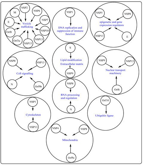
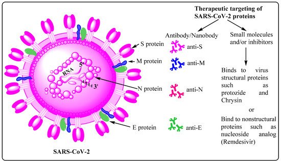
2. Potential Therapeutic Targets on SARS-CoV-2

SARS-CoV-2 primarily relies on the host cell receptor, ACE2, to gain successful cellular entry. Molecular interaction between the SARS-CoV-2’s S protein and ACE2 is accomplished by conformational changes, protease cleavage on the S1/S2 domain, and eventual fusion of viral membrane with host cell membrane [21]. The virus can follow either endocytic or non-endocytic pathways to enter the host cell, and eventually releases its nearly 30 kb genome in the cytosol. Thereafter, a cascade of molecular events leads to synthesis of virus structural and non-structural proteins and multiple copies of viral genomes, which eventually assemble together, forming new viral progenies [22]. Moreover, the S protein is quite immunogenic in nature, leading to the formation of a diverse set of antibodies and memory B cells, each specific to a particular epitope/region/domain of this multidomain structural protein. For instance, a recently published work, employing single-cell sorting, identified 453 neutralizing antibodies and 4277 SARS-CoV-2 spike protein-specific memory B cells from 14 COVID-19 convalescent individuals. This work also shows that there are differential neutralizing capabilities among these antibodies with anti-RBD being the most potent one, followed by the anti-S1 domain, anti-spike protein trimer, and the anti-S2 subunit being the least potent [23]. The most potent antibody may, either in its natural or engineered form, be used as a therapeutic and/or prophylactic measure, and delivered/administered to the patient through intravenous infusion [24][25]. The abovementioned facts are the underlying reason as to why most of the vaccines, irrespective of their type, mainly rely on the S protein as the active antigen ingredient. While choosing therapeutic antibodies, one must also factor in their efficiency and effectiveness against emerging variants of concern (VOC) containing D614G, E484K, and N501Y substitutions [26]. Such variants have been evolving and circulating globally throughout the COVID-19 pandemic, and continue to prevail and show upward infection trend, leading to thousands of death each day around the world. Their rapid global spread is attributed to the acquisition of higher infectivity as a result of favorable mutational changes in the virus genome, especially in the gene encoding S protein. Moreover, diverse antibodies have also been detected against other SARS-CoV-2 structural proteins, such as anti-E, anti-M, and anti-N with varied potential and therapeutic implications [27] (Figure 3). However, the quantities of such antibodies in COVID-19 patients are lower owing to the relatively small molecular sizes of these antigens (M, E, and N proteins), as well as the lesser degree of structural protrusion of their corresponding ectodomains, which are prerequisite for engagement and recognition by B cells and other immune cells. In addition to various structural proteins, non-structural proteins may also acts as antigens as evidenced by the presence of reactive CD4+ T cells against NSP3, NSP4, Orf3a (NS3a), and Orf8 (NS8). Considering all these factors, it may be prudent to design a vaccine, involving both categories of potential antigens, i.e., structural and non-structural proteins, as it may evoke a diverse set of B cells to make antibodies, thereby providing holistic protection against infection. A good vaccine must be able to induce both B and T cells along with formation of corresponding long-term memory cells with minimal or no side effects.
Figure 3. Potential targets on SARS-CoV-2. SARS-CoV-2, the causative organism of COVID-19, possesses four types of structural proteins, namely spike (S), membrane (M), envelope (E), and nucleocapsid (N). Of these, the homotrimeric S protein shows the highest immunogenicity, leading to the production of correspondingly high amount of the anti-S antibody, and generation of memory B and T cells. Anti-M, anti-N and anti-E antibodies, albeit in lesser quantities, are also detected in samples derived from COVID-19 patients.
Apart from therapeutic antibodies, SARS-CoV-2-specific proteins and processes, favoring the infectious virus, may also be targeted using well established small molecules, such as aloxistatin, chloroquine/hydroxychloroquine, anti-viral nucleotide analogs (remdesivir), protease inhibitors (lopinavir and ritonavir), antiviral phytochemicals, and the broad-spectrum antiviral drugs like favipiravir and arbidol. In general, aloxistatin is a cysteine protease inhibitor for calpain and cathepsins, and used as a cancer therapy drug. Since cathepsin L has been reported to play a crucial role in SARS-CoV-2 cell entry as well [28], administration of aloxistatin may be very important in combating infection. Moreover, aloxistatin may also bind SARS-CoV-2 main protease (MPRO), as well as papin-like proteases, albeit with lower specificity, thereby interfering with the proteolysis of polypeptides 1a/ab [29]. Owing to the very high sequence specificity of MPRO, compounds structurally mimicking its substrate cleavage site may be very precise inhibitors with negligible or no adverse effect on host cellular proteases [30]. Furthermore, chloroquine and hydroxychloroquine are well established antimalarial drugs, and are being tested for COVID-19 therapy. Whereas chloroquine inhibits terminal phosphorylation of ACE2, hydroxychloroquine elevates endosomal pH, both being crucial processes prerequisite for successful establishment of SARS-CoV-2 infection. Owing to their involvement in such crucial cellular processes, several clinical trials are underway to establish the efficiency and modalities with respect to these drugs before final approval as candidate drugs against SARS-CoV-2 infection is granted (https://clinicaltrials.gov/; accessed on 6 August 2021). Similarly, RdRp and 3Clpro (also termed MPRO), highly conserved SARS-CoV-1/2 proteins, are also very specific targets to be employed for COVID-19 treatment. Remdesivir and ritonavir/lopinavir, ribonucleotide analogs, have also been found to be capable of interfering with the working of RdRp, and therefore constitute another set of effective candidate drugs against the current pandemic (Figure 3) [31][32][33]. Furthermore, several in silico analyses are also being carried out to find novel drugs and/or bioactive natural compounds to treat COVID-19 [34][35][36][37][38][39].

This entry is adapted from the peer-reviewed paper 10.3390/cells10092427

References

  1. Walls, A.C.; Park, Y.-J.; Tortorici, M.A.; Wall, A.; McGuire, A.T.; Veesler, D. Structure, Function, and Antigenicity of the SARS-CoV-2 Spike Glycoprotein. Cell 2020, 181, 281–292.
  2. Day, C.J.; Bailly, B.; Guillon, P.; Dirr, L.; Jen, F.E.-C.; Spillings, B.L.; Mak, J.; von Itzstein, M.; Haselhorst, T.; Jennings, M.P. Multidisciplinary Approaches Identify Compounds that Bind to Human ACE2 or SARS-CoV-2 Spike Protein as Candidates to Block SARS-CoV-2–ACE2 Receptor Interactions. mBio 2021, 12.
  3. Hoffmann, M.; Kleine-Weber, H.; Schroeder, S.; Krüger, N.; Herrler, T.; Erichsen, S.; Schiergens, T.S.; Herrler, G.; Wu, N.-H.; Nitsche, A.; et al. SARS-CoV-2 Cell Entry Depends on ACE2 and TMPRSS2 and Is Blocked by a Clinically Proven Protease Inhibitor. Cell 2020, 181, 271–280.
  4. Zang, R.; Castro, M.F.G.; McCune, B.T.; Zeng, Q.; Rothlauf, P.W.; Sonnek, N.M.; Liu, Z.; Brulois, K.F.; Wang, X.; Greenberg, H.B.; et al. TMPRSS2 and TMPRSS4 promote SARS-CoV-2 infection of human small intestinal enterocytes. Sci. Immunol. 2020, 5, eabc3582.
  5. Singh, M.; Bansal, V.; Feschotte, C. A Single-Cell RNA Expression Map of Human Coronavirus Entry Factors. Cell Rep. 2020, 32, 108175.
  6. Lambert, S.; Bouttier, M.; Vassy, R.; Seigneuret, M.; Petrow-Sadowski, C.; Janvier, S.; Heveker, N.; Ruscetti, F.W.; Perret, G.; Jones, K.S.; et al. HTLV-1 uses HSPG and neuropilin-1 for entry by molecular mimicry of VEGF165. Blood 2009, 113, 5176–5185.
  7. Wang, H.-B.; Zhang, H.; Zhang, J.-P.; Li, Y.; Zhao, B.; Feng, G.-K.; Du, Y.; Xiong, D.; Zhong, Q.; Liu, W.-L.; et al. Neuropilin 1 is an entry factor that promotes EBV infection of nasopharyngeal epithelial cells. Nat. Commun. 2015, 6, 6240.
  8. Cantuti-Castelvetri, L.; Ojha, R.; Pedro, L.D.; Djannatian, M.; Franz, J.; Kuivanen, S.; Van Der Meer, F.; Kallio, K.; Kaya, T.; Anastasina, M.; et al. Neuropilin-1 facilitates SARS-CoV-2 cell entry and infectivity. Science 2020, 370, 856–860.
  9. Guo, H.-F.; Kooi, C.W.V. Neuropilin Functions as an Essential Cell Surface Receptor. J. Biol. Chem. 2015, 290, 29120–29126.
  10. Llorián-Salvador, M.; González-Rodríguez, S. Painful Understanding of VEGF. Front. Pharm. 2018, 9, 1267.
  11. Jarvis, A.; Allerston, C.K.; Jia, H.; Herzog, B.; Garza-Garcia, A.; Winfield, N.; Ellard, K.; Aqil, R.; Lynch, R.; Chapman, C.; et al. Small Molecule Inhibitors of the Neuropilin-1 Vascular Endothelial Growth Factor A (VEGF-A) Interaction. J. Med. Chem. 2010, 53, 2215–2226.
  12. Wei, J.; Alfajaro, M.M.; DeWeirdt, P.C.; Hanna, R.E.; Lu-Culligan, W.J.; Cai, W.L.; Strine, M.S.; Zhang, S.-M.; Graziano, V.R.; Schmitz, C.O.; et al. Genome-wide CRISPR Screens Reveal Host Factors Critical for SARS-CoV-2 Infection. Cell 2020, 184, 76–91.
  13. Gordon, D.E.; Jang, G.M.; Bouhaddou, M.; Xu, J.; Obernier, K.; White, K.M.; O’Meara, M.J.; Rezelj, V.V.; Guo, J.Z.; Swaney, D.L.; et al. A SARS-CoV-2 protein interaction map reveals targets for drug repurposing. Nature 2020, 583, 459–468.
  14. Kondo, T.; Watanabe, M.; Hatakeyama, S. TRIM59 interacts with ECSIT and negatively regulates NF-κB and IRF-3/7-mediated signal pathways. Biochem. Biophys. Res. Commun. 2012, 422, 501–507.
  15. Li, S.; Wang, L.; Berman, M.; Kong, Y.-Y.; Dorf, M.E. Mapping a Dynamic Innate Immunity Protein Interaction Network Regulating Type I Interferon Production. Immunity 2011, 35, 426–440.
  16. Faria, P.A.; Chakraborty, P.; Levay, A.; Barber, G.N.; Ezelle, H.J.; Enninga, J.; Arana, C.; van Deursen, J.; Fontoura, B.M. VSV Disrupts the Rae1/mrnp41 mRNA Nuclear Export Pathway. Mol. Cell 2005, 17, 93–102.
  17. Nakagawa, K.; Lokugamage, K.; Makino, S. Viral and Cellular mRNA Translation in Coronavirus-Infected Cells. Adv. Virus Res. 2016, 96, 165–192.
  18. Knoops, K.; Kikkert, M.; Worm, S.H.E.V.D.; Zevenhoven-Dobbe, J.C.; Van Der Meer, Y.; Koster, A.; Mommaas, A.M.; Snijder, E.J. SARS-Coronavirus Replication Is Supported by a Reticulovesicular Network of Modified Endoplasmic Reticulum. PLoS Biol. 2008, 6, e226.
  19. Shah, P.; Link, N.; Jang, G.M.; Sharp, P.P.; Zhu, T.; Swaney, D.L.; Johnson, J.; Von Dollen, J.; Ramage, H.R.; Satkamp, L.; et al. Comparative Flavivirus-Host Protein Interaction Mapping Reveals Mechanisms of Dengue and Zika Virus Pathogenesis. Cell 2018, 175, 1931–1945.
  20. Heaton, N.S.; Moshkina, N.; Fenouil, R.; Gardner, T.; Aguirre, S.; Shah, P.; Zhao, N.; Manganaro, L.; Hultquist, J.; Noel, J.; et al. Targeting Viral Proteostasis Limits Influenza Virus, HIV, and Dengue Virus Infection. Immunity 2016, 44, 46–58.
  21. Benton, D.J.; Wrobel, A.G.; Xu, P.; Roustan, C.; Martin, S.R.; Rosenthal, P.B.; Skehel, J.J.; Gamblin, S.J. Receptor binding and priming of the spike protein of SARS-CoV-2 for membrane fusion. Nature 2020, 588, 327–330.
  22. V’Kovski, P.; Kratzel, A.; Steiner, S.; Stalder, H.; Thiel, V. Coronavirus biology and replication: Implications for SARS-CoV-2. Nat. Rev. Genet. 2020, 19, 155–170.
  23. Andreano, E.; Nicastri, E.; Paciello, I.; Pileri, P.; Manganaro, N.; Piccini, G.; Manenti, A.; Pantano, E.; Kabanova, A.; Troisi, M.; et al. Extremely potent human monoclonal antibodies from COVID-19 convalescent patients. Cell 2021, 184, 1821–1835.e16.
  24. Hansen, J.; Baum, A.; Pascal, K.E.; Russo, V.; Giordano, S.; Wloga, E.; Fulton, B.O.; Yan, Y.; Koon, K.; Patel, K.; et al. Studies in humanized mice and convalescent humans yield a SARS-CoV-2 antibody cocktail. Science 2020, 369, 1010–1014.
  25. Liu, L.; Wang, P.; Nair, M.S.; Yu, J.; Rapp, M.; Wang, Q.; Luo, Y.; Chan, J.F.-W.; Sahi, V.; Figueroa, A.; et al. Potent neutralizing antibodies against multiple epitopes on SARS-CoV-2 spike. Nature 2020, 584, 450–456.
  26. Baum, A.; Fulton, B.O.; Wloga, E.; Copin, R.; Pascal, K.E.; Russo, V.; Giordano, S.; Lanza, K.; Negron, N.; Ni, M.; et al. Antibody cocktail to SARS-CoV-2 spike protein prevents rapid mutational escape seen with individual antibodies. Science 2020, 369, 1014–1018.
  27. Lytton, S.; Yeasmin, M.; Ghosh, A.; Bulbul, R.H.; Molla, M.A.; Herr, M.; Duchmann, H.; Sharif, M.; Nafisa, T.; Amin, R.; et al. Detection of Anti-Nucleocapsid Antibody in COVID-19 Patients in Bangladesh Is not Correlated with Previous Dengue Infection. Pathogens 2021, 10, 637.
  28. Heiser, K.; McLean, P.F.; Davis, C.T.; Fogelson, B.; Gordon, H.B.; Jacobson, P.; Hurst, B.; Miller, B.; Alfa, R.W.; Earnshaw, B.A.; et al. Identification of potential treatments for COVID-19 through artificial intelligence-enabled phenomic analysis of human cells infected with SARS-CoV-2. bioRxiv 2020, 054387.
  29. Yousefi, H.; Mashouri, L.; Okpechi, S.C.; Alahari, N.; Alahari, S.K. Repurposing existing drugs for the treatment of COVID-19/SARS-CoV-2 infection: A review describing drug mechanisms of action. Biochem. Pharm. 2020, 183, 114296.
  30. Jin, Z.; Du, X.; Xu, Y.; Deng, Y.; Liu, M.; Zhao, Y.; Zhang, B.; Li, X.; Zhang, L.; Peng, C.; et al. Structure of Mpro from SARS-CoV-2 and discovery of its inhibitors. Nature 2020, 582, 289–293.
  31. Eastman, R.T.; Roth, J.S.; Brimacombe, K.R.; Simeonov, A.; Shen, M.; Patnaik, S.; Hall, M.D. Correction to Remdesivir: A Review of Its Discovery and Development Leading to Human Clinical Trials for Treatment of COVID-19. ACS Cent. Sci. 2020, 6, 1009.
  32. Wang, M.; Cao, R.; Zhang, L.; Yang, X.; Liu, J.; Xu, M.; Shi, Z.; Hu, Z.; Zhong, W.; Xiao, G. Remdesivir and chloroquine effectively inhibit the recently emerged novel coronavirus (2019-nCoV) in vitro. Cell Res. 2020, 30, 269–271.
  33. Gordon, C.J.; Tchesnokov, E.P.; Woolner, E.; Perry, J.K.; Feng, J.Y.; Porter, D.P.; Götte, M. Remdesivir is a direct-acting antiviral that inhibits RNA-dependent RNA polymerase from severe acute respiratory syndrome coronavirus 2 with high potency. J. Biol. Chem. 2020, 295, 6785–6797.
  34. Di Micco, S.; Musella, S.; Scala, M.C.; Sala, M.; Campiglia, P.; Bifulco, G.; Fasano, A. In silico Analysis Revealed Potential Anti-SARS-CoV-2 Main Protease Activity by the Zonulin Inhibitor Larazotide Acetate. Front. Chem. 2021, 8, 628609.
  35. Yadav, R.; Hasan, S.; Mahato, S.; Celik, I.; Mary, Y.; Kumar, A.; Dhamija, P.; Sharma, A.; Choudhary, N.; Chaudhary, P.K.; et al. Molecular docking, DFT analysis, and dynamics simulation of natural bioactive compounds targeting ACE2 and TMPRSS2 dual binding sites of spike protein of SARS CoV-2. J. Mol. Liq. 2021, 116942.
  36. Yadav, R.; Imran, M.; Dhamija, P.; Suchal, K.; Handu, S. Virtual screening and dynamics of potential inhibitors targeting RNA binding domain of nucleocapsid phosphoprotein from SARS-CoV-2. J. Biomol. Struct. Dyn. 2021, 12, 4433–4448.
  37. Yadav, R.; Imran, M.; Dhamija, P.; Chaurasia, D.K.; Handu, S. Virtual screening, ADMET prediction and dynamics simulation of potential compounds targeting the main protease of SARS-CoV-2. J. Biomol. Struct. Dyn. 2020, 1–16.
  38. Yadav, R.; Parihar, R.D.; Dhiman, U.; Dhamija, P.; Upadhyay, S.K.; Imran, M.; Behera, S.K.; Prasad, T.S.K. Docking of FDA Approved Drugs Targeting NSP-16, N-Protein and Main Protease of SARS-CoV-2 as Dual Inhibitors. Biointerface Res. Appl. Chem. 2020, 11, 9848–9861.
  39. Azam, F.; Eid, E.E.M.; Almutairi, A. Targeting SARS-CoV-2 main protease by teicoplanin: A mechanistic insight by docking, MM/GBSA and molecular dynamics simulation. J. Mol. Struct. 2021, 131124.
More
This entry is offline, you can click here to edit this entry!