Exercise attenuates pathological changes in the liver induced by high-fat diets. The underlying mechanisms might be related to NRF2 and mediated by SIRT1/AMPK signaling.
1. Introduction
Nonalcoholic fatty liver disease (NAFLD) is one of the most prevalent causes of chronic liver disease, and its morbidity continues to rise worldwide in the context of the obesity epidemic. NAFLD is a chronic liver disease with excessive fat accumulation in liver and accompanied by other complications, such as insulin resistance, type 2 diabetes, and cardiopathy [
1]. As a continuum of liver abnormalities, NAFLD has an intricate pathway from nonalcoholic fatty liver (NAFL) to nonalcoholic steatohepatitis (NASH) [
2,
3]. NASH is a more aggressive and serious stage of inflammation and steatohepatitis, and is accompanied by fibrosis and cirrhosis [
3].
Reactive oxygen species (ROS) play a vital role in regulating cell growth and hepatocyte death in NAFLD [
7]. Prolonged high-fat diets induce ROS accumulation in the liver and impair the normal physiological function of hepatocytes [
8]. Antioxidant therapy for NAFLD, such as treatment with glutathione, could significantly decrease ALT levels in patients [
9]. NADPH oxidase 4 (NOX4) is the main source of ROS generation [
10]. Targeting NOX4- or NOX4-derived ROS could be a potential therapeutic strategy for NAFLD [
11,
12]. Nuclear factor erythroid-2-related factor 2 (NRF2) is an important endogenous transcription factor that can defend cells against oxidative stress [
13]. During NAFLD, a reduction in NRF2 levels is observed with redox upregulation [
14,
15]. Pharmacological activation of NRF2 restores injured liver tissue and attenuates oxidative stress in NAFLD animal models [
16,
17,
18]. Therefore, antioxidative interventions in patients with NAFLD could prevent oxidative stress by activating Nrf2 and its downstream genes.
Regular exercise is an effective non-pharmaceutical therapy for NAFLD, and the underlying mechanism is related to the metabolic sensors SIRT1/AMPK [
19]. A previous study revealed that exercise could stimulate SIRT1/AMPK signaling, which can both improve the pathogenesis and prevent the development of NAFLD [
20]. The activation of SIRT1/AMPK signaling induced by exercise could decrease lipid accumulation, increase energy expenditure, limit de novo lipogenesis (DNL), and upregulate fatty acid metabolism [
21,
22]. SIRT1/AMPK signaling also attenuates inflammation [
23], and may be a potential therapeutic target for reducing antioxidative stress and ameliorating oxidative impairment. SIRT1/AMPK signaling regulates NRF2 in pneumonia pathogenesis, cardiovascular diseases, and diabetic nephropathy [
24,
25,
26].
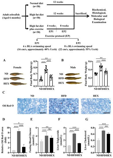
2. Swimming Exercise Reduces Body Weight Gain and Lipid Accumulation in HFD Zebrafish
After 12 weeks, both female and male HFD zebrafish had significantly increased body weight compared to normal diet (ND) zebrafish. However, high-fat diet plus exercise (HEX) inhibited excessive body weight gain (Figure 1A,B). Oil Red O staining revealed that HFD zebrafish livers had more lipid droplets than ND zebrafish livers, and that the HEX group had fewer lipid drops than the HFD group (Figure 1C,D). The HEX group also had improved fasting blood glucose, liver triglyceride, and cholesterol levels compared to the HFD group (Figure 1E–G). These results suggest that exercise can mitigate lipid homeostasis disorders caused by excessive fat intake.
Figure 1. Zebrafish receiving a high exercise regimen (HEX) had reduced body weight gain and lipid accumulation compared to high-fat diet (HFD) zebrafish. Normal diet (ND) zebrafish represent the control group. (A,B) Morphology and body weight of zebrafish. (C) Oil Red O staining of zebrafish livers (n = 3). (D) Quantitation of Oil Red O staining. (E) Fasting blood glucose (ND, n = 13; HFD, n = 10; HEX, n = 11). (F) Liver triglyceride (n = 5). (G) Liver cholesterol (n = 5). The above experiments were carried out using 9-month-old zebrafish. *, p < 0.05, **, p < 0.01, ***, p < 0.001. Data represent the mean, and error bars represent SEM. Scale bar, 20 μm. NAFLD, non-alcoholic fatty liver disease; ND, normal diet; HFD, high fat diet; HEX, high-fat diet plus exercise.
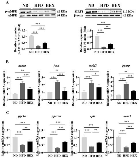
3. Swimming Exercise Activates SIRT1/AMPK Signaling and Improves Lipid Metabolism Disorders in HFD Zebrafish Livers
The SIRT1/AMPK signaling pathway is closely related to lipid metabolism, and previous research has suggested that it may contribute to the pathogenesis of NAFLD [
33,
34]. First, we showed that HFD zebrafish had strong inhibition of SIRT1 expression and AMPK phosphorylation compared to ND zebrafish, indicating that a high-fat diet significantly suppressed SIRT1/AMPK signaling. Conversely, swimming exercise upregulated SIRT1 expression and AMPK phosphorylation in HEX zebrafish (
Figure 2A). Excessive body weight gain and fat accumulation induced by an HFD can be attributed to lipid metabolism disorders. We therefore next measured the mRNA expression of lipid metabolism genes regulated by SIRT1/AMPK signaling. HEX zebrafish had comparatively lower levels of lipogenesis genes (
acaca,
fasn,
srebf1,
pparg) (
Figure 2B) and higher level of fatty acid β-oxidation genes (
pgc1α,
pparab,
acox1,
cpt1a) (
Figure 2C) than HFD zebrafish.
Figure 2. SIRT1/AMPK signaling and lipid metabolism in zebrafish livers. (A) Western blot representing SIRT1/AMPK signaling in 9-month-old zebrafish livers (n = 8). (B) Expression of lipogenesis-related genes (n = 6). (C) Expression of β-oxidation-related genes in 9-month-old zebrafish livers (n = 6). *, p < 0.05, **, p < 0.01, ***, p < 0.001. Data represents the mean, and error bars represent SEM. NAFLD, non-alcoholic fatty liver disease; ND, normal diet; HFD, high fat diet; HEX, high-fat diet plus exercise.
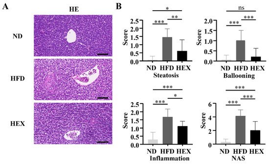
4. Swimming Exercise Ameliorates Liver Pathological Changes in HFD Zebrafish
Zebrafish livers were stained with HE to investigate pathological changes associated with NAFLD. After 12 weeks of HFD, the zebrafish livers developed macrovesicular steatosis, lobular inflammation, and hepatocellular ballooning, and had significantly increased NAS scores, indicating the development of NAFLD (Figure 3A,B).
Figure 3. Quantitation of pathological changes in zebrafish livers. (A) H&E staining of 9-month-old zebrafish livers (n = 3). (B) NAFLD activity scores. *, p < 0.05, **, p < 0.01, ***, p < 0.001. Data represent the mean and error bars represent SEM. Scale bar, 20 μm. NAFLD, non-alcoholic fatty liver disease; ND, normal diet; HFD, high fat diet; HEX, high-fat diet plus exercise.
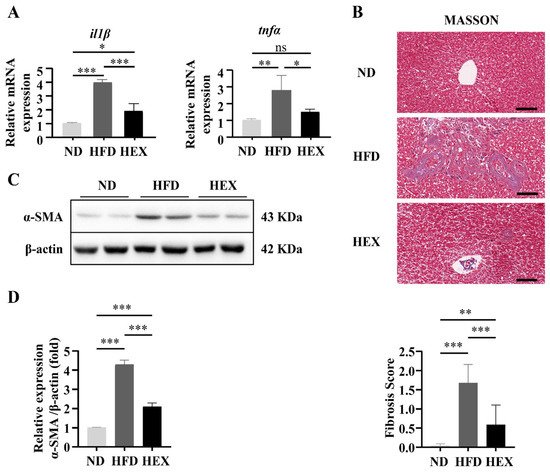
Moreover, proinflammatory cytokines (il1β and tnfα) were upregulated significantly in HFD group (Figure 4A). Swimming treatment attenuated these pathological changes in liver tissue, reducing the incidence of steatosis and cell ballooning and suppressing il1β and tnfα expression. The collagen content of the zebrafish livers was quantified, along with the protein levels of α-smooth muscle actin (α-SMA), to indicate the levels of fibrosis. These fibrotic markers were significantly increased in the livers of the HFD group. Swimming exercise prevented the HFD-induced fibrotic response in the HEX zebrafish livers, which had a lower collagen content and α-SMA protein expression compared to the HFD group (Figure 4B–D). These data suggest that HFD zebrafish could develop the pathology of NAFLD, and that swimming exercise could attenuate the pathogenic effects caused by the HFD.
Figure 4. Quantitation of pathological changes in 9-month-old zebrafish livers. (A) Gene expression of il1β and tnfα (n = 6). (B) Masson’s staining and fibrosis score of zebrafish livers (n = 3). (C,D) The protein levels of α-smooth muscle actin (α-SMA) (n = 8). *, p < 0.05, **, p < 0.01, ***, p < 0.001. Data represent the mean and error bars represent SEM. Scale bar, 20 μm. NAFLD, non-alcoholic fatty liver disease; ND, normal diet; HFD, high fat diet; HEX, high-fat diet plus exercise.
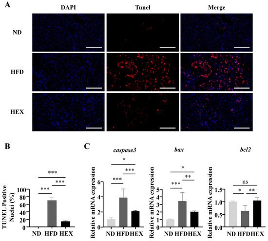
5. Swimming Exercise Attenuates Hepatocyte Apoptosis in HFD Zebrafish
TUNEL staining was used to detect hepatocyte apoptosis. TUNEL-positive hepatic cells increased significantly in the HFD group compared to in the other two groups, and swimming exercise decreased the number of apoptotic cells compared to the HFD group (Figure 5A,B). Moreover, zebrafish in the HFD group had upregulated genes related to apoptosis (caspase3, Bax) and a downregulated anti-apoptosis gene (bcl2), while exercise appeared to reduce the rate of apoptosis (Figure 5C). Taken together, these data suggest that swimming protected hepatocytes from apoptosis.
Figure 5. Hepatocyte apoptosis in 9-month-old zebrafish livers. (A) Tunel staining of zebrafish livers, and (B) Quantitation of Tunel-positive cells (n = 3). (C) Gene expression of caspase3, bax, and bcl2 (n = 6). *, p < 0.05 **, p < 0.01, ***, p < 0.001. Data represent the mean, and error bars represent SEM. Scale bar, 20 μm. NAFLD, non-alcoholic fatty liver disease; ND, normal diet; HFD, high fat diet; HEX, high-fat diet plus exercise.
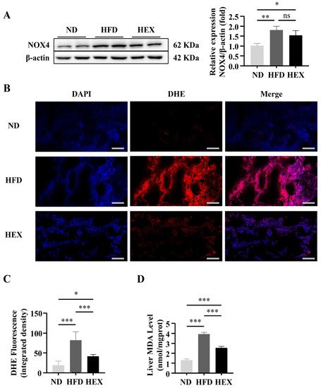
6. Swimming Exercise Protects HFD Zebrafish Livers from Oxidative Stress
Oxidative stress affects many physiological processes under pathological conditions, including cell survival [
35]. The progression of simple steatosis to NASH can be prevented by decreasing ROS production [
36]. As NOX4 is the main source of ROS, we examined the protein levels of NOX4 in the HFD and HEX groups. NOX4 was significantly upregulated in the HFD and HEX groups compared with the ND group, while the expression of NOX4 was slightly decreased in the HEX group compared to in the HFD group (
Figure 6A). We next investigated the antioxidative effects of swimming exercise in HFD zebrafish. Dihydroethidium (DHE) staining revealed that swimming exercise significantly decreased the high hepatic ROS levels associated with a HFD (
Figure 6B,C). In addition, the elevated malondialdehyde (MDA) level induced by the HFD were also decreased in fish in the HEX group (
Figure 6D). The above results indicate that exercise can alleviate oxidative stress in the livers of zebrafish.
Figure 6. Oxidative stress in 9-month-old zebrafish livers. (A) NOX4 protein expression (n = 8). (B) Dihydroethidium (DHE) staining of zebrafish livers (n = 3), and (C) quantitation of DHE staining. (D) MDA levels in zebrafish livers (n = 5). *, p < 0.05, **, p < 0.01, ***, p < 0.001. Data represent the mean, and error bars represent SEM. Scale bar, 20 μm. NAFLD, non-alcoholic fatty liver disease; ND, normal diet; HFD, high fat diet; HEX, high-fat diet plus exercise.
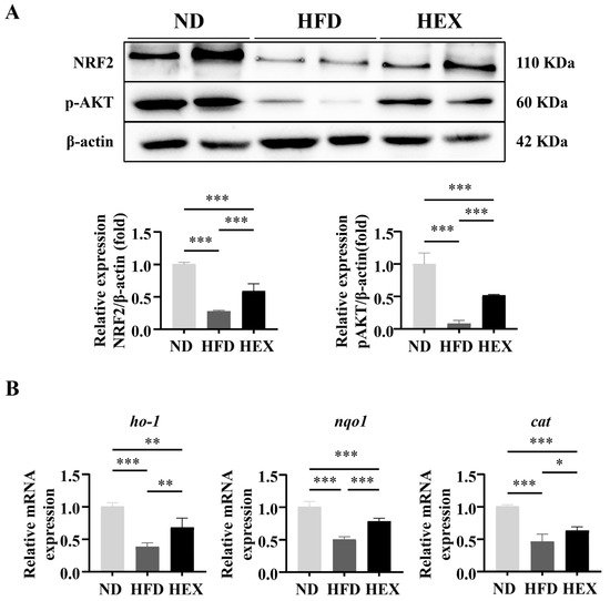
7. Swimming Exercise Upregulates the Expression and Function of NRF2 in HFD Zebrafish
Accumulating evidence indicates that enhanced APMK activity is related to increased NRF2 expression and its antioxidative function. AMPK phosphorylation can directly promote NRF2 nuclear accumulation [
37] or upregulate AKT phosphorylation and activate NRF2 [
38]. In the present study, significantly lower expression levels of p-AKT and NRF2 were detected in the livers of HFD zebrafish compared to in ND and HEX zebrafish livers. Swimming attenuated the degradation of p-AKT and NRF2 induced by HFD (
Figure 7A). Consistent with the observed downregulation of NRF2 expression, NRF2 downstream antioxidant genes
ho-1,
nqo1, and
cat expression were significantly decreased in HFD zebrafish livers compared to in ND and HEX zebrafish livers (
Figure 7B). These results suggest that exercise attenuates oxidative stress that is associated with the activation of NRF2 signaling in HFD zebrafish.
Figure 7. NRF2 signaling in 9-month-old zebrafish livers. (A) Western blot of p-AKT and NRF2 expression in zebrafish livers (n = 8). (B) Gene expression of ho-1, nqo1, and cat (n = 6). *, p < 0.05, **, p < 0.01, ***, p < 0.001. Data represent the mean, and error bars represent SEM. NAFLD, non-alcoholic fatty liver disease; ND, normal diet; HFD, high fat diet; HEX, high-fat diet plus exercise.
8.Conclusion
In summary, a HFD zebrafish model is applied of NAFLD to investigate the antioxidative effects of exercise on NAFLD. Zebrafish had similar histopathology compared to human and rodent models. Swimming exercise ameliorated the pathogenesis of NAFLD in the livers of model zebrafish. Exercise suppressed excessive ROS production in NAFLD model zebrafish via upregulation of NRF2, mediated by SIRT1/AMPK signaling. In view of the fact that exercise mimetics, such as AIACR, metformin, and resveratrol, also have the effect of activating SIRT1/AMPK signaling, it is worthy of further study to investigate and compare the alternative or combined effect of these drugs with exercise in the treatment of NAFLD.
This entry is adapted from the peer-reviewed paper 10.3390/ijms222010940