Lactococcosis, caused by
L. garvieae, is a systemic hyperacute bacterial disease causing general hemorrhagic symptoms in susceptible aquatic organisms [
24,
25]. Based on their ability to agglutinate serum raised against
L. garvieae, there are two groups of bacterial serotypes: non-agglutinating (KG−) and agglutinating (KG+) phenotypes [
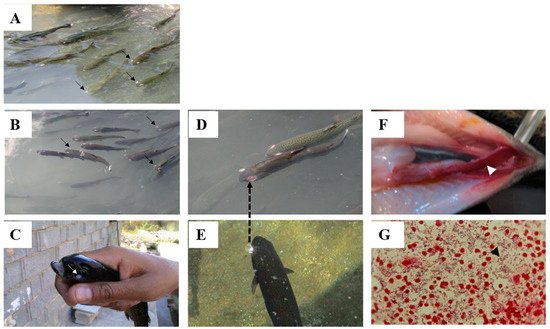
26]. The affected fish first become relatively anorexic, with a visible darkening of skin color, showing sluggish movement and abnormal behaviors, such as erratic and spiral swimming [
11,
24]. In the later stages of the disease the affected fish display signs of swollen abdomen, anal prolapsus, lateral or bilateral exophthalmia (
Figure 1A,B), cataracts (
Figure 1C), congestion of the internal organs, spleen and liver enlargement, accumulation of turbid ascitic fluid in the peritoneal cavity, and the presence of exudates in the brain [
11,
22,
27,
28]. Acute hyperemia and or extensive hemorrhage and petechiae of the organs, including the mucosal layers of the intestine, can also be seen in the diseased fish (
Figure 1F), and in some cases the diseased fish show signs of explosion in the eyes prior to the loss of their eyes (
Figure 1D,E) [
11,
22,
29]. In advanced forms of the disease, a Gram stain preparation of hematopoietic tissue imprints, including spleen and kidney, can exhibit huge numbers of Gram-positive coccoid cells in single or chain forms (
Figure 1G).
Figure 1. Rainbow trout growing in race ways and affected by L. garvieae showing: (A) typical bilateral exophthalmia and no change in the color skin, (B) typical bilateral exophthalmia and beginning of skin color change, (C) typical cataract and dark color, (D) bilateral exophthalmia and a complete loss of the eye, (E) darkening of body and an explosion of the eye, (F) hemorrhage of intestine, (G) direct Gram stain preparation of spleen of diseased fish showing huge numbers of Gram-positive coccoid cells confirmed as L. garvieae by phenotyping and molecular works. (photos by Professor Mehdi Soltani).
Pericarditis, peritonitis and meningitis, diffuse hemorrhage in the sclera of the eye, focal necrosis in the spleen and clumps of bacteria, hemorrhage in serosa of the swim bladder and in the interstitium of the skeletal muscles, degeneration and necrosis in epithelia of the stomach glands and their lumens full of necrotic material are among the identified histopathological findings in lactococcosis infection caused by
L. garvieae [
30,
31,
32]. Vascular changes in spleen and kidneys [
33] and degenerations in the tubular epithelium with an increase in the melano-macrophage centers, hemorrhage in the form of a hematoma covering the myocardium and the bulbus arteriosa, petechial hemorrhage, vascular change, degeneration and necrosis are major histopathological findings. Lipid and ell infiltration in the liver, hemorrhage and vascular change in muscles, and petechial hemorrhage and edema in the gills are further microscopic changes reported in the infected fish by
L. garvieae [
31]. The severity of such pathological changes is, however, varied and depended on various factors, including level of virulent of bacterial strain, fish species and size and level of health management criteria, such as water temperature.
Evidence of bacterial cells in fish macrophage in tissues of spleen, kidney, heart (endothelial), and peritoneum are evidence of a septicemic condition, suggesting that macrophages play a key role in the host immune response to L. garvieae infection. However, intra-macrophage resistance of the bacteria can cause a spread of the pathogen to all tissues of fish by macrophages. Further, as in the young fish phagocytosis by macrophage activation may not be sufficient, thus, pathogenesis is a determinant factor, and the disease can progress.
Several factors play roles in the virulence of
L. garvieae. Polysaccharide capsule is the major virulence factor in
L. garvieae infection [
29]. The capsulated strains resist to phagocytosis, but some non-capsulated strains are pathogenic in fish causing high mortality in rainbow trout [
34], thus, the bacterial capsule may not the sole determinant of the bacterial pathogenicity. Haemolytic toxin is known to cause mortality in fish via intramuscular injection and an intracellular toxin with a low leukocidal activity reported by the bacterial isolates recovered from the diseased fish [
35]. Plasmids of the virulent isolates contain a protein with an enzymatic domain corresponding to the family of actin-ADP-ribosyltransferases [
36] that can kill eukaryotic cells by transferring ADP-ribose to essential proteins [
37]. The toxicity of this protein in fish however, warranted future research works. The presence of a putative set of virulence factor genes (hly1, hly2, hly3, nox, sod, pavA, psaA), and proteins of enolase, lactate dehydrogenase phosphoenolpyruvate-protein phosphotransferase with roles in adhesion, cytolytic activity, oxidative stress tolerance, and metal homeostasis have been detected in strains of
L. garvieae, including the avirulent reference strains ATCC
® 49156 and ATCC
® 4392, isolates from diseased rainbow trout in Turkey, France, Iran, Spain, and Italy [
38], and fish pathogenic non-capsulated strains in South Africa [
6]. These virulence lifestyle factors can indirectly contribute to host tissue damage through aiding in the infection process by evasion of the host’s innate immunity, systemic invasion, cofactor homeostasis, and spreading in the host and adhesion to host tissues. Further research works need to be directed studying the differential expression of virulence lifestyle and true virulence genes during growth in the host environment. Additionally, more studies need to assess the specific virulence factors responsible for the pathogenicity of
L. garvieae, as putative virulence factor genes are present in both the fish pathogenic isolates and the avirulent isolates.
2.2. Diseases Caused by Other Species of Lactococcus Genus
L. lactis strains are genetically classified into four subspecies of lactis, cremoris, tractae, and hordniae [13]. It is not a common veterinary pathogen, although it can cause cattle mastitis and be involved in septic arthritis of the neonatal calf. For example, several variants of L. lactis have been associated with bovine mastitis [79]. In humans, it has been reported as a cause of endocarditis, arthritis, and septicemia in patients, although this requires more clarification [80,81,82,83,84].
Up to date, there are only four reports of lactococcosis by L. lactis in an aquatic organisms. The first report was an outbreak of white tail disease in cultured giant freshwater prawn (Macrobrachium rosenbergii) in Taiwan [85]. The affected prawns were cloudy and whitish in the muscles, showing remarkable edema and necrosis and inflammation in the muscles and hepatopancreas. In subsequent report by Chen et al. [23] L. lactis subsp. Lactis was isolated from affected hybrid sturgeon, Bester (Huso huso x Acipenser ruthenus) with signs of anorexia, pale body color, reddish spots on the abdomen, enteritis, enlarged abdomen, rapid respiration rate ascites, and 70%–100% mortality. Microscopically, the affected sturgeons demonstrated extensive haemorrhagic multifocal necrotic foci of spleen and liver with degeneration of hepatic cells, lipid droplets and glycogen granules, necrosis and renal tubule epithelial swelling and hydropic degeneration in kidney, skin ulcers deep in underling muscles, appearance of present of immunocompetent cells in the stomach, and small focus on tips of gills and on the myocardium [23]. No histopathological changes were, however seen in the eyeball, cerebrum and meninges of affected fish. The third report was from silver carp (Hypophthalmichthys molitrix) with extensive skin lesions near the caudal peduncle and musculoskeletal lesion in the USA [86]. The fourth outbreak of infection by L. lactis has been reported as the cause of endocarditis valvularis, parientalis thromboticans in mature allis shad (Alosa alosa) in Europe in 2018 that could be associated with the stressors, such as capturing, transport, breeding, and low oxygen level [87]. Although, in some cases the disease was reproduced experimentally, the mechanisms of pathogenesis by L. lactis in aquatic animals warranted future research works.
3. Phytotherapy of Lactococcosis in Aquaculture
3.1. In Vitro Studies
Almost all in vitro studies with vegetable and lichens extractives were performed against L. garvieae. For convenience, details of in vitro and in vivo studies have been included in Table 1 and Table 2. Overall, extracts do not show strong antibacterial activity against L. garvieae, but essential oils are more effective, mainly those that contain thymol or carvacrol. There are some differences on minimum inhibitory and bactericidal concentrations for the same extractive in different studies (Table 1).
3.2. In Vivo Studies
All in vivo studies were related to survival against L. garvieae infection, and, in most cases, the extractives of medicinal herbs and plants were added to the diets for various periods before the treated fish being challenged with L. garvieae infection. Overall, the essential oils that showed the best in vitro antibacterial activity against L. garvieae (Table 1) were not tested for the in vivo bioassays yet. The extractives tested under in vivo conditions presented moderate in vitro antibacterial activity against this bacterium or even were not tested in vitro. However, the dietary supplementation with all tested extractives reduced mortality of infected animals (Table 2), probably because they improved immune parameters before challenging the treated fish with L. garvieae. A 12-day feeding giant freshwater prawn (Macrobrachium rosenbergii) with hot-water extract of water hyacinth (Eichhornia crassipes) leaves at 1, 2, and 3 g kg−1 diet induced significantly higher survival rate after challenge with L. garvieae infection, but higher disease resistance was seen in the prawn treated with higher concentration of the extract [108]. In addition, the treated animals exhibited an enhancement in the immune responses including respiratory burst, phenoloxidase activity, superoxide dismutase activity, glutathione peroxidase, total hemocyte value, differential hemocyte count, transglutaminase activity, and phagocytic activity towards L. garvtieae. In the subsequent research work by Chang and Cheng [109], dietary addition of three tested water hyacinth extracts (Table 2) for 120 days increased survival and immune parameters, i.e., total hemocyte count, semi-granular and granular cells counts of giant freshwater prawn while phenoloxidase activity, respiratory bursts of hemocytes were not observed only with dietary addition of powder of this plant to the diet.
4. Conclusions
Disease outbreaks by Lactococcus species specially L. garvieae is one of the major concerns faced in the aquaculture production worldwide, and various biological and environmental variables, as well as the aquaculture practices and husbandry can affect the quantity and impacts of the morbidity and mortality. Data influencing the economic losses can, thus, assist to develop policies and strategies to reduce the losses by lactococcosis outbreaks in aquaculture industry. Lactococcosis outbreaks especially by L. garvieae are increasingly recognized as a significant and re-emerging bacterial disease in aquaculture, but there is no an estimation of its economic impacts. Data describing antagonistic activity and disease resistance efficacy of potential medicinal herbs and plants towards lactococcosis caused by L. garvieae, L. lactis, L. piscium and L. raffinolactis in finfish are not very much. Almost all in vitro studies with vegetable and lichens extractives were performed against L. garvieae. Despite no strong antibacterial activity by herb extracts against L. garvieae, essential oils especially those that contain thymol and carvacrol are more effective against L. garvieae strains. The exhibited differences on minimum inhibitory and bactericidal values for the same extractive in different studies could be due to the use of different bacterial strains or parts or chemotypes of the same plant. Despite best anti-L. garvieae activity by the essential oils under in vitro assays, the in vivo bioassays need be assessed yet. The extractives tested under in vivo conditions presented moderate antibacterial activity against this bacterium or even were not tested in vitro. However, the dietary supplementation with all tested extractives reduced mortality of infected animals, probably because they improved immune parameters before challenging the treated fish with L. garvieae.