Therapy of Cardiac-Atrial-Appendage-Stem-Cells and Pyridoxamine: History
Please note this is an old version of this entry, which may differ significantly from the current revision.
Contributor:

Myocardial infarction (MI) occurs when the coronary blood supply is interrupted. As a consequence, cardiomyocytes are irreversibly damaged and lost. Unfortunately, current therapies for MI are unable to prevent progression towards heart failure. As the renewal rate of cardiomyocytes is minimal, the optimal treatment should achieve effective cardiac regeneration, possibly with stem cells transplantation. In that context, our research group identified the cardiac atrial appendage stem cells (CASCs) as a new cellular therapy. However, CASCs are transplanted into a hostile environment, with elevated levels of advanced glycation end products (AGEs), which may affect their regenerative potential. In this study, we hypothesize that pyridoxamine (PM), a vitamin B6 derivative, could further enhance the regenerative capacities of CASCs transplanted after MI by reducing AGEs’ formation. Methods and Results: MI was induced in rats by ligation of the left anterior descending artery. Animals were assigned to either no therapy (MI), CASCs transplantation (MI + CASCs), or CASCs transplantation supplemented with PM treatment (MI + CASCs + PM). Four weeks post-surgery, global cardiac function and infarct size were improved upon CASCs transplantation. Interstitial collagen deposition, evaluated on cryosections, was decreased in the MI animals transplanted with CASCs. Contractile properties of resident left ventricular cardiomyocytes were assessed by unloaded cell shortening. CASCs transplantation prevented cardiomyocyte shortening deterioration. Even if PM significantly reduced cardiac levels of AGEs, cardiac outcome was not further improved. Conclusion: Limiting AGEs’ formation with PM during an ischemic injury in vivo did not further enhance the improved cardiac phenotype obtained with CASCs transplantation. Whether AGEs play an important deleterious role in the setting of stem cell therapy after MI warrants further examination. 

  • stem cells
  • CASCs
  • advanced glycation end products
  • glycated proteins
  • myocardial infarction
  • transplantation
  • remodeling
  • cardiomyocytes
  • aldehyde dehydrogenase

1. Introduction

Our research group discovered a new type of cardiac stem cells, cardiac atrial appendage stem cells (CASCs) [1]. Identification of these stem cells is based on high aldehyde dehydrogenase (ALDH) enzyme activity. In vitro experiments have shown that the differentiation capacity of CASCs towards cardiomyocytes is superior to other CSCs types [1]. In addition, we have shown that autologous CASCs transplantation after MI improves global LV function [2]. This better cardiac outcome was associated with cell engraftment and CASCs’ differentiation in a cardiomyogenic phenotype [2]. Altogether, these data suggest a true high potential for using CASCs to repair lost cardiac tissue.
CASCs are transplanted after MI in a hostile environment of inflammation, fibrosis, and increased levels of advanced glycation end products (AGEs) [3]. AGEs are proteins and lipids that become glycated and oxidized after persistent contact with reducing sugars or short-chain aldehydes and/or a high degree of oxidative stress [4]. Next, to be abundantly present in our Western diet, accumulation of AGEs in the body is a natural process. This occurs with aging when the turnover rate of proteins is reduced. There is growing evidence reporting that AGEs contribute to the development and progression of cardiovascular dysfunction [5]. Indeed, increased circulating AGEs have been described to arise at an early lifetime in patients with cardiovascular diseases [6][7]. In ischemic heart disease patients, high levels of AGEs can also result from increased oxidative stress [5][8]. In addition, it is reported that immune cells (like neutrophils and macrophages) are mobilized to the ischemic area as a result of inflammation and cell death. These cells were shown to be major contributors to AGEs’ production [9][10]. These contribute to increased AGEs levels in patients suffering from MI. Recently, systematic review analysis [11] revealed that AGEs affect the viability and proliferation capacity of multiple types of stem cells in vitro, including CASCs [12], thereby affecting their therapeutic potential. These effects occur throughout several underlying mechanisms including excessive reactive oxygen species (ROS) generation, activation of the receptor for AGEs (RAGE), or via apoptotic pathways. As AGEs are increased in MI, we tested whether the regenerative capacities of CASCs could be further enhanced when combined with pyridoxamine (PM). PM is a compound able to reduce AGEs’ formation, a co-enzyme associated with multiple oxidative stress and inflammatory pathways and a strong iron chelator [13][14][15]. Using PM could thus potentially improve the efficiency of CASCs transplantation with no need to genetically modify them, in order to observe their full potential [16].

2. Analysis on Results

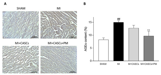
2.1. AGEs’ Levels Are Reduced with PM Treatment

Total AGEs’ levels were measured in heart tissues from SHAM, MI, MI + CASCs, and MI + CASCs + PM, and representative images are provided in Figure 1A. AGEs’ content was significantly increased in MI animals compared with SHAM (Figure 1B; 15% ± 0.6 in MI vs. 8.2% ± 0.7 in SHAM) and PM significantly decreased the AGEs’ content compared with MI (Figure 1B; 9.7% ± 1.8 in MI + CASCs + PM).
Figure 1. AGEs’ content in heart tissue is significantly decreased by PM. (A) Representative examples of transverse heart sections 4 weeks after surgery. The AGEs’ content (brown) was immunohistologically determined with DAB staining. Scale bar = 100 µm. (B) Quantification of AGEs’ content in hearts from SHAM (n = 4), MI (n = 11), MI + CASCs (n = 10), and MI + CASCs + PM (n = 5). Data are expressed as mean ± SEM. ** denotes p < 0.01 vs. MI and ## denotes p < 0.01 vs. SHAM.

2.2. CASCs Transplantation Prevents Loss of LV Function after MI

In vivo cardiac function was assessed by echocardiographic and hemodynamic measurements. Representative examples of echocardiographic images of SHAM, MI, MI + CASCs, and MI + CASCs + PM 4 weeks post-operative are shown in Figure S1. Echocardiographic parameters of the different groups, SHAM, MI, MI + CASCs, and MI + CASCs + PM, 4 weeks post-operative are summarized in Table 1. Additional echocardiographic parameters are summarized in Table S1. MI animals undergoing CASCs transplantation with or without additional PM treatment displayed a significantly increased ejection fraction (EF) compared with MI (Table 1; 59% ± 4 in MI vs. 79% ± 3 in MI + CASCs; vs. 72% ± 3 in MI + CASCs + PM, p = 0.051).
Table 1. Echocardiographic characteristics.
Parameters 4 Weeks Post-Operative
SHAM MI MI + CASCs MI + CASCs + PM
EF (%) 80 ± 4 59 ± 4 ## 79 ± 3 *** 72 ± 3
HR (bpm) 333 ± 14 318 ± 12 332 ± 12 327 ± 13
SV (µL) 170 ± 16 162 ± 17 172 ± 17 192 ± 17
CO (mL/min) 57 ± 4 51 ± 5 59 ± 6 65 ± 6
EDV (µL) 215 ± 26 298 ± 51 217 ± 17 267 ± 22
ESV (µL) 45 ± 13 136 ± 36 44 ± 5 * 77 ± 11
AWT (mm) 1.74 ± 0.13 1.55 ± 0.16 1.65 ± 0.12 1.80 ± 0.16
PWT (mm) 1.54 ± 0.16 1.63 ± 0.15 1.57 ± 0.09 1.62 ± 0.17
Echocardiographic characteristics 4 weeks post-surgery in SHAM (n = 5), MI (n = 11), MI + CASCs (n = 10), and MI + CASCs + PM (n = 9) animals. Data are expressed as mean ± SEM. * denotes p < 0.05, *** denotes p < 0.001 vs. MI, and ## denotes p < 0.01 vs. SHAM. EF: ejection fraction, HR: heart rate, SV: stroke volume, CO: cardiac output, EDV: end-diastolic volume, ESV: end-systolic volume, AWT: anterior wall thickness, PWT: posterior wall thickness.
 
Hemodynamic measurements of SHAM, MI, MI + CASCs, and MI + CASCs + PM were performed 4 weeks after surgery. Compared with MI, additional PM treatment significantly reduced the time constant for isovolumetric relaxation (Table 2; Tau; 0.0499 s ± 0.017 in MI vs. 0.0117 s ± 0.001 in MI + CASCs + PM).
Table 2. Hemodynamic characteristics.
Parameters 4 Weeks Post-Operative
SHAM MI MI + CASCs MI + CASCs + PM
Max LV pressure (mmHg) 99 ± 3 90 ± 2 96 ± 3 103 ± 4 *
dP/dtmax (mmHg/s) 6773 ± 529 6038 ± 242 6923 ± 340 6551 ± 257
dP/dtmin (mmHg/s) −7269 ± 683 −6550 ± 705 −6816 ± 354 −6917 ± 273
Tau (s) 0.0130 ± 0.001 0.0499 ± 0.017 0.0148 ± 0.002 0.0117 ± 0.001 **
Hemodynamic characteristics 4 weeks post-op of SHAM (n = 5), MI (n = 9), MI + CASCs (n = 10), and MI + CASCs + PM (n = 9) animals. Data are expressed as mean ± SEM. * denotes p < 0.05 and ** denotes p < 0.01 vs. MI. LV: left ventricular, dP/dtmax: peak rate of pressure rise, dP/dtmin: peak rate of pressure decline, Tau: time constant for isovolumetric relaxation.


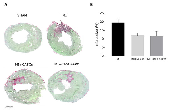
2.3. CASCs Transplantation Tended to Reduce Infarct Size

Figure 2A demonstrates representative examples of Sirius Red/Fast Green stained cryosections from SHAM, MI, MI + CASCs, and MI + CASCs + PM 4 weeks after surgery. Infarct size tended to decrease in animals undergoing CASCs transplantation (Figure 2B; 19% ± 2 in MI vs. 12% ± 2 in MI + CASCs, p = 0.07). Additional PM treatment did not further reduce infarct size (Figure 2B; 12% ± 3 in MI + CASCs + PM, p = 0.16).
Figure 2. Assessment of infarct size. (A) Representative examples of hearts from SHAM, MI, MI + CASCs, and MI + CASCs + PM. Fibrotic tissue, as a surrogate for infarct size, is stained red, while viable tissue is stained green. Scale bar = 2000 µm. (B) Quantification of infarct size in transversal sections 4 weeks post-surgery. MI (n = 11), MI + CASCs (n = 10), and MI + CASCs + PM (n = 5). Data are expressed as mean ± SEM.

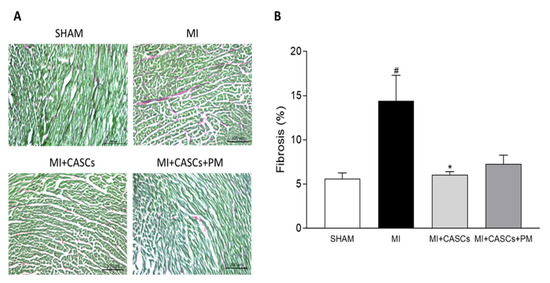
2.4. CASCs Transplantation Prevents the Increased Interstitial Collagen Deposition Seen with MI

Representative images of interstitial collagen obtained with Sirius Red/Fast Green staining in LV sections from the groups are provided in Figure 3A. Interstitial collagen deposition was significantly lower in CASCs transplanted animals compared with MI (Figure 3B; 14% ± 3 in MI vs. 6% ± 0.4 in MI + CASCs). Fibrosis tended to be lower in PM animals (Figure 3B; 7% ± 1 in MI + CASCs + PM).
Figure 3. Interstitial collagen deposition in the LV. (A) Representative examples of collagen deposition (red) in the LV. Scale bar = 200 µm. (B) Quantification of collagen content in LV transversal sections 4 weeks after surgery of SHAM (n = 4), MI (n = 11), MI + CASCs (n = 10), and MI + CASCs + PM (n = 5). Data are expressed as mean ± SEM. * denotes p < 0.05 vs. MI, # denotes p < 0.05 vs. SHAM.

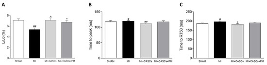
2.5. CASCs Transplantation Prevents Resident Cardiomyocyte Functional Remodeling

Unloaded cell shortening was measured in freshly isolated cardiomyocytes isolated from SHAM, MI, MI + CASCs, and MI + CASCs + PM animals 4 weeks post-surgery. As shown in Figure 4A, cells isolated from the border zone of infarcts displayed altered functional properties, namely reduced and slower unloaded cell shortening. CASCs transplantation, with or without PM treatment, could prevent the deterioration of the cardiomyocyte contractile properties (Figure 4A; L/L0, 5% ± 0.3 in MI vs. 7% ± 0.5 in MI + CASCs; vs. 7% ± 0.4 in MI + CASCs + PM). The kinetics of cell contraction and cell relaxation, i.e., TTP and RT50, were significantly better in MI + CASCs and tended to be improved in MI + CASCs + PM animals (Figure 4B TTP; 120 ms ± 1 in MI vs. 112 ms ± 2 in MI + CASCs; vs. 118 ms ± 3 in MI + CASCs + PM; Figure 4C RT50; 197 ms ± 3 in MI vs. 184 ms ± 4 in MI + CASCs; vs. 190 ms ± 4 in MI + CASCs + PM).
Figure 4. Resident cardiomyocyte shortening during field stimulation at 4 Hz. Quantification of (A) unloaded cell shortening normalized to diastolic cell length (L/L0), (B) time to peak of contraction (TTP), and (C) time to half-maximal relaxation (RT50) of resident cardiomyocytes from SHAM (ncells = 80; nanimals = 7), MI (ncells = 60; nanimals = 5), MI + CASCs (ncells = 41; nanimals = 4), and MI + CASCs + PM (ncells = 31; nanimals = 3). Data are expressed as mean ± SEM. * denotes p < 0.05; ** denotes p < 0.01 vs. MI, # denotes p < 0.05 vs. SHAM, and ## denotes p < 0.01 vs. SHAM.

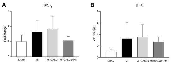
2.6. PM Treatment Tended to Reduced Tissue Pro-Inflammatory Cytokine Levels

Gene expressions of pro-inflammatory cytokines (IFN-γ and IL-6) were evaluated in the four groups of animals, 4 weeks post-surgery. As shown in Figure 5, the expression of inflammatory cytokines tended to be lower in MI + CASCs + PM animals compared with MI and MI + CASCs animals (Figure 5A IFN-γ; 1.60 ± 0.27 in MI vs. 1.83 ± 0.30 in MI + CASCs; vs. 1.06 ± 0.14 in MI + CASCs + PM; Figure 5B IL-6; 3.28 ± 1.06 in MI vs. 3.56 ± 0.81 in MI + CASCs; vs. 2.77 ± 0.48 in MI + CASCs + PM).
Figure 5. Gene expression of inflammatory cytokines. Quantification of gene expression of (A) IFN-γ and (B) IL-6 in LV tissue from SHAM (n = 4), MI (n = 7), MI + CASCs (n = 7), and MI + CASCs + PM (n = 3). Data are expressed as mean ± SEM.

3. Current Insights

In our study, we have shown that, after MI, CASCs transplantation is able to improve the cardiac phenotype by limiting cellular remodeling. However, preventing AGEs’ formation did not further enhance the positive outcome provided by CASCs alone.

3.1. Combining CASCs and PM to Enhance Cardiac Repair

Oxidative stress is one of the main factors inducing AGEs’ synthesis, by formation of reactive carbonyl compounds and glycoxidation of Amadori products in the Maillard reaction. In patients with MI, AGEs’ levels are significantly increased [8][17] and have potential deleterious effects on cardiac function [18][19]. Moreover, in our study, AGEs are significantly increased in MI animals. In addition to increased oxidative stress, the inflammatory process induced after MI activates neutrophils and macrophages. These immune cells are known to further secrete AGEs and are reported to be key inducers of AGEs’ formation in MI [9][10].
Even if they provide new insights into tissue regeneration, stem cells are transplanted in the border zone of MI with increased oxidative stress, inflammation, and AGEs’ levels. Previous studies have shown that increased levels of AGEs affect stem cells’ properties, i.e., by reducing their proliferation and migration properties [11]. Recently, we have demonstrated that the same adverse effects of AGEs apply to CASCs’ properties in vitro [12]. In that context, reducing AGEs’ formation could potentially enhance CASCs’ regenerative properties upon in vivo transplantation. To find out whether such an approach would offer new therapeutic insights, was the goal of our study. In the context of improving stem cell therapy by targeting AGEs, we evaluated the effect of CASCs transplantation in combination with PM in a rat model of MI. This vitamin B derivate is an effective and safe AGEs-lowering therapy [20], which has different mechanisms of action [21][22]. First, PM can bind with catalytic redox metal ions, which are needed for glycoxidation of Amadori products. As such and related to its iron-chelator properties, Amadori-to-AGEs conversion is blocked. Secondly, PM can scavenge reactive carbonyl compounds, the latter being major AGEs’ precursors. In addition, studies have demonstrated that PM is a co-enzyme associated with multiple inflammatory pathways, thereby potentially inhibiting inflammation [15]. Finally, by inhibiting ROS formation or scavenging oxygen radicals, PM has been shown to be a potent antioxidant. Previous studies have shown that, even independent of stem cell transplantation, PM alone improves survival and reduces extracellular remodeling after MI, by reducing AGEs’ levels [23]. Moreover, in clinical trials, PM has been demonstrated as a safe and effective drug in diabetic patients [21]. However, owing to financial issues, a clinical trial of NephroGenex in 2014, testing PM as an anti-diabetic treatment, was stopped [24]. No other clinical trials are currently investigating PM as a therapy.
In our study, PM succeeded in reducing total AGEs’ tissue levels. However, PM did not further improve cardiac function obtained with CASCs transplantation alone, which was, at first sight, not expected in our study. This could be partially explained by the recent discovery of Vagnozzi et al. [25]. In their article, the authors show that cellular therapy itself induces an inflammatory response after MI that could be the primary beneficial effect underlying stem cell treatment. Pro-inflammatory macrophages, mobilized and activated by transplanted stem cells, could indeed rejuvenate the mechanical properties of the injured cardiac area. By affecting fibroblast activity, the ECM content and area occupied by scar tissue could be reduced. The precise underlying mechanisms responsible for the repair response of these immune cells are unclear and require further investigation. In our study, it is thus likely that, by reducing local inflammation, PM could not further enhance the repair process, when combined with CASCs transplantation. Inflammatory cytokines such as IFN-γ and IL-6 tended to be reduced by PM treatment in our study. As an immune reaction is thus needed as a base for would healing with stem cells, PM could counteract the positive effects of CASCs therapy by reducing local tissue inflammation. This could explain the lack of additive value of PM in the context of MI and stem cell transplantation.
Even if our data do not demonstrate an additional effect of PM to CASCs transplantation, the potential of other anti-AGEs therapies still needs to be investigated. It has been shown in Alzheimer’s, Parkinson’s, and rheumatoid arthritis disease animal models that stem cell survival was prolonged, migration capacity was enhanced, and the MSCs were better protected against apoptosis, when sRAGE-secreting MSCs were transplanted. By scavenging AGEs with sRAGE, the effectiveness of MSCs transplantation was improved, thus suggesting a role of AGEs in regenerative approaches with stem cells [26][27][28]. However, using genetically modified stem cells is still highly experimental and needs to be investigated in vivo before any possible translation into the clinical setting is possible. Therefore, one cannot exclude that other anti-AGEs therapy approaches such as RAGE inhibitors, sRAGE, or ALT-711 could potentially succeed in further lowering AGEs’ concentrations in MI and potentially have an additive effect on cardiac outcome. These therapeutic options need to be investigated in both pre-clinical and clinical studies in combination with CASCs therapy.

3.2. CASCs Alone Are an Effective Therapy for MI

Independent of PM treatment, our data confirm that transplantation of CASCs can prevent worsening of cardiac function after an ischemic injury, as shown by Fanton et al. in the minipig model [2]. Indeed, we have shown that EF significantly increased up to 20% after CASCs transplantation compared with non-treated animals. Meta-analysis of other CSCs therapies for the treatment of MI in mice showed an overall increase in EF of 9.9% [29]. Therefore, CASCs do have more effective regenerative effects compared with other CSCs and are remarkable candidates for cellular therapy. Other parameters of global cardiac function, such as dP/dtmax, SV, CO, and ESV, even if not significantly affected, followed the same trend, indicating an overall improvement of systolic function upon CASCs transplantation. In addition, infarct size tended to decrease after CASCs transplantation. Furthermore, as shown by the prevention of adverse remodeling at the cardiomyocyte level, our data suggest that mechanical load subjected to the resident myocytes of the ischemic area was reduced with CASCs transplantation. The prevention of collagen deposition seen in our study is also in line with a potentially reduced mechanical load with CASCs transplantation. Indeed, an important pathway in post-MI remodeling and scar formation is the TGF-β1 signaling pathway. In this study, we did not evaluate the underlying mechanisms resulting in reduced fibrosis with CASCs. However, TGF-β1 could be an essential contributor. Indeed, increased TGF-β1 is detected after MI and is known to decrease the expression and function of enzymes responsible for matrix degradation and increase the inhibitors of proteases [30][31]. Whether CASCs transplantation results in a reduced lysyl oxidase expression and/or PI3K/Akt, Smad3, and MAPK signaling pathway as a consequence of increased TGF-β1 activation [32], remains to be confirmed. However, other studies have shown that MSCs transplantation is able to ameliorate cardiac fibrosis by decreasing TGF-β1 levels [33]. Therefore, it seems likely that this TGF-β1 pathway is also involved in our study as it is a common pathway found in many diseases, but this has to be confirmed. In addition, we have demonstrated that CASCs transplantation can prevent adverse cellular remodeling of resident cardiomyocytes, isolated from the border zone of the infarct. Indeed, we show that, compared with MI animals, the amplitude and kinetics of cardiomyocyte shortening isolated from the border zone of MI in transplanted animals are improved. In that context, it has been described that the extent of mechanical load determines the extent of remodeling in both peri-infarct and remote regions [34]. It is then very likely that even a small decrease in infarct size, which we observe upon CASCs transplantation, will reduce adverse cellular remodeling in the resident cardiomyocytes.
However, whether the beneficial cardiac outcome is solely attributed to new cardiomyocytes differentiated from CASCs or to the paracrine factors secreted by these stem cells remains to be investigated. This then raises the question of whether CASCs were still present 4 weeks after transplantation. Indeed, pre-clinical studies have shown that most of the stem cells injected at the site of injury are cleared out within seconds, resulting in only 1–3% of the injected cells persisting at the site of injury [35]. Yet, previously, the presence of differentiated CASCs 8 weeks post-MI was demonstrated by immunostainings [2]. In addition, engraftment of the CASCs after 8 weeks of acute MI has been shown to be 19%, a value higher than that previously described with other stem cells [2]. It is thus very likely that CASCs are still present in the cardiac tissue 4 weeks post-transplantation. In addition, previous studies have shown that stem cells are able to secrete paracrine factors to promote survival and proliferation or have immunomodulatory effects on resident cardiomyocytes [36]. The strong paracrine effects of stem cells have been well documented and are, for a part, related to the limited improvement of cardiac function seen in some studies, as those may compensate for the lack of cardiomyocyte differentiation [37][38]. Whether the differentiation of CASCs to new cardiomyocytes and/or the paracrine factors secreted by the CASCs are the reason for the improvements seen after MI, remains to be clarified.

This entry is adapted from the peer-reviewed paper 10.3390/ijms22179266

References

  1. Koninckx, R.; Daniels, A.; Windmolders, S.; Mees, U.; Macianskiene, R.; Mubagwa, K.; Steels, P.; Jamaer, L.; Dubois, J.; Robic, B.; et al. The cardiac atrial appendage stem cell: A new and promising candidate for myocardial repair. Cardiovasc. Res. 2013, 97, 413–423.
  2. Fanton, Y.; Robic, B.; Rummens, J.L.; Daniels, A.; Windmolders, S.; Willems, L.; Jamaer, L.; Dubois, J.; Bijnens, E.; Heuts, N.; et al. Cardiac atrial appendage stem cells engraft and differentiate into cardiomyocytes in vivo: A new tool for cardiac repair after MI. Int. J. Cardiol. 2015, 201, 10–19.
  3. Kralev, S.; Zimmerer, E.; Brueckmann, M.; Lang, S.; Kalsch, T.; Rippert, A.; Lin, J.; Borggrefe, M.; Hammes, H.P.; Suselbeck, T. Elevation of the glycoxidation product N(epsilon)-(carboxymethyl)lysine in patients presenting with acute myocardial infarction. Clin. Chem. Lab. Med. 2009, 47, 446–451.
  4. Singh, R.; Barden, A.; Mori, T.; Beilin, L. Advanced glycation end-products: A review. Diabetologia 2001, 44, 129–146.
  5. Hartog, J.W.; Voors, A.A.; Bakker, S.J.; Smit, A.J.; van Veldhuisen, D.J. Advanced glycation end-products (AGEs) and heart failure: Pathophysiology and clinical implications. Eur. J. Heart Fail. 2007, 9, 1146–1155.
  6. Ramasamy, R.; Yan, S.F.; Schmidt, A.M. Stopping the primal RAGE reaction in myocardial infarction: Capturing adaptive responses to heal the heart? Circulation 2008, 117, 3165–3167.
  7. Liu, Y.; Qu, Y.; Wang, R.; Ma, Y.; Xia, C.; Gao, C.; Liu, J.; Lian, K.; Xu, A.; Lu, X.; et al. The alternative crosstalk between RAGE and nitrative thioredoxin inactivation during diabetic myocardial ischemia-reperfusion injury. Am. J. Physiol. Endocrinol. Metab. 2012, 303, E841–E852.
  8. Fishman, S.L.; Sonmez, H.; Basman, C.; Singh, V.; Poretsky, L. The role of advanced glycation end-products in the development of coronary artery disease in patients with and without diabetes mellitus: A review. Mol. Med. 2018, 24, 59.
  9. Yang, J.; Zhang, F.; Shi, H.; Gao, Y.; Dong, Z.; Ma, L.; Sun, X.; Li, X.; Chang, S.; Wang, Z.; et al. Neutrophil-derived advanced glycation end products-Nepsilon-(carboxymethyl) lysine promotes RIP3-mediated myocardial necroptosis via RAGE and exacerbates myocardial ischemia/reperfusion injury. FASEB J. 2019, 33, 14410–14422.
  10. Son, M.; Kang, W.C.; Oh, S.; Bayarsaikhan, D.; Ahn, H.; Lee, J.; Park, H.; Lee, S.; Choi, J.; Lee, H.S.; et al. Advanced glycation end-product (AGE)-albumin from activated macrophage is critical in human mesenchymal stem cells survival and post-ischemic reperfusion injury. Sci. Rep. 2017, 7, 11593.
  11. Evens, L.; Belien, H.; Deluyker, D.; Bronckaers, A.; Gervois, P.; Hendrikx, M.; Bito, V. The Impact of Advanced Glycation End-Products (AGEs) on Proliferation and Apoptosis of Primary Stem Cells: A Systematic Review. Stem Cells Int. 2020, 2020, 8886612.
  12. Evens, L.; Heeren, E.; Rummens, J.L.; Bronckaers, A.; Hendrikx, M.; Deluyker, D.; Bito, V. Advanced Glycation End Products Impair Cardiac Atrial Appendage Stem Cells Properties. J. Clin. Med. 2021, 10, 2964.
  13. Ramis, R.; Ortega-Castro, J.; Caballero, C.; Casasnovas, R.; Cerrillo, A.; Vilanova, B.; Adrover, M.; Frau, J. How Does Pyridoxamine Inhibit the Formation of Advanced Glycation End Products? The Role of Its Primary Antioxidant Activity. Antioxidants 2019, 8, 344.
  14. Voziyan, P.A.; Hudson, B.G. Pyridoxamine as a multifunctional pharmaceutical: Targeting pathogenic glycation and oxidative damage. Cell. Mol. Life Sci. 2005, 62, 1671–1681.
  15. Paul, L.; Ueland, P.M.; Selhub, J. Mechanistic perspective on the relationship between pyridoxal 5’-phosphate and inflammation. Nutr. Rev. 2013, 71, 239–244.
  16. Lemcke, H.; Voronina, N.; Steinhoff, G.; David, R. Recent Progress in Stem Cell Modification for Cardiac Regeneration. Stem Cells Int. 2018, 2018, 1909346.
  17. Qiu, H.; Li, W.P.; Shen, X.H.; Guo, X.Y.; Hua, B.; Li, H.W. Dynamic fluctuations of advanced glycation end products and its C-terminal truncated receptor level in patients with acute ST-segment elevation myocardial infarction and undergoing diabetes or not: A retrospective study. Medicine 2018, 97, e11278.
  18. Greven, W.L.; Smit, J.M.; Rommes, J.H.; Spronk, P.E. Accumulation of advanced glycation end (AGEs) products in intensive care patients: An observational, prospective study. BMC Clin. Pathol. 2010, 10, 4.
  19. Celec, P.; Hodosy, J.; Jani, P.; Janega, P.; Kudela, M.; Kalousova, M.; Holzerova, J.; Parrak, V.; Halcak, L.; Zima, T.; et al. Advanced glycation end products in myocardial reperfusion injury. Heart Vessel. 2012, 27, 208–215.
  20. Dwyer, J.P.; Greco, B.A.; Umanath, K.; Packham, D.; Fox, J.W.; Peterson, R.; Broome, B.R.; Greene, L.E.; Sika, M.; Lewis, J.B. Pyridoxamine dihydrochloride in diabetic nephropathy (PIONEER-CSG-17): Lessons learned from a pilot study. Nephron 2015, 129, 22–28.
  21. Voziyan, P.A.; Hudson, B.G. Pyridoxamine: The many virtues of a maillard reaction inhibitor. Ann. N. Y. Acad. Sci. 2005, 1043, 807–816.
  22. Williams, M.E. Clinical studies of advanced glycation end product inhibitors and diabetic kidney disease. Curr. Diabetes Rep. 2004, 4, 441–446.
  23. Deluyker, D.; Ferferieva, V.; Driesen, R.B.; Verboven, M.; Lambrichts, I.; Bito, V. Pyridoxamine improves survival and limits cardiac dysfunction after MI. Sci. Rep. 2017, 7, 16010.
  24. Borg, D.J.; Forbes, J.M. Targeting advanced glycation with pharmaceutical agents: Where are we now? Glycoconj. J. 2016, 33, 653–670.
  25. Vagnozzi, R.J.; Maillet, M.; Sargent, M.A.; Khalil, H.; Johansen, A.K.Z.; Schwanekamp, J.A.; York, A.J.; Huang, V.; Nahrendorf, M.; Sadayappan, S.; et al. An acute immune response underlies the benefit of cardiac stem cell therapy. Nature 2020, 577, 405–409.
  26. Oh, S.; Son, M.; Choi, J.; Lee, S.; Byun, K. sRAGE prolonged stem cell survival and suppressed RAGE-related inflammatory cell and T lymphocyte accumulations in an Alzheimer’s disease model. Biochem. Biophys. Res. Commun. 2018, 495, 807–813.
  27. Park, M.J.; Lee, S.H.; Moon, S.J.; Lee, J.A.; Lee, E.J.; Kim, E.K.; Park, J.S.; Lee, J.; Min, J.K.; Kim, S.J.; et al. Overexpression of soluble RAGE in mesenchymal stem cells enhances their immunoregulatory potential for cellular therapy in autoimmune arthritis. Sci. Rep. 2016, 6, 35933.
  28. Lee, J.; Bayarsaikhan, D.; Arivazhagan, R.; Park, H.; Lim, B.; Gwak, P.; Jeong, G.B.; Lee, J.; Byun, K.; Lee, B. CRISPR/Cas9 Edited sRAGE-MSCs Protect Neuronal Death in Parkinsons Disease Model. Int. J. Stem Cells 2019, 12, 114–124.
  29. Lang, C.I.; Wolfien, M.; Langenbach, A.; Muller, P.; Wolkenhauer, O.; Yavari, A.; Ince, H.; Steinhoff, G.; Krause, B.J.; David, R.; et al. Cardiac Cell Therapies for the Treatment of Acute Myocardial Infarction: A Meta-Analysis from Mouse Studies. Cell. Physiol. Biochem. 2017, 42, 254–268.
  30. Biernacka, A.; Dobaczewski, M.; Frangogiannis, N.G. TGF-beta signaling in fibrosis. Growth Factors 2011, 29, 196–202.
  31. Frangogiannis, N.G. The role of transforming growth factor (TGF)-beta in the infarcted myocardium. J. Thorac. Dis. 2017, 9, S52–S63.
  32. Voloshenyuk, T.G.; Landesman, E.S.; Khoutorova, E.; Hart, A.D.; Gardner, J.D. Induction of cardiac fibroblast lysyl oxidase by TGF-beta1 requires PI3K/Akt, Smad3, and MAPK signaling. Cytokine 2011, 55, 90–97.
  33. Jin, L.; Zhang, J.; Deng, Z.; Liu, J.; Han, W.; Chen, G.; Si, Y.; Ye, P. Mesenchymal stem cells ameliorate myocardial fibrosis in diabetic cardiomyopathy via the secretion of prostaglandin E2. Stem Cell Res. Ther. 2020, 11, 122.
  34. Richardson, W.J.; Clarke, S.A.; Quinn, T.A.; Holmes, J.W. Physiological Implications of Myocardial Scar Structure. Compr. Physiol. 2015, 5, 1877–1909.
  35. Kanda, P.; Davis, D.R. Cellular mechanisms underlying cardiac engraftment of stem cells. Expert Opin. Biol. Ther. 2017, 17, 1127–1143.
  36. Baraniak, P.R.; McDevitt, T.C. Stem cell paracrine actions and tissue regeneration. Regen. Med. 2010, 5, 121–143.
  37. Wu, S.Z.; Li, Y.L.; Huang, W.; Cai, W.F.; Liang, J.; Paul, C.; Jiang, L.; Wu, Z.C.; Xu, M.; Zhu, P.; et al. Paracrine effect of CXCR4-overexpressing mesenchymal stem cells on ischemic heart injury. Cell Biochem. Funct. 2017, 35, 113–123.
  38. Dulak, J.; Szade, K.; Szade, A.; Nowak, W.; Jozkowicz, A. Adult stem cells: Hopes and hypes of regenerative medicine. Acta Biochim. Pol. 2015, 62, 329–337.
More
This entry is offline, you can click here to edit this entry!