Metabolomics in Asthma: History
Please note this is an old version of this entry, which may differ significantly from the current revision.
Subjects: Biology
Contributor:

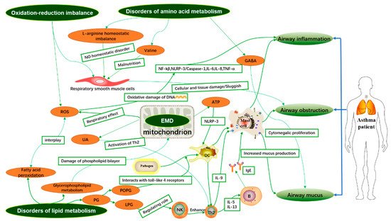
Asthma is a highly heterogeneous disease, but the pathogenesis of asthma is still unclear. It is well known that the airway inflammatory immune response is the pathological basis of asthma. Metabolomics is a systems biology method to analyze the difference of low molecular weight metabolites (<1.5 kDa) and explore the relationship between metabolic small molecules and pathophysiological changes of the organisms. 

  • metabolomics
  • asthma
  • inflammation
  • pathogenesis

1. Introduction

Asthma is a highly heterogeneous disease characterized by an inflammatory response in the airways. With nearly 300 million people in the world suffering from asthma, precise and personalized treatment of asthma patients is extremely important [1]. Some of the current traditional clinical tests for asthma, such as quantitative sputum cytometry, blood eosinophil count, a fraction of exhaled nitric oxide (FeNO), and serum IgE, are almost impossible to determine in early asthma [2]. Asthma is a complex disease composed of different endotypes with different inflammatory and clinicopathological characteristics, so the clinical phenotype of asthma is extremely complex. It is difficult to make accurate judgments in clinical diagnosis and identification, so it is almost impossible to provide precise and personalized treatment for asthma patients [1,3]. Metabolomics, as an emerging method of research, can better reflect the phenotypes of complex diseases and their pathophysiological changes, and to some extent, even elucidate the pathogenesis of diseases from a metabolic perspective. In a highly heterogeneous and severely phenotypically complex disease such as asthma, metabolomic study tools from studies of large clinical cohorts may become a better research method to obtain more comprehensive information than simple cytometric metrics [4,5]. Comprehensive understanding of asthma-related metabolomic data can provide powerful clues or evidence for precise and personalized diagnosis, treatment, and prognosis of asthma patients, which can further help clinical implementation of personalized patient treatment plans [4]. In addition, differential metabolites or called biomarkers screened by a large amount of metabolomics data are likely to be one of the most important pieces of evidence to explain the mechanism of asthma pathogenesis.

2. Integrative Analysis of Asthma-Related Metabolites and Metabolic Pathways in Different Samples

High-throughput sequencing technology has been widely applied to the phenotypic identification, diagnosis, and intervention of highly heterogeneous and complex diseases such as asthma. The metabolomic analysis of various samples. i.e., blood serum plasma samples, urine samples, local tissue samples, exhaled breath condensate (EBC) samples, bronchoalveolar lavage fluid (BALF) samples, induced sputum, and stool samples showed that the different metabolites associated with asthma in different samples and the integration of disordered metabolic pathways may provide evidence for the pathogenesis of asthma. This review explores the potential association of different asthma metabolites in various samples with airway inflammation, airway obstruction, and mucus secretion during the development of asthma, and explains the possible pathogenesis of asthma from a metabolic perspective as shown in Figure 1.
Figure 1. Mechanism of asthma based on metabolites. Disturbances in lipid metabolism, amino acid metabolism, energy metabolism, and oxidative–oxidative imbalance lead to inflammation and oxidative stress, which may be involved in the development of asthma. GABA: gamma-aminobutyric acid; NF-κB: Enhanced κ-light chain in nuclear factor-activated B cells. NLRP-3: nod-like receptor family pyrin domain containing 3; IL-6: interleukin-6; IL-8: interleukin-8; IL-5: interleukin-5; IL-13: interleukin-13; IL-9: interleukin-9; IgE: Immunoglobulin E; TNF-α: Tumor Necrosis Factor α; ROS: reactive oxygen species; NK: natural killer cell; Th2: T helper 2 cell.
The keywords “asthma” and “metabolomics” were searched in PubMed, and the following criteria were used for screening: (1) clinical studies or cohort studies with human samples in the last 5 years; and (2) studies must include asthma patients and normal controls. We obtained a total of 19 references, of which 9 studies were blood/serum/plasma samples, 6 studies were urine samples, 1 study was cellular samples, 2 studies were EBC samples, and 1 study was sputum samples. In addition to this, one clinical control study of BALF samples was manually searched and added. We screened 20 research papers on asthma metabolomics and integrated the asthma-related differential metabolites into Table 1, which can more visually present the asthma metabolomics research in recent years and provide readers with a comprehensive report on asthma metabolomics analysis. Apart from that, the biomarkers in Table 1 are likely to be one of the significant evidence to explain the pathogenesis of asthma, and their disordered metabolic pathways after integration may, to some extent, better explain the pathogenesis of asthma from a metabolic perspective.
Table 1. A summary of asthma-associated metabolomic studies.
Author and Year Subjects Sample/Methods Significant Metabolites
Up Down
Pang, Z. et al. (2018) [6] eosinophilic asthmatics (EA, n = 13), noneosinophilic asthmatics (NEA, n = 16), and healthy controls (HC, n = 15) Serum/UPLC-MS/MS Monosaccharides, LysoPC(18:1), Retinyl ester, PC(18:1/2:0), LysoPC(o-18:0), Arachidonic acid, PE(18:3/14:0), PC(16:0/18:1) Glycerophosphocholine, PS(18:0/22:5), Cholesterol glucuronide, Phytosphingosine, Sphinganine, LysoPC(p-18:1), Retinols, PC(20:4/16:1)
Guo, C. et al. (2021) [7] 51 asthma patients and 9 healthy individuals Serum/LC-MS No report SM 34:2, SM 38:1, SM 40:1
Chiu, C.-Y. et al. (2020) [8] Asthma (n = 28) and healthy controls (n = 26) Plasma and urine/NMR Histidine 1-methylnicotinamide, trimethylamine N-oxide (TMAO)
Turi, K.N. et al. (2021) [9] 600 infants from 3 independent cohorts Plasma/LC-MS Succinate, N-(2-furoyl)glycine Iminodiacetate (IDA)
Jiang, T. et al. (2021) [10] 28 healthy controls and 33 outpatients with asthma Plasma/LC-MS/MS Phosphatidylethanolamine (PE) (18:1p/22:6), PE (20:0/18:1), PE (38:1), sphingomyelin (SM) (d18:1/18:1), triglyceride (TG) (16:0/16:0/18:1) Phosphatidylinositol (PI) (16:0/20:4), TG (17:0/18:1/18:1), phosphatidylglycerol (PG) (44:0), ceramide (Cer) (d16:0/27:2), lysophosphatidylcholine (LPC) (22:4)
Bian, X. et al. (2017) [11] 15 healthy human and 15 asthma patients Serum/UHPLC-Q-TOF-MS Ursodeoxycholic acid, Deoxycholic acid, Isodeoxycholic acid, EPA Palmitic acid, Lauric acid
Matysiak, J. et al. (2020) [12] asthmatic children (n = 13) and the control group (n = 17) Blood/LC-MS/MS l-Arginine, Β-Alanine, Ƴ-Amino-N-Butyric Acid, l-Histidine, Hydroxy-l-Proline d,l-Β-Aminoisobutyric Acid, Taurine, l-Tryptophan, l-Valine
Ghosh, N. et al. (2020) [13] (i) controls = 33 (ii) asthma = 34 (iii) COPD = 30 and (iv) ACO = 35 Serum/GC-MS 2-palmitoylglycerol, cholesterol, serine, threonine, Ethanolamine, Glucose, Stearic acid, Linoleic acid, d-Mannose, Succinic acid Lactic acid, 2-palmitoylglycerol
Liang, Y. et al. (2019) [14] A total of 17 patients with mildly persistent asthma, 17 patients with stable COPD, and 15 healthy subjects Serum/LC-MS Hypoxanthine, P-chlorophenylalanine, Inosine, Theophylline, Bilirubin, Palmitic acid l-Glutamine, Glycerophosphocholine, Succinate, Xanthine, Arachidonic Acid, l-Pyroglutamic acid, Indoxyl sulfate, l-Valine, l-Norleucine, l-Leucine, l-Phenylalanine
Chiu, C.-Y. et al. (2018) [15] Asthma (n = 30) and healthy controls (n = 30) Urine/NMR Guanidoacetic acid 1-methylnicotinamide, allantoin
Li, S. et al. (2020) [16] Asthmatic children (n = 30) and healthy controls (n = 30) Urine/GC-MS l-allothreonine 1, stearic acid, succinic acid, 2-hydroxybutanoic acid, azelaic acid, gentiobiose 2, tyramine, leucine, d-altrose 1, d-erythrosphingosine 1, citraconic acid 4 Valine, uric acid, methionine 1, 3,4-dihydroxycinnamic acid, purine riboside, malonic acid 1, cysteine, erythrose 1, lactamide 1
Chawes, B.L. et al.(2018) [17] 171 and 161 healthy neonates born from mothers with asthma Urine/UPLC-MS bile acid taurochenodeoxycholate-3-sulfate, fatty acid 3-hydroxytetradecanedioic acid glucoronidated steroid compound
Carraro, S. et al. (2018) [18] Children for transient wheezing (n = 16) and early-onset asthma (n = 16) Urine/UPLC-MS 4-(4-deoxy-α-d-gluc-4-enuronosyl)-d-galacturonate, Glutaric acid, 4-hydroxynonenal, Phosphatidyl glycerol, 3-methyluridine, Steroid O-sulfate, 5-hydroxy-l-tryptophan, 3-indoleacetic acid, Tiglylglycine, Indole, Cytosine, N-acetylputrescine, Indole-3-acetamide, 6-methyladenine, 5-methylcytosine, N-acryloylglycine, Hydroxyphenyllactic acid Oxoadipic acid, (-)-epinephrine, l-tyrosine, 3-hydroxyhippuric acid, Benzoic acid,3-hydroxy-sebacic acid, Dihydroferulic acid 4-sulfate, p-cresol, Indolelactic acid, N-acetyl-l-phenylalanine, N2-acetyl-ornithine
Tao, J.-L. et al. (2019) [19] Children for healthy control (n = 29), uncontrolled asthma (n = 37) or controlled asthma (n = 43) Urine/GC-MS Aspartic acid, Xanthosine, Hypoxanthine,
N-acetylgalactosamine
Stearic acid, Heptadecanoic acid, Uric acid, d-threitol
Adamko, D.J. et al. (2015) [20] Adults with asthma (n = 58) and COPD (n = 24) Urine/NMR Glutamine, succinate, uracil, pantothenate Arginine, dimethylamine, 3-Hydroxyisovalerate, betaine, choline, glucose, 1-methylnicotinamide
Ravi, A. et al. (2021) [21] Healthy controls (n = 7) and patients with severe asthma (n = 9) BECs/UPLC-MS Phosphatidylcholines, lysophosphatidylcholines, lysophosphatidylethanolamines, bis(monoacylglycero)phosphates No report
Chang-Chien, J. et al.(2020) [22] stable asthma (n = 92) and non-asthmatic controls (n = 73) EBC/NMR lactate, formate, butyric acid, isobutyrate No report
Ferraro, V.A. et al. (2020) [23] asthmatic children (n = 26) and healthy children (n = 16) EBC/UPLC-MS 9-amino-nonanoic acid, 12-amino-dodecanoic acid, lactone of PGF-MUM, N-linoleoyl taurine, 17-phenoxy trinor PGF2α ethyl amide, lysoPC (18:2(9Z,12Z)) No report
Kang, Y.P. et al. (2014) [24] 38 asthma patients and 13 healthy subjects BALF/HPLC-QTOF-MS lysophosphatidylcholine (LPC), phosphatidylcholine (PC), phosphatidylglycerol (PG), phosphatidylserine (PS), sphingomyelin (SM), triglyceride (TG) No report
Tian, M. et al. (2017) [25] 15 healthy controls and 20 asthma patients Sputum/UHPLC-QTOF-MS Glycerol 1-stearate_1, 1-Hexadecanoyl-sn-glycerol_1, Cytidine 2′,3′-cyclic phosphate, 1-Hexadecanoyl-2-(9Z-octadecenoyl)-sn-glycero-3-phospho-(1′-rac-glycerol), 1-Octadecanoyl-2-(9Z-octadecenoyl)-sn-glycero-3-phosphoserine His-Pro, Thr-Phe_1, Arg-Phe_1, Adenine_1, Phe-Tyr_1, Phe-Gln_1, Tyr-Ala_2, Phe-Ser_1, Urocanic acid

Up: metabolites are higher in asthmatics than in normal people; Down: metabolites are lower in asthmatics than in normal people. UPLC-MS/MS: ultra-performance liquid chromatography-tandem mass spectrometry; LC-MS: liquid chromatography-mass spectrometry; NMR: nuclear magnetic resonance; LC-MS/MS: liquid chromatography-tandem mass spectrometry; GC-MS: Gas chromatography-mass spectrometry; UPLC-MS: ultra-performance liquid chromatography-mass spectrometry; HPLC-QTOF-MS: high performance liquid chromatography tandem quadrupole time-of-flight mass spectrometry; UHPLC-QTOF-MS: ultra-performance liquid chromatography tandem quadrupole time-of-flight mass spectrometry.

2.1. Analysis of Biomarkers Associated with Asthma in Blood/Serum/Plasma Samples

Metabolomics of blood/serum/plasma samples reflects changes in organic global metabolites. The levels of metabolites in blood/serum/plasma may change when the organism has a local disease or dysregulation. Metabolomic studies of blood serum plasma samples clearly demonstrate that the body’s stress response to this local disease or local dysfunction is reflected by the level of metabolites in the sample. Metabolomic studies of blood/serum/plasma provide a more comprehensive picture of global changes in the body system and help us to explore the pathogenesis of asthma disease from a metabolic perspective.

2.1.1. Phenotypic Identification and Treatment of Asthma

Traditional asthma-phenotyping criteria are based on a set of features and clinical characteristics that are considered to be a syndrome rather than a particular disease diagnosis, such as proportion of eosinophils in sputum, obesity, age, and presence of severe airflow obstruction with bronchodilator responsiveness [26]. Researchers have proposed a classification of inflammatory phenotypes of asthma based on the characteristics of airway inflammatory cells: the eosinophilic asthma and non-eosinophilic asthma [27]. Neutrophilic asthma, also known as steroid-resistant asthma, is not sensitive to glucocorticoid therapy, and the clinical treatment options for asthma patients with different inflammatory phenotypes are also different.
The mechanisms of airway inflammation in asthmatics are complicated, involving various types of cells and a large number of metabolic pathways, and the various metabolites produced by cells involved in metabolic processes may play a major role in the pathogenesis of asthma. Most asthmatic patients suffer from type 2 inflammation, which is associated with certain cytokine profiles (IL-4, IL-5 and IL-13) and inflammatory cells (eosinophils, mast cells, basophils, type 2 T helper lymphocytes, and immunoglobulin E producing plasma cells) [28]. The release of cytokines from epithelial cells, especially interleukin-33 (IL-33), induces the expression of OX 40 ligand on dendritic cells (DCs), which in turn activates the migration of primitive CD4+ T cells to the B-cell area and promotes the maturation of Th2 cells [29]. Th2 cells migrate to the airway epithelium, and cells of the epithelial mucosa secrete IL-5 and IL-13 and mediate changes in airway mucosal inflammation and remodeling [28,29]. In addition, bronchial smooth muscle contraction causes airway narrowing. Smooth muscle also causes bronchial inflammation by secreting a series of inflammatory mediators that recruit and activate inflammatory cells, such as mast cells or T lymphocytes [30].
Serum glycerophospholipid metabolic profile is significantly different between eosinophilic and non-eosinophilic asthmatic patients [31]. The levels of Lysophosphatidylglycerol (LPG) were significantly elevated in the metabolic profile of patients with eosinophilic asthma, and LPG may be an important biomarker for asthma typing [32]. The metabolomics can be further used to effectively classify asthma patients according to the level of glycerophospholipids, which can be used for subsequent clinical treatment. There are different metabolic profiles for various clinical inflammatory phenotypes of asthma in northeastern China. The findings of the serum metabolic profile showed significant changes in three metabolic pathways: glycerophospholipid, retinol, and sphingolipid metabolism [6]. The metabolic physiological activities of glycerophospholipids, phospholipids, and retinol are closely related to the pathogenesis of the inflammatory phenotype of asthma. Serum metabolic profile also differs in patients with mild, moderate, and severe asthma, with oleic acid ethanolamine increasing with asthma severity [33]. Unconjugated bilirubin was strongly associated with childhood asthma and recurrent wheezing in early childhood [9]. The metabolomic signature of childhood patients with co-existing food allergies and asthma was also markedly altered in lipid metabolites [34], most notably sphingolipids and ceramides. There was evidence that dust mite sensitization in asthmatic children is associated with microbial carbohydrate, amino acid, and lipid metabolism [35]. Clinical mild, moderate, and severe asthma substyles can be distinguished by plasma metabolic analysis of patients. Of even greater concern is the fact that worsening asthma is associated with severe childhood morbidity and mortality. Repeated asthma attacks can lead to the progressive loss of lung function, which is sometimes fatal or near fatal [36]. In recent decades, there has been increasing evidence that metabolic changes are associated with immune inflammation and clinical outcomes in obese asthma [37,38,39,40]. The metabolic characteristics of obese asthma are different from those of lean asthma, and the metabolic spectrum of serum showed that the contents of valine, uric acid, and N-methyl-DL-alanine β-glycerophosphate in serum of obese patients with asthma were higher than those of patients with lean asthma, while the contents of asparagine 1 and D-glyceric acid were decreased [41]. Furthermore, a recent study suggested that the relative proportion of acetic acid in obese children with asthma was significantly lower than in children with normal weight asthma [42]. The difference in the pathway of bioenergy metabolism between thin and obese asthmatic patients is partly due to the different sensory effects of NO signals [43]. In summary, the potential metabolic characteristics of these different phenotypic asthma reveal their immune metabolic mechanism, and metabolomics can be used to help the clinical phenotypic diagnosis and treatment of asthma.
Currently, short-acting beta agonists (e.g., salbutamol) are the most frequently used drugs for the medical treatment of asthma [44]. The serum metabolome revealed that sustained albuterol β2 receptor activation in normal healthy subjects promoted lactate production and altered aerobic glycolysis, gluconeogenesis, and free fatty acid production, whereas arachidonic acid metabolism and linoleic acid metabolic pathways were altered during asthma control with albuterol, and two metabolites -monoHETE_0863 and sphingosine-1-phosphate (S1P) were significantly modified before and after asthma control [45]. S1P is a potent leukocyte chemokine that organizes the migration of lymphocytes and is involved in several major symptoms of asthma, such as airway hyper-reactivity and pulmonary eosinophil sequestration [46,47], and S1P has been identified as a possible drug target for the treatment of asthma. The above studies suggested that lipid mediators played an essential role in airway inflammation, and the sphingolipid metabolites were new molecular candidates for future functional validation studies. In clinical trial research, levels of dehydroepiandrosterone sulfate, cortisone, cortisol, prolylhydroxyproline, pipecolate, and N-palmitoyl taurine were found to be significantly correlated with inhaled doses of glucocorticoids [33].
In recent years, traditional Chinese medicine and its prescriptions have attracted enormous interest for their low side effects in asthma treatment. Modified Kushen Gancao Formula (MKG) extracted from traditional Chinese medicine exerted beneficial therapeutic effects on experimental allergic asthma by regulating the disorders of fatty acid metabolism, sphingolipid metabolism, glycerophospholipid metabolism, and arachidonic acid metabolism [48]. After the treatment of asthma of rats with dry ginger and Linggan Wuwei Jiangxin decoction, most of the metabolites and metabolic pathways disrupted by asthma can be restored to normal levels [49]. Gu-Ben-Fang-Xiao decoction regulates the protein kinase (AMPK) pathway to regulate fatty acid metabolism and thereby alleviate asthma [50]. The main metabolic change we observed was an altered glycerophospholipid metabolic pathway in patients with asthma [12,13,33]. In addition, Rhodiola wallichiana var. cholaensis (RWC) significantly improved steroid resistance in neutrophilic asthma [51]. The combination of RWC and dexamethasone treatment in asthma model mice affected the metabolic profile of the asthma phenotype [52], and the combination of the two drugs treated steroid-resistant asthma through significant modulation of linoleic acid metabolism, glycerophospholipid metabolism, and primary bile acid biosynthetic metabolism, which was significant for modern clinical drug use research.
The serum metabolic profile of the animal model of OVA-induced asthma is distinct from that of normal controls [53], and this change is also mainly a disturbance of the glycerophospholipid metabolic pathway, which is very similar to that of clinical patients. However, the major metabolic disorder in asthma models can be reversed by surfactant protein A (SPA), and, from the metabolic point of view, SPA is highly likely to improve the condition of asthma patients and may be a potential drug for the treatment of asthma [53]. 12-OH-17,18-Epoxyeicosatetraenoic acid ameliorates eosinophilic airway inflammation in mice and is a potential target for the treatment of asthma [54].

2.1.2. Diagnosis of Asthma

We can distinguish asthmatic patients from healthy individuals based on their different plasma metabolomic profiles, and plasma metabolomic analysis of asthmatic patients can, to some extent, point to the activation pathways of their inflammatory and immune pathways [55]. Serum sphingolipid metabolism was significantly different between asthmatics and healthy controls, with plasma sphingomyelin (SM) levels being significantly lower in asthmatics than in healthy controls [7], and this study suggested that SM may be a protective factor in asthma and may be involved in the pathogenesis of asthma. SM is also a human CD300f physiological receptor ligand that inhibits receptor-mediated activation of high-affinity IgE mast cells [56]. Plasma histidine levels were significantly higher in children with asthma than in normal children [8,57], and there were significant alterations in lipid metabolic pathways and purine metabolic pathways [58]. In contrast, the metabolomic profile of childhood asthma with airway hyperresponsiveness was distinctly different compared to other types of asthma, with associated metabolites being polar and nonpolar lipids [59], suggesting a change in the lipid composition of plasma in childhood asthma patients.
Serum metabolomics results suggested that lipid metabolism in asthmatic patients was significantly different from that in healthy subjects, with a significantly altered glycerophospholipid metabolite profiles in the plasma of adult asthmatics [60]. Furthermore, abnormal lipid metabolism is associated with severity and IgE levels in asthmatics [10]. It is obvious to show that asthma attacks are closely associated with related lipid metabolites which may have a strong correlation with the diagnosis, treatment, and prognosis of asthma. Long chain-free fatty acids (LCFFAs), a large class of biomarkers for asthma [11], play significant roles in physiological activities such as inflammation occurrence, tissue repair, and immune cell behavior [61], which may be closely involved in the airway inflammatory response in asthma.
However, the degree of bronchodilator response (BDR) in treated asthmatic patients was different at each age level, and the age level of asthmatic patients was negatively correlated with BDR [62,63]. In addition, the serum metabolomic of asthmatic patients suggest that the increased levels of cholesterol ester, gammaminobutyric acid, and ribothymidine may attenuate the age-associated BDR decline [64]. In this follow-up study, phosphatidylethanolamine (PE) and sphingomyelin (SM) metabolites were identified to be associated with circulating fibrinolytic enzymes [65]. Fibrinolytic enzymes may alter the structural properties of lung surfactant and thus affect BDR. In further studies on lipid metabolism associated with asthma, there have also been correlations between nicotinamide and pyrimidine metabolism, bile salt production, heme catabolism, and microbial-related secondary metabolism, and the occurrence of asthma [66].
A serum amino acid metabolomics study in asthmatics showed that serum levels of taurine, l-valine, and DL-β-aminoisobutyric acid were lower in asthmatics than in healthy controls, while levels of ƴ-amino-n-butyric acid and l-arginine were higher in asthmatics than in controls, and plasma tryptophan levels were elevated in asthmatics [12,67,68]. Amino acids exert antioxidant and immune activities related to asthma pathogenesis through metabolic activities [69], suggesting that amino acid metabolism also plays an important role in the development and progression of asthma.

This entry is adapted from the peer-reviewed paper 10.3390/metabo11090567

This entry is offline, you can click here to edit this entry!