Laser ablation is a technique by which a laser beam is focused on a substrate to remove part of the material from the irradiated surface. Under the right conditions, nanomaterials of the desired size and in the absence of additional substances are obtained. This green methodology applied to a silicon substrate produces silicon-based nanomaterials, which include the characteristic advantages of these materials and reduce the potential toxicity. The applicability of these nanomaterials for nanomedicine is incalculable. This review highlights the latest advances in the treatment of bacterial infection with silicon-based nanomaterials and points out the future challenges in this field.
- laser ablation
- silicon
- silica
- nanoparticles
- nanomaterials
- bacteria
- infection
- biofilm
1. Definition
Nanomaterials have unique properties and characteristics derived from their shape and small size that are not present in bulk materials. If size and shape are decisive, the synthesis method used, which determines the above parameters, is equally important. Among the different nanomaterial’s synthesis methods, we can find chemical methods (microemulsion, sol-gel, hydrothermal treatments, etc.), physical methods (evaporation-condensation, laser treatment, etc.) and biosynthesis. Among all of them, the use of laser ablation that allows obtaining non-toxic nanomaterials (absence of foreign compounds) with a controlled 3D size, has emerged in recent years as a simple and versatile alternative for the synthesis of a wide variety of nanomaterials with numerous applications. This manuscript reviews the latest advances in the use of laser ablation for the synthesis of silicon-based nanomaterials, highlighting its usefulness in the prevention of bacterial infection.
2. Antibacterial Effect of Silicon-Based Nanomaterials
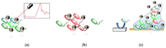
Regarding the antibacterial effect of the nanocoating, both of them presented stronger inhibitory effect on the S. aureus and P. aeruginosa biofilm formation compared to the non-ablated silicon sample. The authors attribute this antimicrobial effect to the generation of reactive oxygen species formed on the surface of Si nanoparticles during their production. Lately, an study in terms of productivity for nanosecond-laser plasma-mediated ablation regimes of these nanoparticles in water was performed[2].
Sometimes the laser ablation of the substrate does not lead to the formation of nanomaterials but to certain periodically aligned nanostructures that are called LIPSS (Laser-Induced Periodic Surface Structures). Controversy exists over the process by which these structures are formed. Some authors attribute them to a process of interference between the incident electromagnetic wave and that reflected and scattered by the material that gives rise to a pattern that affects only certain parts of the surface[3].
Others propose that it is the result of self-organization of the material surface during relaxation after application of the pulses[4]. Kudryashov et al. used picosecond IR-laser pulses to irradiate a Si wafer surface in liquid CS2 to produce Si ripples nanosheet arrays via nanoplasmonic ablative self-organization[5]. The antifouling capacity of the material was tested by culturing the nanosheet with Staphylococcus aureus bacteria for 18 h. “Live/Dead Biofilm Viability Kit” was used to study the viability of bacteria, showing the absence of the biofilm and the death of almost all the bacteria. Contrary, the appearance of a biofilm was observed for both controls: smooth Si wafer and silica glass slide.
In addition to the usefulness of pure silicon-based nanomaterials in preventing bacterial infection, some articles have reported that their combination with other materials or small molecules (such as antibiotics) that have bactericidal properties improves its action against microorganisms. For instance, it was observed that the coating of Si ripples nanosheets with Se, TeO2, Sb2O3, and Ag NPs nanoparticles, capable of damaging the bacterial DNA by the production of ROS, increased the antibacterial properties of the surfaces. Being the sample that combined Si nanoripples and TeO2 the most effective for biofilm prevention[6].
The addition of antibiotics to the silicon-based nanostructures is another strategy to increase the antibacterial effect. For most of the materials the ablation temperature is lower than the decomposition temperature, however, for some polymers, biomolecules, proteins, antibiotics, etc. this standard is not followed. These materials are very sensitive to temperature and degrade easily.
In order to achieve a fine coating of these organic materials, a modification called matrix-assisted pulsed laser evaporation (MAPLE) is used. In MAPLE, the sensitive compound is dissolved or dispersed in an inert solvent and then frozen to form a solid substrate. During laser ablation, the volatile solvent absorbs most of the laser energy whereas the intact molecule of interest acquires enough kinetic energy to be transported and form a uniform film on the desired surface. This variation was used by Mihaiescu et al. to create magnetite/salicylic acid/silica shell/antibiotics thin films[7]. The authors reported that despite the differences found for both types of bacteria (S. aureus and P. aeruginosa), the thin films exhibited an inhibitory effect for the biofilm formation.
3.Conclusions and Future Perspectives
This entry is adapted from the peer-reviewed paper 10.3390/nano10081443
References
- N A Smirnov; Sergey Kudryashov; A A Nastulyavichus; A A Rudenko; I N Saraeva; E R Tolordava; S A Gonchukov; Yu M Romanova; A A Ionin; D A Zayarny; et al. Antibacterial properties of silicon nanoparticles. Laser Physics Letters 2018, 15, 105602, 10.1088/1612-202x/aad853.
- Sergey Kudryashov; Alena A. Nastulyavichus; Anastasiya K. Ivanova; Nikita A. Smirnov; Roman A. Khmelnitskiy; Andrey A. Rudenko; Irina N. Saraeva; Etery R. Tolordava; Alexander Yu. Kharin; Irina N. Zavestovskaya; et al. High-throughput laser generation of Si-nanoparticle based surface coatings for antibacterial applications. Applied Surface Science 2019, 470, 825-831, 10.1016/j.apsusc.2018.11.201.
- Jörn Bonse; Sandra Hohm; Sabrina V. Kirner; Arkadi Rosenfeld; Jorg Kruger; Laser-Induced Periodic Surface Structures— A Scientific Evergreen. IEEE Journal of Selected Topics in Quantum Electronics 2016, 23, 1-1, 10.1109/jstqe.2016.2614183.
- J Reif; Florenta Costache; Matthias Henyk; Stanislav V. Pandelov; Ripples revisited: non-classical morphology at the bottom of femtosecond laser ablation craters in transparent dielectrics. Applied Surface Science 2002, 197, 891-895, 10.1016/s0169-4332(02)00450-6.
- Sergey Kudryashov; Luong V. Nguyen; Demid A. Kirilenko; Pavel N. Brunkov; Andrey A. Rudenko; Nikolay I. Busleev; Alexander L. Shakhmin; Alexander V. Semencha; Roman A. Khmelnitsky; Nikolay N. Melnik; et al. Large-Scale Laser Fabrication of Antifouling Silicon-Surface Nanosheet Arrays via Nanoplasmonic Ablative Self-Organization in Liquid CS2 Tracked by a Sulfur Dopant. ACS Applied Nano Materials 2018, 1, 2461-2468, 10.1021/acsanm.8b00392.
- Irina N Saraeva; Eteri R Tolordava; Alyona A Nastulyavichus; Anastasiya K Ivanova; Sergey I Kudryashov; Andrey A Rudenko; Nikolay N Melnik; Dmitriy A Zayarny; Andrey A Ionin; Yulia M Romanova; et al. A bacterial misericorde: laser-generated silicon nanorazors with embedded biotoxic nanoparticles combat the formation of durable biofilms. Laser Physics Letters 2020, 17, 025601, 10.1088/1612-202x/ab5fca.
- Dan Eduard Mihaiescu; R. Cristescu; G Dorcioman; C E Popescu; C Nita; G Socol; I N Mihailescu; Alexandra Elena Stoica; D Tamas; Monica Enculescu; et al. Functionalized magnetite silica thin films fabricated by MAPLE with antibiofilm properties. Biofabrication 2012, 5, 015007, 10.1088/1758-5082/5/1/015007.
- Martin Kogler; Yu. V. Ryabchikov; Sanna Uusitalo; Alexey Popov; Anton Popov; Gleb Tselikov; Anna-Liisa Välimaa; Ahmed Al-Kattan; Jussi Hiltunen; Riitta Laitinen; et al. Bare laser-synthesized Au-based nanoparticles as nondisturbing surface-enhanced Raman scattering probes for bacteria identification. Journal of Biophotonics 2018, 11, e201700225, 10.1002/jbio.201700225.